Review of Medical Physiology - William F. Ganong 2002
Blood Circulation
Cardiovascular Homeostasis in Health and Disease
Physical Exercise
Physical exercise is associated with significant Changes in the cardiovascular and respiratory systems. The adjustments of the Circulatory system are examined in this chapter, whereas the adaptation of the Respiratory system is discussed in Chapter 37. However, it should be noted that these systems develop simultaneously and in an integrated manner as parts of a unified homeostatic response that enables the body to withstand both moderate and strenuous physical exertion.
Resting BLOOD FLOW IN Skeletal Muscle is low (2-4 mL/100 g/min). Muscle contraction at 10% of maximum tension causes vascular compression; if contraction reaches 70%, blood flow ceases altogether (Fig. 33-2). However, a marked surge in blood flow occurs between contractions: during rhythmic muscle activity, blood flow per unit of time increases up to 30-fold. Occasionally, blood flow increases at the onset of or even prior to physical exercise. This initial elevation is neurally mediated, likely via a system of sympathetic vasodilators. Resting muscle blood flow doubles following sympathectomy, suggesting that basal vascular tone is reduced through the relaxation of vasoconstrictors. Yet once exercise is underway, local mechanisms maintain high blood flow at levels that are comparable between normal conditions and sympathectomized animals.
Class="center">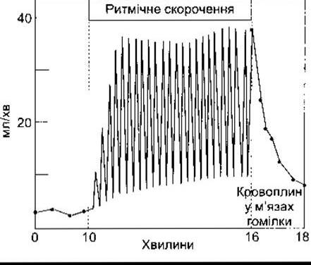
Fig. 33-2. Blood flow through the calf muscle during rhythmic contraction (reprinted with permission from Barcroft H, Swann HJC: Sympathetic Control of Human Blood Vessels. Arnold, 1953).
Local mechanisms maintaining high blood flow include a decrease in tissue PO2, an increase in tissue PCO2, the accumulation of K+ and other vasodilator metabolites (see Chapter 31). Muscle Temperature rises during activity, which further promotes vasodilation. The dilatation of arterioles and precapillary sphincters increases the number of open (functional) capillaries 10- to 100-fold. Consequently, the mean distance between the blood and active Cells—and thus the diffusion distance for O2 and metabolic byproducts—is significantly reduced. Vasodilation increases the cross-sectional area of the vascular bed, leading to a decrease in blood flow velocity. Capillary pressure rises until it matches the oncotic pressure along the entire length of the capillary bed. Furthermore, the rapid accumulation of osmotically active metabolites relative to their clearance diminishes the osmotic gradient across the Capillary Wall. As a result, fluid transudation into the interstitial space increases markedly. Lymphatic drainage is also substantially enhanced, which limits fluid accumulation in the interstitium and accelerates Lymph flow. A decrease in pH and an elevation in temperature shift the Hemoglobin dissociation curve to the right, facilitating greater oxygen unloading by the blood. The concentration of 2,3-DPG in erythrocytes increases, which decreases the affinity of hemoglobin for oxygen (see Chapters 27 and 35). The net result is a threefold increase in the arteriovenous oxygen difference and accelerated CO2 Transport from the tissue. The combination of all these changes enables up to a 100-fold increase in skeletal muscle oxygen consumption during exercise. Even greater short-term energy output is possible, where energy reserves are replenished via anaerobic glucose METABOLISM and the muscle incurs an oxygen debt (see Chapter 3). Overall, the alterations in Intermediary Metabolism during exercise are described in Chapter 17.
The presence of K+ dilates arterioles during muscle exercise, particularly at the onset of contraction. Muscle blood flow during exercise is less markedly increased in individuals with depleted K+ stores, a condition that tends to be associated with a propensity for severe muscle injury (exertional rhabdomyolysis).
Systemic Circulatory Responses
The generalized cardiovascular manifestations of exercise depend on whether the primary muscular contraction is isotonic or isometric. If the process begins with isometric muscle contraction, Heart rate accelerates. This acceleration occurs even when muscle shortening is prevented by the local administration of neuromuscular blockers. Similar changes are recorded simply upon the thought of the impending muscle contraction. In this case, The Heart rate changes result from central mental stimuli acting on the Medulla Oblongata. The deceleration of the rhythm is primarily associated with enhanced vagal tone, although Action Potential generation in cardiac sympathetic nerves also plays a role. Within a few seconds after the onset of isometric contraction, systolic and diastolic blood pressures rise sharply. Cardiac Output changes relatively little, and blood flow to the contracting Muscles decreases due to vascular compression.
The response to exercise initiated by isotonic muscle contraction resembles the previous one in terms of a rapid increase in heart rate, but it differs by a substantial augmentation in cardiac output. Consequently, There is a decrease in total peripheral resistance (Fig. 33-3) associated with vasodilation in the exercising muscles (Table 33-2). Systolic blood pressure rises moderately, whereas diastolic pressure usually remains unchanged or decreases. The difference in responses between isometric and isotonic muscle contraction can be explained in part by the fact that active muscles are tonically contracted during isometric exercise and thus contribute to the overall increase in peripheral resistance. In addition, there is a generalized increase in muscle sympathetic nerve activity, which is apparently triggered by signals originating from the contracting muscles. However, because cholinergic sympathetic vasodilation occurs in inactive skeletal muscles, the Functional Significance of this enhanced sympathetic activity remains unclear.
Cardiac output increases during isotonic exercise and can reach 35 L/min; this volume changes in proportion to enhanced O2 consumption. The underlying mechanisms are described in Chapter 29. The acceleration of heart rate is sustained by autonomic adjustments and the stimulatory effect of elevated PCO2 levels on the medulla oblongata. Maximum heart rate (HR) during exercise decreases with age. In children, HR increases to 200 beats per minute or more; in adults, it reaches about 195, and in elderly individuals, the increase is even smaller.
Fig. 33-3. Effects of varying levels of isotonic exercise on cardiovascular function (reprinted with permission from Berne RM, Levy MN: Cardiovascular Physiology, 5th ed. Mosby, 1986).
Table 32-2. Cardiac Output and Regional Blood Flow in a Sedentary Man1
Standing Resting State |
Exercise |
|
Cardiac output |
5900 |
24 000 |
Blood flow to: heart |
250 |
1000 |
750 |
750 |
|
active skeletal muscles |
650 |
20 850 |
inactive skeletal muscles |
650 |
300 |
500 |
500 |
|
3100 |
600 |
|
1 Values are given in mL/min at rest and during isotonic contraction at maximal oxygen uptake.
There is a marked increase in venous return, although this is not the primary cause of the increased cardiac output. Venous return is augmented by skeletal muscle contraction and the action of the thoracic pump (during inspiration, pleural pressure decreases while abdominal pressure increases, thereby raising central venous pressure), the mobilization of blood from visceral Organs, the transmission of elevated pressures through dilated arterioles to Veins, and noradrenaline-mediated vasoconstriction that reduces blood volume within the veins. The volume of blood mobilized from Internal Organs and other reservoirs during strenuous exercise can increase The amount of blood in the arterial tree by up to 30%.
Following exercise, blood pressure may gradually fall to subnormal levels, primarily due to the accumulation of metabolic byproducts that sustain muscle vasodilation for a short period. However, blood pressure returns to baseline levels more rapidly than heart rate normalizes.
Temperature Regulation
Quantitative indicators of temperature elevation during exercise are shown in Fig. 33-4. Blood reaches many areas of the skin from branches of muscular Arteries; consequently, a portion of the blood warmed in the muscles flows directly to the skin, where some of the heat is dissipated into the environment. There is a marked increase in pulmonary ventilation (see Chapter 37), resulting in additional heat loss via expired air. As body temperature rises, hypothalamic centers regulating heat-dissipating mechanisms are activated. This temperature increase is partly related to the inability of heat-dissipating mechanisms to fully cope with marked heat production. However, there is evidence for a thermostat-like effect, whereby heat-dissipating mechanisms are actively triggered in response to an elevation in core body temperature. Under such conditions, sweating is greatly enhanced, and its evaporation serves as the primary route of heat loss. Cutaneous vasodilation also occurs, driven mainly by the inhibition of vasoconstrictor tone, although the local release of vasodilators may also contribute (see Chapter 31).
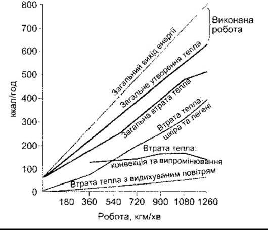
Fig. 33-4. Energy exchange during physical muscle exercise. The shaded area represents the excess of heat production over heat loss. Total energy output equals the sum of heat production and external work performed (reprinted with permission from Nielsen M: Die Regulation der Körpertemperatur bei Muskelarbeit. Scand Arch Physiol 1938;79:193).
Training
In trained athletes, both at rest and during exercise, there is a higher stroke volume and a lower heart rate compared to untrained individuals (see Chapter 29). Furthermore, heart dimensions are increased in trained subjects. Physical conditioning raises the level of maximal oxygen uptake (VO2max). Mean VO2max levels can reach 38 mL/kg/min in active healthy men and 29 mL/kg/min in active healthy women. These values are lower in older individuals. The level of VO2max is determined by maximal cardiac output and maximal tissue oxygen extraction, both of which increase as a result of training.
Structural and functional changes in skeletal muscles induced by training include an increase in the number of Cell/35.html">Mitochondria and the concentrations of Enzymes involved in oxidative metabolism. There is also an increase in capillary density and improved Blood supply to muscle fibers. The net effect is more complete oxygen extraction and, consequently, a less pronounced lactate accumulation during exercise. The increase in muscle blood flow will also be less marked; therefore, changes in heart rate and cardiac output will be less dramatic than in untrained individuals. This provides one of the explanations for THE BENEFICIAL ROLE of physical exercise in patients with cardiovascular disease.
Given the popularity of health-improving running and Other forms of physical exercise today, It is worth noting that one of the crucial conditions for successful regulated training is the psychological factor. Patients who exercise regularly feel better. Consistent physical activity increases the likelihood of maintaining an active lifestyle even in retirement. Furthermore, evidence suggests that systematic training, even with moderate loads, reduces the incidence and mitigates the severity of myocardial infarction.
Last update: 10/08/2026
Editorial and Educational Adaptation: This material has been compiled based on the primary/original source text. The project team performed an editorial review, corrected technical inaccuracies, structured sections, and adapted the content for an educational format.
What was processed:
- elimination of formatting defects (OCR errors, structural breaks, corrupted characters);
- editorial organization of content;
- standardization of terminology in accordance with academic sources;
- verification of factual statements against the original source text.
All mentions of the author, publication year, and origin of the primary text have been preserved in accordance with the source.