Review of Medical Physiology - William F. Ganong 2002
Blood Circulation
Cardiovascular Homeostasis in Health and Disease
Inflammation and Wound Healing
Local Tissue Injury
Inflammation is a complex local response to harmful agents such as Bacteria, or occasionally to compounds generated within the body. It involves a sequence of reactions featuring the initial activation of cytokines, neutrophils, adhesion molecules, Complement, and IgG. PAF (platelet-activating factor), a compound that amplifies the inflammatory response, also plays a role (see Chapter 27). Monocytes and lymphocytes join the process at a later stage. The affected area exhibits arteriolar dilation and increased Capillary Wall permeability (see Chapters 31 and 32). When inflammation develops in or beneath the Skin (Fig. 33-5), classic signs include redness, Swelling, tenderness, and pain. In other Organs, inflammation serves as a key pathogenetic factor in asthma, Ulcerative Colitis, and many other disorders.
Research data indicate that the METABOLISM/31.html">Transcription factor nuclear factor kB (NF- kB) plays a crucial role in the inflammatory response. NF- kB is a heterodimer normally sequestered in the Cell Cytoplasm in a complex with IkBa, which keeps it in an inactive state. Stimuli such as cytokines, Viruses, and oxidants cause NF-kB to dissociate from IkBa followed by degradation of the latter; NF- kB then translocates to The Nucleus, where it binds to the DNA of genes encoding numerous inflammatory Transmitters. Consequently, the synthesis and release of these transmitters are upregulated. Glucocorticoids inhibit the activation of NF-kB by reducing The formation of IkBa. This effect is likely the primary mechanism underlying the anti-inflammatory action of glucocorticoids (see Chapter 20).
Systemic Response to Injury
Cytokines produced during inflammation and Other types of injury also trigger systemic manifestations, which include changes in acute-phase reactant levels (Proteins whose concentrations increase or decrease by at least 25% following injury). A significant proportion of these proteins are synthesized in The Liver and are listed in Table 27-10. Their plasma levels are illustrated in Fig. 33-6. While the precise mechanisms governing the changes in concentration of these proteins are not yet fully understood, most of them appear to serve important homeostatic Functions. For example, an elevated level of C-reactive protein activates monocytes and induces the subsequent production of cytokines.
Class="center">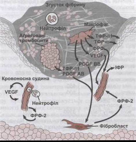
Fig. 33-5. Skin wound three days post-injury. Numerous cytokines and growth factors regulate the healing process; VEGF stands for vascular endothelial growth factor. For other Abbreviations, see the Appendix. Note the epithelium proliferating beneath the fibrin clot to progressively restore the skin (modified from Singer AJ, Clark RAF: Cutaneous wound healing. N Engl J Med 1999; 341: 738).
Other systemic responses to injury include somnolence, negative nitrogen balance, and fever.
Wound Healing
Following tissue injury, platelets adhere to the exposed Extracellular matrix via integrin-Collagen and integrin-Laminin interactions (see Fig. 33-5). The clotting cascade generates Thrombin, which enhances platelet aggregation and degranulation. Compounds contained within platelet granules further drive the inflammatory response. Leukocytes are recruited by selectins and bind to endothelial cell Integrins, promoting their migration across the vessel wall. Cytokines released by leukocytes stimulate integrins on macrophages, prompting them to migrate into the site of inflammation. Receptors on fibroblasts and epithelial Cells are also activated, participating in tissue repair and scar formation. Plasmin facilitates healing by clearing excess fibrin, which enables keratinocyte migration into the wound and re-epithelialization beneath the scab. Collagen synthesis and scar formation ensue. Wounds regain 20% of their ultimate tensile strength by three weeks. Although the remodeling process continues thereafter, the tensile strength of a healed wound never exceeds 70% of that of normal skin.

Fig. 33-6. Time course of changes in major acute-phase Plasma Proteins; C3 indicates complement component 3 (modified and reprinted with permission from Gitlin JD, Colten HR: Molecular biology of acute phase plasma proteins. In Pick E et al [editors]: Lymphokines, vol 14, pages 123-153. Academic Press, 1987).
Last update: 10/08/2026
Editorial and Educational Adaptation: This material has been compiled based on the primary/original source text. The project team performed an editorial review, corrected technical inaccuracies, structured sections, and adapted the content for an educational format.
What was processed:
- elimination of formatting defects (OCR errors, structural breaks, corrupted characters);
- editorial organization of content;
- standardization of terminology in accordance with academic sources;
- verification of factual statements against the original source text.
All mentions of the author, publication year, and origin of the primary text have been preserved in accordance with the source.