Review of Medical Physiology - William F. Ganong 2002
Circulation
Cardiovascular Homeostasis in Health and Disease
Shock
Shock is a syndrome that has always been surrounded by considerable controversy. Part of the difficulty stems from the inappropriate use of the term by physiologists, clinicians, and, above all, laypeople. For instance, electrical and spinal shock have very little in common with the state that arises during Hemorrhage and its associated cardiovascular disorders. In a narrow sense, hemodynamic shock is understood as a constellation of various conditions sharing certain common features. The most characteristic sign of all these states is inadequate tissue perfusion with a relative or absolute insufficient cardiac output. Cardiac output may be inadequate if the fluid volume within The Vascular System is insufficient to fill it adequately (hypovolemic shock). Furthermore, a relative insufficiency of cardiac output can occur due to an expansion of the vascular capacity resulting from vasodilation, even with a normal Blood volume (redistributive, vasogenic, or low-resistance shock). Shock can also be caused by inadequate Pumping Function of The Heart associated with myocardial damage (cardiogenic shock), as well as by insufficient cardiac output resulting from the obstruction of BLOOD FLOW IN the Lungs and heart (obstructive shock). These forms of shock and Examples of conditions that may cause them are listed in Table 33-3.
Hypovolemic Shock
Hypovolemic shock is also referred to as "cold shock." Its manifestations include hypotension, a rapid thready pulse, cold and pale clammy Skin, pronounced thirst, rapid breathing, fatigue, or conversely, lethargy. However, none of these signs is mandatory. Hypotension can be relative; for example, a patient with Hypertension and a usual blood pressure of 240/140 may experience severe shock at a pressure of 120/90.
Hypovolemic shock is often subdivided into several types according to its underlying cause. For example, The Use of terms such as hemorrhagic, traumatic, surgical, or burn shock is justified because, despite similarities in the general clinical picture, there are important features specific to each particular type.
Hemorrhagic Shock
Let us examine the processes of hemorrhage in greater detail, as they reflect features common to all types of hypovolemic shock along with the multiple compensatory reactions that develop to maintain circulating blood volume. The main reactions are summarized in Table 33-4.
The reduction in blood volume caused by bleeding impairs venous return and leads to a decrease in cardiac output. In moderate hemorrhage (5–15 mL/kg of body weight), pulse pressure drops, while mean arterial pressure may remain normal. Blood pressure changes are highly individualized even with identical blood loss. The skin is cold and pale, and may acquire a grayish tint due to capillary stasis and mild cyanosis. Respiration becomes rapid, and conscious patients often experience marked thirst.
In hypovolemic and Other forms of shock, inadequate tissue perfusion leads to increased anaerobic Glycolysis with The production of significant amounts of lactic acid. In severe cases, blood lactate levels rise to as high as 9 mmol/L (normal is 1 mmol/L). Consequently, lactic acidosis depresses the myocardium, reduces the sensitivity of peripheral vessels to catecholamines, and may cause coma.
Class="center">Table 33-3. Types of shock and examples of conditions and diseases that may cause them
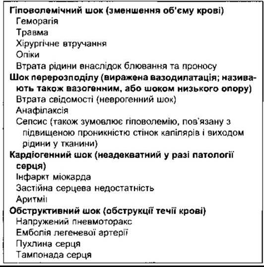
Table 33-4. Compensatory reactions activated during hemorrhage

Immediate Compensatory Reactions
With the reduction in blood volume and venous return, arterial baroreceptors undergo less stretch, leading to an enhanced sympathetic influence. Even if the mean arterial pressure does not drop, the decrease in pulse pressure results in a lowered frequency of Action Potential generation in the arterial baroreceptors, thereby triggering reflex tachycardia and vasoconstriction. Interestingly, as blood loss increases, tachycardia transitions into bradycardia. This can be observed as long as the shock remains reversible (see below). As bleeding continues, the heart rate increases again. Bradycardia is primarily associated with the unmasking of a depressor reflex mediated by the Vagus nerve. This reaction can serve as a mechanism to halt further blood loss.
Vasoconstriction is generalized, sparing only the cerebral and coronary vessels. The vasoconstrictor innervation of cerebral arterioles is probably functionally insignificant, while coronary vessels are dilated due to increased myocardial METABOLISM resulting from the elevated heart rate (see Chapter 32). Vasoconstriction is most pronounced in the skin—where it causes a feeling of coolness and pallor—as well as in the Kidneys and visceral Organs.
Hemorrhage also triggers widespread venoconstriction—a venous response that helps maintain cardiac filling pressure, although the receptors responsible for venoconstriction have not yet been fully identified. Pronounced vascular spasm in visceral organs leads to a redistribution of blood from visceral reservoirs into the systemic Circulation. Blood mobilization also occurs from subcutaneous areas and Pulmonary Veins. Splenic contraction releases "depleted" or stored blood into the circulation, although the volume of blood mobilized by this pathway in humans is insignificant.
In the kidneys, a spasm of both afferent and efferent arterioles is observed, though the efferent vessels constrict to a greater degree. The Glomerular Filtration rate is reduced, but renal plasma flow is decreased even more; consequently, the filtration fraction (glomerular filtration rate divided by renal plasma flow) increases. Blood may be shunted through the renal medulla bypassing the cortical glomeruli; very little urine is produced; and there is marked retention of Na+ along with an accumulation of nitrogenous Metabolic waste products in the blood (azotemia or uremia). In cases of prolonged hypotension, significant tubular damage (ACUTE RENAL FAILURE) may occur.
Blood loss acts as a potent stimulus for the secretory activity of the adrenal cortex (see Chapter 20). Circulating adrenaline levels also rise due to increased firing frequency in sympathetic noradrenergic Neurons. The elevated content of circulating catecholamines has relatively little effect on generalized vasoconstriction, but it can stimulate the reticular formation (see Chapter 11). This may explain why some patients with hemorrhagic shock are restless and anxious. Others are quiet and apathetic, with a clouded consciousness, likely resulting from cerebral ischemia and acidosis. Restlessness increases motor activity, and enhanced respiratory movements augment the muscular and thoracic pumping of venous blood.
The loss of erythrocytes diminishes the blood's O2-carrying capacity, and Blood flow through the carotid and aortic bodies is also reduced. Anemia and stagnant Hypoxia (see Chapter 37), along with acidosis, stimulate the chemoreceptors. Enhanced chemoreceptor activity is presumably the primary driver of respiratory stimulation during shock. Activation of chemoreceptors leads to the excitation of vasomotor centers in the Brainstem, thereby amplifying the vasoconstrictor effect. Indeed, in dogs subjected to hemorrhage resulting in arterial pressures down to 70 mmHg, sectioning of the nerves from carotid baroreceptors and chemoreceptors is more likely to cause a further drop in pressure than an increase. This paradoxical response is related to the fact that at pressures down to 70 mmHg, baroreceptor firing ceases, and the activity originating from the carotid chemoreceptors is transmitted to the vasomotor centers very weakly against the Background of diminishing baroreceptor inhibition.
Elevated circulating levels of angiotensin II, resulting from increased plasma renin during hemorrhage, induce thirst by acting on the subfornical organ (see Chapter 32); fluid intake helps restore circulating blood volume. Increased angiotensin II also helps maintain blood pressure. The drop in blood pressure caused by the loss of blood volume is more pronounced in animals administered angiotensin II receptor blockers compared to controls. Vasopressin also causes an increase in blood pressure when administered in large doses to normal animals; however, administration of doses that produce plasma vasopressin levels comparable to those seen in hemorrhage leads to only a slight rise in blood pressure because of a compensatory decrease in cardiac output (see Chapter 31). At the same time, administration of Peptides with opposing effects to vasopressin following hemorrhage results in a drop in blood pressure. Thus, vasopressin appears to play a significant role in maintaining blood pressure. The elevation of circulating angiotensin II and ACTH levels enhances aldosterone secretion, while increased amounts of aldosterone and vasopressin promote the retention of Na+ and Water, helping to restore blood volume. However, The Effect of aldosterone takes about 30 min to develop, and the initial reduction in urine output and Na+ excretion is primarily related to Hemodynamic changes in the kidneys.
During arteriolar spasm and decreased venous pressure resulting from reduced blood volume, a drop in capillary pressure is observed. Fluid shifts into the capillaries to sustain circulating blood volume. This leads to a decrease in interstitial fluid volume, which in turn causes fluid to move out of the Cells.
Long-Term Compensatory Reactions
Following moderate hemorrhage, the circulating plasma volume is restored within 12–72 hours (Fig. 33-7). There is also a rapid influx of previously synthesized albumin from extravascular stores, although the majority of the mobilized tissue fluid is protein-poor. This fluid dilutes Plasma Proteins and Blood Cells; however, in the event of whole-blood loss, the hematocrit may not decrease for several hours after the onset of bleeding. Following the initial influx of preformed albumin, the remaining lost plasma proteins are replenished through hepatic synthesis over the course of three to four days. Erythropoietin appears in the blood, and the reticulocyte count increases, reaching a peak by the tenth day. The erythrocyte count returns to normal within four to eight weeks. The low hematocrit is well compensated by The Influence of various physiological mechanisms. One of these is an increase in erythrocyte 2,3-DPG concentration, which enhances oxygen release from Hemoglobin to the Tissues (see Chapter 27). In chronic anemia in previously healthy individuals, exertional dyspnea does not appear until hemoglobin concentration falls to about 7.5 g/dL. Weakness becomes pronounced at 6 g/dL; resting dyspnea appears at 3 g/dL; and cardiac failure develops when the hemoglobin level drops to 2 g/dL.
Refractory Shock
Depending on the volume of blood loss, some patients die immediately following hemorrhage, while others recover thanks to compensatory mechanisms enhanced by appropriate Treatment, which leads to the gradual restoration of normal circulation. In an intermediate group of patients, shock persists for hours, progressively advancing to a state where the body fails to respond to vasopressor administration, and in which cardiac output remains diminished even if circulating blood volume is restored. This condition is known as refractory shock. It occurs not only in hemorrhagic shock but in Other types of shock as well. It is also referred to as irreversible shock, resulting in patient mortality despite intensive care. However, an increasing number of patients are successfully rescued as a better understanding of pathophysiological mechanisms improves therapeutic approaches. Therefore, refractory shock is arguably a more appropriate term.
Spasm of precapillary sphincters and venules, particularly in Internal Organs, is a hallmark of this stage. Impaired capillary perfusion resulting from precapillary sphincter constriction leads to hypoxic tissue injury. After 3-5 hours, the precapillary sphincters dilate, whereas the venules remain spastic. At this stage, blood re-enters the capillaries and stagnates within these vessels, meaning tissue hypoxia persists. Capillary hydrostatic pressure rises, and fluid escapes the vascular system in large quantities. Granulocytes adhere to the damaged vascular wall and release free radicals, particularly O2, causing subsequent tissue damage. Evidence indicates that Antibodies against Structure/178.html">Protein Complexes linking neutrophils to tissue significantly improve survival in severe shock. Gastrointestinal mucosal injury leads to bacterial translocation into the vascular bed.
Various positive feedback mechanisms play a critical role in The Development of refractory shock. For instance, severe cerebral ischemia ultimately leads to the depression of vasomotor and cardiac centers in the Brain, causing vasodilation and a reduced heart rate. Both processes contribute to a drop in blood pressure, followed by a further decrease in cerebral blood flow and additional depression of the vasomotor and cardiac centers.
Another important example of this type of positive feedback is myocardial depression. In cases of pronounced shock, coronary blood flow is impaired due to hypotension and tachycardia (see Chapter 32), even despite coronary vasodilation. Myocardial insufficiency worsens the course of shock and intensifies acidosis, which in turn inhibits myocardial function. If this functional impairment is significant and prolonged, myocardial injury may reach a point where cardiac output can no longer be restored to normal levels, regardless of volume replacement therapy.

Fig. 33-7. Changes in erythrocyte volume (dark), plasma volume (light), and total plasma protein following hemorrhage in a normal individual.
A late and potentially fatal complication of shock is lung injury culminating in acute respiratory distress syndrome (ARDS, adult respiratory distress syndrome; see Chapter 37). This syndrome is characterized by Acute Respiratory Failure and high mortality, and can be triggered not only by shock but also by Sepsis, pulmonary contusion, and other trauma or critical conditions. A common feature of this injury is damage to capillary endothelial cells and alveolar epithelial cells with the release of cytokines.
Other Forms of Hypovolemic Shock
Traumatic shock occurs As a result of extensive Muscle and bone injury. This type of shock is frequently observed in victims of physical assaults and automobile accidents. Significant Hemorrhage into the injury site is the primary cause of shock, although some plasma also extravasates into the tissue. The amount of blood that can be lost from a relatively minor injury is striking: up to 1 L of extravasated blood can accumulate in the thigh Muscles, for example, with an increase in thigh diameter of just 1 cm.
Skeletal Muscle destruction (rhabdomyolysis) presents an additional challenge when shock is accompanied by severe muscle crushing (Crush syndrome). Once tissue pressure drops and perfusion is restored, free radicals are generated, leading to further tissue damage (reperfusion injury). During ischemia, substantial amounts of free radicals are produced as tissue xanthine dehydrogenase is converted into xanthine oxidase, and upon the restoration of blood flow, this enzyme promotes the generation of O2. Leukocyte adhesion to tissue elements is observed, which also contributes to free radical formation. The xanthine oxidase inhibitor allopurinol and antibodies preventing neutrophil adhesion mitigate reperfusion injury. Another cause of tissue damage in reperfusion areas is excessive intracellular Ca2+ accumulation, primarily linked to the exchange of accumulated intracellular Na+ for extracellular Ca2+. In crush syndrome, renal impairment is common. This is associated with the accumulation of Myoglobin and other metabolic byproducts from reperfusion zones within the kidneys, where glomerular filtration is already compromised by shock. These metabolic products damage and obstruct the tubules, precipitating anuria, which can be fatal.
Surgical shock typically results from a combination of external hemorrhage, internal bleeding, and dehydration. In burn shock, the most prominent alteration is the loss of plasma as exudate from the burned areas. Because such situations predominantly involve the loss of plasma rather than whole blood, the hematocrit rises, and
hemoconcentration is observed. Burns also induce complex, poorly understood metabolic changes In addition to fluid loss. For example, a 50% increase in metabolic rate, independent of thyroid function, has been documented in some patients, while others develop hemolytic anemia. Given these complications, along with the severity of shock, infection risks, and renal damage, the mortality rate for third-degree burns (exceeding 75% of the body surface area) approaches 100%.
Hypovolemic shock is a complication of various metabolic and infectious diseases. For instance, although the underlying mechanisms vary in each case, adrenal insufficiency, diabetic ketoacidosis, and severe diarrhea are accompanied by the loss of Na+ from the blood. The resulting reduction in Blood Plasma volume can be the deciding factor in precipitating cardiovascular collapse.
Distributive Shock
As noted above, distributive shock occurs when blood volume is normal, but the capacity of the vascular system increases due to marked vasodilation. It is also referred to as warm shock because the skin is not cold and clammy as it is in hypovolemic shock. An example is anaphylactic shock—a severe, rapidly developing allergic reaction that occasionally occurs upon the repeated administration of an antigen to previously sensitized individuals. The resulting Antigen-Antibody Reaction is accompanied by the release of substantial amounts of histamine, causing increased capillary permeability and widespread dilation of arterioles and capillaries.
Another frequent form of distributive shock is septic shock. In this condition, Bacterial toxins trigger vasodilation. Furthermore, myocardial depression occurs and capillary permeability increases, causing plasma to leak into the tissues and reducing blood volume. Consequently, septic shock is simultaneously cardiogenic, hypovolemic, and distributive in nature.
The course of shock is more severe in febrile patients because cutaneous Blood Vessels are frequently dilated (see Chapter 32), which exacerbates the mismatch between vascular capacity and circulating blood volume.
Gram-negative Bacteria, which frequently cause septic shock, release endotoxin, a lipopolysaccharide component of the MICROBIAL Cell wall. This compound activates macrophages and stimulates the release of elevated levels of cytokines. Antibodies against cytokines or portions of the endotoxin molecule can be used to treat shock, though clinical outcomes have not been overly optimistic. Glucocorticoids are effective therapeutic agents in animals, but not in humans.
Another type of distributive shock is neurogenic shock, in which sudden autonomic dysfunction leads to vasodilation and blood pooling in the veins. An example is fainting triggered by intense emotional stress, such as fear or grief.
Fainting (Syncope)
Fainting, or syncope, is a sudden, transient loss of consciousness. It may be associated with metabolic or neurological disorders, but it more frequently arises from peripheral vascular or cardiac disturbances that cause inadequate cerebral blood flow. The clinical course is typically benign and linked to sudden vasodilation leading to hypotension, which is usually accompanied by bradycardia. The term vasovagal syncope is used to describe this condition.
Postural syncope refers to fainting caused by blood pooling in gravity-dependent PARTS OF THE body. Micturition syncope—fainting that occurs during urination—is observed in patients with orthostatic hypotension. It is associated with orthostatic and reflex bradycardia induced by voiding. Pressure on the carotid sinus, caused, for example, by a tight collar, can provoke marked bradycardia and vasodilation, resulting in fainting (carotid sinus syncope). Rarely, vasodilation and bradycardia may be associated with swallowing (swallowing syncope). Cough syncope occurs when increased intrathoracic pressure during straining or coughing reaches a level sufficient to obstruct venous return to the heart. Exertional syncope denotes fainting during exertion due to the inability to increase cardiac output to meet heightened metabolic demands, and is typically characteristic of patients with aortic or pulmonary stenosis.
Syncope can be associated with more serious pathologies. Approximately 25% of cardiac syncope episodes are caused either by temporary obstruction of blood flow through the heart or by a sudden reduction in cardiac output resulting from various Cardiac Arrhythmias. Fainting associated with bradycardia, heart block, or sinus node dysfunction is termed neurocardiogenic syncope. Additionally, fainting is the presenting symptom in 7% of patients with myocardial infarction. Therefore, all episodes of syncope warrant a thorough investigation to determine the underlying cause.
Cardiogenic and Obstructive Shock
When the pumping function of the heart is impaired to such an extent that tissue perfusion is inadequate for normal metabolic needs, a condition known as cardiogenic shock ensues. It typically occurs in The Setting of a massive left ventricular infarction, but it can also be triggered by other disorders that compromise ventricular function. Symptoms characteristic of shock appear, along with pulmonary and visceral congestion, because the heart is unable to pump out all the venous blood returning to it. For this reason, the condition is sometimes referred to as congestive shock. The incidence of such shock in patients with myocardial infarction is approximately 10%, with a mortality rate ranging from 60% to 90%.
A clinical picture similar to stagnant shock is also observed in obstructive shock. If the obstruction is caused by a tension pneumothorax with compression of the large veins (see Chapter 37) or by hemorrhage into the Pericardium resulting in external cardiac compression (cardiac tamponade), immediate surgical intervention is essential to prevent fatal outcomes.
Treatment of Shock
The management of shock aims to eliminate the underlying cause and stimulate physiological compensatory mechanisms to restore adequate tissue perfusion. In hemorrhagic, traumatic, and surgical shock, for example, the primary cause is blood loss, and treatment involves early and rapid transfusion of adequate amounts of compatible whole blood. Physiological saline can be used in a limited capacity—only as a temporary measure. The paramount objective is to restore an adequate circulating blood volume; because saline distributes into the ECF, only 25% of the administered fluid remains within the vascular system. In burn shock and other conditions accompanied by blood hemoconcentration, plasma is the treatment of choice to correct the primary defect—plasma loss. Plasma protein fraction preparations, high-molecular-weight carbohydrate solutions, and related substances that do not cross the Capillary Wall can provide some therapeutic benefit. Concentrated human serum albumin and other hypertonic solutions increase circulating blood volume by drawing interstitial fluid into the vasculature. Although they can be used for emergency resuscitation, their drawback is that they may exacerbate tissue dehydration.
In anaphylactic shock, adrenaline is a highly effective and somewhat specific agent whose action is more complex than simply constricting dilated blood vessels. Across all types of shock, maintaining an adequate blood pressure plays a crucial role in supporting Coronary Circulation. To this end, vasoconstrictors such as noradrenaline can be used, though its administration should be discontinued as soon as possible, as it induces renal vasodilation and positive inotropic effects while simultaneously causing vasoconstriction in other vascular beds (see Chapter 20). In traumatic and cardiogenic shock, dopamine exhibits beneficial therapeutic effects.
Certain interventions employed during shock may suppress physiological compensatory mechanisms. Sedatives and other Central Nervous system depressants must be used with caution, as they inhibit The activity of the vasomotor center. Alcohol is particularly dangerous because it suppresses the central nervous system and causes cutaneous vasodilation (via a central mechanism). Overheating, which leads to cutaneous vasodilation, must be avoided. Changes in patient posture, such as sitting or standing up, can have hazardous circulatory consequences. Gravity should be utilized to assist rather than hinder compensatory mechanisms. Elevating the FOOT of the bed by 15–30 cm to place the patient in a modified position with slightly raised legs is a simple yet vital therapeutic measure designed to increase venous return from the lower body to The Heart and improve cerebral circulation. However, a steep HEAD-down (Trendelenburg) position causes the abdominal viscera to press against the Diaphragm, impairing respiration and predisposing the patient to pulmonary complications. Therefore, this method should not be maintained for extended periods.
Last update: 10/08/2026
Editorial and Educational Adaptation: This material has been compiled based on the primary/original source text. The project team performed an editorial review, corrected technical inaccuracies, structured sections, and adapted the content for an educational format.
What was processed:
- elimination of formatting defects (OCR errors, structural breaks, corrupted characters);
- editorial organization of content;
- standardization of terminology in accordance with academic sources;
- verification of factual statements against the original source text.
All mentions of the author, publication year, and origin of the primary text have been preserved in accordance with the source.