Chemistry and Biology of Proteins - F. Haurowitz 1953
The Role of Proteins in Immune Responses
Antigen-Antibody Reaction
At The Heart of all immunological reactions lies the same primary process: the Combination of an antigen with an antibody. Precipitation, agglutination, cytolysis, and other more complex reactions are secondary expressions of this process. The simplest of all antigen-antibody reactions is the precipitation of a soluble antigen by its corresponding antibody. By using Antigens tagged with colored groups, isotopes, or easily traceable chemical elements, one can quantitatively analyze the precipitate and determine its composition under various conditions. Such investigations have been conducted by numerous authors using labeled antigens such as Hemoglobin, iodinated Proteins, Phosphoproteins, azoproteins, and hemocyanin.
The earliest analyses of this kind were performed by By [93]. Studies employing more refined Methods have demonstrated that the antibody-to-antigen ratio in the precipitate increases as this ratio increases in the antigen-antibody mixture prior to precipitation [46, 94–96]. The antibody-to-antigen ratio in the precipitate also depends on the particle size of the antigen used [97]. If the precipitating antigen is a Cell, the layer of antibody molecules bound to its surface is very small relative to the volume of The Cell. A different picture emerges when low-molecular-weight substances are used as antigens; in some precipitates, only 2–3% of the sediment consists of the antigen, while the bulk of the precipitate is made up of Antibodies.
Knowing the molecular weights of the antigen and antibody, one can calculate the molecular antibody-to-antigen ratio within the precipitate.
Class="center">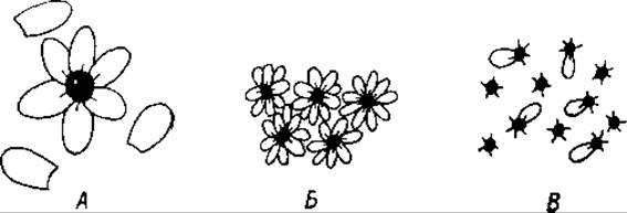
Fig. 50. Combination of antigen molecules (black) with antibody molecules (white).
A — formation of an antigen-antibody complex between a multivalent antigen and a univalent antibody (with the antibody in excess); B — formation of precipitates via nonspecific aggregation of antigen-antibody complexes; C — soluble complex in the presence of antigen excess.
This ratio is sometimes extremely high. For instance, a thyroglobulin–antithyroglobulin precipitate contains 60 antibody molecules per single thyroglobulin molecule [98]. Arsanilazoglobulin is capable of binding approximately 50 antibody molecules for each antigen molecule [95]. It is conceivable that, in the simplest case, an antigen-antibody complex consists of a single antigen molecule acting as a core at the center of the complex, surrounded by numerous attached antibody molecules (Fig. 50). When these resulting complexes reach a certain size, they become unstable and form insoluble aggregates, much like edestin and euglobulin molecules do. It should also be noted that The formation of these aggregates is inhibited by the same agents that suppress globulin aggregation—namely, neutral salts, acids, and bases [95]. Ninhydrin and certain other nonspecific Reagents promote precipitation [99].
Precipitates composed of antigen-antibody complexes generally dissolve in an excess of antigen. This is attributed to the formation of numerous small complexes containing only a single antibody molecule per antigen molecule [81] (see Fig. 50). Precipitates formed by horse antibodies dissolve in an excess of these antibodies. In contrast, precipitates formed by rabbit antibodies do not redissolve in an excess of the corresponding rabbit antiserum. The results of these studies proved particularly unexpected when foreign serum globulin was used for immunization. In such cases, both the antigen and the antibody are serum globulins sharing very similar physicochemical properties. This marked difference between the action of horse antibodies and rabbit antibodies indicates a fundamental distinction between these Two Types of antibodies. Some authors attribute the inability of rabbit antibodies to dissolve the precipitate to their univalency. It may be assumed that univalent rabbit antibodies are incapable of binding to more than one antigen molecule and therefore cannot form a lattice composed of alternating antigen-antibody units. According to these authors, the formation of such a lattice is characteristic of multivalent antibodies. The difference between the lattice formed by multivalent antibodies and the aggregates of univalent antibodies is illustrated in Fig. 50. It essentially consists in the fact that in the aggregate formed by univalent antibodies, the antigen-antibody complexes are linked together via nonspecific bonds [100], whereas lattice formation occurs exclusively through specific bonds. The distinction between these two types becomes blurred when dealing with precipitates such as thyroglobulin–antithyroglobulin, where approximately 60 antibody molecules correspond to each thyroglobulin molecule. Obviously, in such cases, only a very few of the bonds forming the spatial network can be specific. Since antigen-antibody precipitates formed by horse antibodies dissolve in an antigen excess [101, 102], it is reasonable to assume that horse antibodies are multivalent. However, it must be noted that The properties of horse antibodies can depend heavily on the method of immunization [101].
As can be seen from the above, the question of antibody valency remains definitively unresolved. Regarding the valency of the antigen, however, there is no doubt that antigen molecules are multivalent—that is, they possess multiple groups with which an antibody can combine. This is evidenced by the fact that low-molecular-weight haptens, whose molecules contain only a single determining group, are unable to form precipitates with antibodies directed against those groups. Nevertheless, precipitates are formed by certain divalent or trivalent haptens [103]. A hapten bearing both R and X groups is precipitated only by a mixture of anti-R and anti-X, but by neither of these antibodies individually (where R denotes $n$-azophenylazophenylarsonic acid and X denotes $n$-azophenylazobenzoic acid) [104]. The interpretation of these experiments presents major difficulties because some haptens can form aggregates consisting of A large number of molecules, thereby acquiring multiple determining groups [105, 106].
The formation of an insoluble precipitate depends not only on the number of reactive groups in the antigen and antibody molecules, but also on the spatial arrangement of these groups, their polarity, and their hydrophilic or hydrophobic properties [107]. Clearly, antibodies can combine only with those antigen groups that are located On the surface of the antigen molecule, rather than with internal end groups [95].
Attempts have been made to elucidate The Nature of the antigen-antibody reaction by applying the law of mass action [108]. The primary difficulty encountered in these attempts is that the antigen-antibody reaction is only partially reversible. Some precipitates do not redissolve in an antigen excess, even though such an excess inhibits the precipitation process itself [109]. Denoting the antigen molecule by G and the antibody molecule by B, the antigen-antibody reaction can be represented as follows [83]:
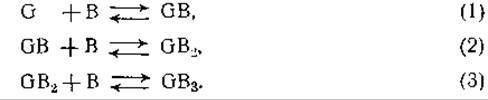
In subsequent phases, Reactions of the type $2GB_3 \rightarrow G_2B_6$ may occur, or ring structures may form through the end-to-end joining of chains [110]. If antigen-antibody aggregates connect with one another via nonspecific bonds, bonds of the B–B type arise. In certain cases, the existence of such nonspecific bonds has been confirmed by the formation of mixed agglutinates consisting of several different antigen-antibody pairs. Such coagglutination, however, is not a general phenomenon [110]. The attachment of virus particles to The surface of antibody molecules has been successfully observed using an Electron microscope [111].
The interaction between antigen and antibody is mediated by electrostatic forces arising between polar groups. This issue was already discussed in Chapter X, where it was emphasized that these forces act only over very short distances, as their intensity drops off proportionally to $r^6$—that is, inversely to the sixth power of the distance between the two poles [112]. Although the force exerted by each individual polar group is very weak, the total interaction force can reach a significant magnitude if a large number of polar groups interact simultaneously. This is possible only when the polar groups are situated on two large molecules whose surface configurations are geometrically complementary to each other. The distance between the antigen and antibody surfaces is approximately 4 Å; consequently, a change of even 0.1 Å significantly affects the mutual attractive force. This explains why the subtle difference existing between the $o$-, $m$-, and $p$-isomers of the same compound is of paramount importance for the mutual attraction of antigen and antibody molecules [113].
The Free energy change accompanying the binding of a hapten to an antibody is approximately 1,510 cal/mol [114]. Direct calorimetric Determination of the heat released As a result of the reaction between hemocyanin and antihemocyanin yielded a value on the order of 3 cal per gram of nitrogen, or 40,000 cal per mole of bound antibody [115].
Precipitation is inhibited by high pressure [116]. This indicates that the binding of antigen to antibody is accompanied by the release of Water. Both the antigen and antibody molecules apparently undergo partial dehydration at the precise site where they make direct contact with one another. As a result, the two monomolecular water layers that originally surrounded each of the reacting components are replaced by a single shared layer common to both components. The free energy change for the reaction $GB_n \rightarrow GB_{n-1} + B$ can be calculated from the Equilibrium Constant $K$ using the van 't Hoff equation:
$\Delta F = -RT \ln K$.
For the reaction $GB_8 \rightarrow GB_7 + B$, the value of $\Delta F$ was found to be 9,800 cal per mole of antibody (where G is sheep serum pseudoglobulin complexed with aniline-$m$-sulfonic acid, and B is the corresponding antibody) [117].
A number of studies have focused on INVESTIGATING THE REACTION between antibody and antigen within monomolecular layers. If egg albumin is adsorbed onto the surface of a chromium plate and this plate is subsequently immersed in an antibody solution, a monomolecular layer of antibody forms on the antigen surface [118]. However, in the reverse process—that is, upon adsorbing the antibody onto the metal and immersing the plate in an antigen solution—no deposition of antigen occurs on the antibody layer surface [118]. Using this technique, one can prepare films featuring alternating layers of toxin and antitoxin [119]. Reports [120] stating that antigen and antibody combine via forces acting through plastic films over distances exceeding 100 Å appear to be erroneous. This error is most likely attributable to the diffusion of reacting particles through cracks or pores in these films [121, 122].
Since both antigens and antibodies are proteins, the reaction taking place between these two compounds is inhibited by all factors that affect protein molecules. Consequently, erroneous results are obtained if the reaction is not carried out at a neutral pH [92], if merthiolate1 or similar compounds are used as antiseptics [123], or in cases where the antigen is masked by other colloids. For instance, the precipitation of virus by antiviruses becomes impossible after heating the virus with serum albumin [124]; however, if the serum albumin layer protecting the virus particles is subsequently digested with Pepsin, the virus regains its ability to be precipitated by the corresponding antiserum.
1 An organic mercury compound with antiseptic properties. — Ed. note
Last update: 06/08/2026
Editorial and Educational Adaptation: This material has been compiled based on the primary/original source text. The project team performed an editorial review, corrected technical inaccuracies, structured sections, and adapted the content for an educational format.
What was processed:
- elimination of formatting defects (OCR errors, structural breaks, corrupted characters);
- editorial organization of content;
- standardization of terminology in accordance with academic sources;
- verification of factual statements against the original source text.
All mentions of the author, publication year, and origin of the primary text have been preserved in accordance with the source.