Medical Genetics - V. M. Zaporozhan 2005
Prevention of Hereditary Diseases
Prenatal Diagnosis of Hereditary Diseases and Congenital Malformations
Invasive Methods
Invasive Diagnostics involves obtaining embryonic, fetal, and provisional organ Cells and Tissues during Pregnancy for subsequent analysis using cytogenetic, molecular cytogenetic, biochemical, cytochemical, and other Methods. It is performed when There is a high risk of giving birth to children with monogenic and Chromosomal Disorders, with the aim of diagnosing (confirming) a hereditary condition in the fetus.
Indications for Invasive Prenatal Diagnostics
1. Maternal age — under 18 and over 35 years. These age groups carry a high risk of having a child with Down syndrome and other chromosomal abnormalities. The Procedure is performed to diagnose chromosomal
pathology in the fetus. However, today it is possible to initially screen for chromosomal abnormalities in the first trimester of pregnancy using non-invasive methods (at 10–14 weeks), and then, in cases of high individual maternal risk, use invasive methods to confirm the Diagnosis.
2. A family history of a child (or fetus) with a chromosomal disorder or multiple Congenital Malformations. If the parental karyotypes are normal, the same approach as in item 1 can be applied.
3. Presence of chromosomal pathology or chromosomal rearrangements in the parents.
4. Biochemical and ultrasound screening results suggesting a chromosomal disorder in the fetus.
5. High risk of having a child with a monogenic disorder based on MEDICAL Genetic Counseling or screening programs, or the detection of heterozygous carrier status, provided the disease Gene has been mapped and molecular genetic testing is available.
6. Clarification of the diagnosis of congenital malformations (for example, amniocentesis followed by determination of AFP concentration in the Amniotic Fluid is performed to clarify the diagnosis of a neural tube defect).
7. Diagnosis of fetal infections and maternal-fetal immunological incompatibility.
8. Use of cytostatic pharmacological agents by the woman or her partner, or exposure of either spouse to radiation shortly before pregnancy, which carries an increased risk of chromosomal pathology.
Contraindications: 1) genital tract infections; 2) threatened Miscarriage; 3) acute infectious diseases; 4) large uterine myoma.
Since the procedure of invasive prenatal diagnosis itself carries risks and, in the event of a positive test result, implies Termination of the pregnancy, it should be performed only after the couple has been informed about the risk of complications and has consented to a potential early termination of pregnancy.
The primary method of targeted invasive prenatal diagnosis in the first trimester of pregnancy is chorionic villus sampling (chorionic villus biopsy). In the second trimester, amniocentesis, placentocentesis, and cordocentesis are performed.
Chorionic villus sampling involves obtaining chorionic tissue. The chorion is the villous membrane that forms from the trophoblast during pregnancy; therefore, chorionic cells share the same genotype as embryonic cells. It is recommended to perform this procedure at 10–14 weeks of gestation. Although it can be done earlier, early chorionic villus sampling (before 9 weeks) has been associated with an increased incidence of limb reduction defects. Chorionic tissue is obtained transabdominally through the anterior abdominal wall or transcervically under ultrasound guidance (Fig. 11.4). Using a special needle, 15–20 mg of material is aspirated. The tissue is used for cytogenetic diagnosis or DNA extraction for molecular genetic testing. Complications (risk of pregnancy loss) are 2.5–3%.
Table 11.6. Changes in serum markers (triple test) in fetal Down and Edwards syndromes
|
Chromosomal |
Alpha-fetoprotein |
Unconjugated estriol |
Human chorionic gonadotropin |
|
syndromes |
(AFP) |
(UE) |
(hCG) |
|
Down syndrome |
Decreased |
Decreased |
Increased |
|
Edwards syndrome |
Decreased |
Decreased |
Decreased |
Class="center">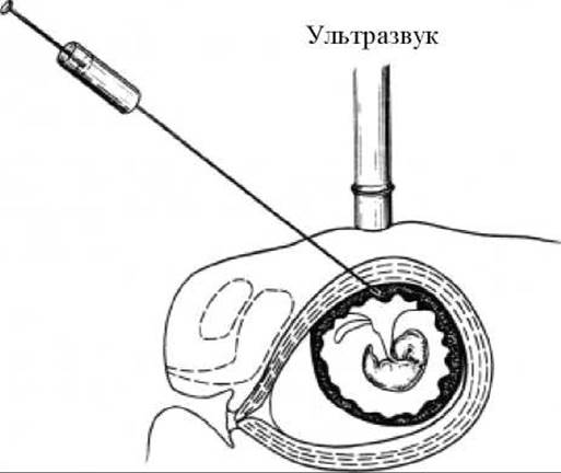
Fig. 11.4. Transabdominal chorionic villus or placental biopsy
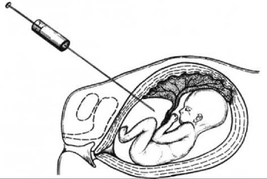
Fig. 11.5. Amniocentesis

Fig. 11.6. Cordocentesis
Placentocentesis involves obtaining placental tissue and is performed from the 14th week of gestation onward (see Fig. 11.4).
Fifteen to twenty milligrams of placental tissue are aspirated. This tissue is used for the same purposes as chorionic tissue.
Amniocentesis involves obtaining amniotic fluid containing desquamated fetal and amniotic cells. Early amniocentesis is performed at the 13th–14th week, while late amniocentesis is typically done at the 16th–20th week (preferably at the 16th week). Under ultrasound guidance, 10–20 mL of amniotic fluid is collected through the anterior abdominal wall (Fig. 11.5). The fluid can be analyzed for AFP levels as well as The activity of certain Enzymes. The recovered cells are suitable for cytogenetic or DNA diagnostics. Complication rates range from 0.5% to 1%.
Cordocentesis is the sampling of fetal Blood from the umbilical cord under ultrasound guidance via the anterior abdominal wall (Fig. 11.6). Approximately 1–1.5 mL of blood is obtained. It is performed starting from the 20th week of gestation. Complications do not exceed 2%. The blood sample is analyzed using cytogenetic, molecular genetic, and Biochemical Methods. Cordocentesis is used to diagnose inherited hematological disorders (hemoglobinopathies, coagulopathies, thrombocytopenias), immunodeficiencies, intrauterine infections, maternal-fetal immunological incompatibility, and for intrauterine Gene Therapy.
Fetoscopy allows direct visualization of the fetus using endoscopic equipment. It is performed between the 18th and 23rd weeks of gestation, with complication rates of 7–8%. Following the widespread adoption of ultrasound, this method is rarely used today, as virtually all structural anomalies detectable by endoscopy are reliably diagnosed via ultrasound scanning.
Fetal tissue biopsy involves obtaining a sample of fetal Skin or Muscle under ultrasound guidance. It is performed in the second trimester of pregnancy to diagnose skin disorders (such as ichthyosis or epidermolysis) and Duchenne muscular dystrophy. The collected material is examined using cytological, cytochemical, and immunofluorescence techniques.
Challenges in Invasive Prenatal Diagnosis
Invasive prenatal diagnosis can be complicated by difficulties in Cell culturing. In some cases, it is impossible to obtain a sufficient number of cells for subsequent analysis or to establish a cell culture. The probability of such complications generally does not exceed 1%.
Difficulties related to result interpretation may also arise. Chorionic villus sampling and placentocentesis can sometimes reveal mosaicism. While this may indicate a mosaic form of a chromosomal disorder in the fetus, in some instances, mosaicism stems from other factors. It can result from maternal cell contamination or be confined to the placental membranes (placental mosaicism occurs more frequently than in embryonic tissues and may have no clinical consequences for the developing Organism). Counseling couples in these situations is exceptionally challenging.
In certain cases, predicting the phenotypic consequences of chromosomal or genomic Mutations is ambiguous. This typically occurs in the following situations:
1. Sex chromosome aneuploidy in the fetus may not necessarily be accompanied by intellectual disability. Females with trisomy X can present with a practically normal phenotype (although clinically pronounced forms of the condition are also possible).
2. A balanced chromosomal aberration (such as a translocation or inversion) may be diagnosed in the fetus. If one of the parents carries the exact same mutation and has a normal phenotype, it is highly probable that the child will also have a normal phenotype. However, if the mutation is de novo, Gene Expression can occasionally be disrupted due to a 'position effect,' leading to a chromosomal disorder in the child. Consequently, result interpretation becomes difficult.
3. Marker Chromosomes in the fetus also present significant evaluation challenges. A marker chromosome is an extra chromosome (specifically, a centromere-containing fragment of a chromosome). If the marker chromosome consists solely of heterochromatin, the phenotype remains unaffected. Conversely, if it contains euchromatin (expressed genes), it is associated with The Development of a chromosomal disorder. If a parent with a normal phenotype carries the same marker chromosome, a normal phenotype in the child can be anticipated.
Last update: 11/08/2026
Editorial and Educational Adaptation: This material has been compiled based on the primary/original source text. The project team performed an editorial review, corrected technical inaccuracies, structured sections, and adapted the content for an educational format.
What was processed:
- elimination of formatting defects (OCR errors, structural breaks, corrupted characters);
- editorial organization of content;
- standardization of terminology in accordance with academic sources;
- verification of factual statements against the original source text.
All mentions of the author, publication year, and origin of the primary text have been preserved in accordance with the source.