HUMAN MEDICAL BIOLOGY, ANATOMY, PHYSIOLOGY AND PATHOLOGY - Y.I. Fedoniuk 2010
ANATOMY, PHYSIOLOGY, PATHOLOGY
CHAPTER 7. NUTRITION PROCESS AND ITS PATHOLOGY
ANATOMY AND PHYSIOLOGY OF THE DIGESTIVE ORGANS
ABDOMINAL CAVITY AND PERITONEAL CAVITY
The Abdominal cavity is bounded superiorly by the Diaphragm, extends inferiorly into the pelvic cavity, its lateral and posterior walls are formed by the lumbar spine and Muscles, and its anterior wall is formed by the broad Abdominal muscles and their aponeuroses. The abdominal cavity contains such Organs as The Stomach, small and large intestines, Liver, Pancreas, Spleen, as well as the Organs of the Urogenital System (Fig. 7.12, 7.13). The inner surface of the abdominal cavity is lined with a serous membrane called the Peritoneum, which consists of two layers: the parietal layer (which lines the cavity walls) and the visceral layer (which covers the Internal Organs to varying degrees). If the visceral layer covers an organ on all sides, it is referred to as an intraperitoneal position (stomach); if an organ is covered by the peritoneum on three sides, it is in a mesoperitoneal position (ascending and descending colons); and finally, if an organ is covered by the peritoneum on only one side, it lies extraperitoneally (pancreas). Between the visceral and parietal layers lies a slit-like potential space filled with serous fluid (1-2 mL), which constitutes the peritoneal cavity. The smooth, moistened peritoneum facilitates the movement of organs relative to each other by reducing friction between the layers.
Class="center">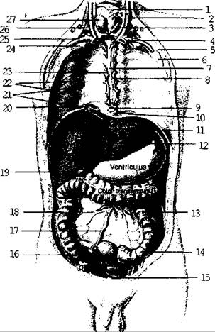
Fig. 7.12. Thoracic and abdominal cavities

Fig. 7.13. Thoracic and abdominal cavities
Furthermore, as the abdominal organs develop between the peritoneum and the posterior abdominal wall, they migrate away from the wall, pulling the serous membrane along with them to form serous folds that suspend the organs. These folds consist of two layers of peritoneum and are called mesenteries.
Passing from one organ to another or from the wall to an organ, the peritoneum forms ligaments and omenta. The greater and lesser omenta are distinguished. The greater omentum hangs down from the greater curvature of the stomach like an apron and consists of four layers of peritoneum with adipose tissue deposited between them. The lesser omentum is represented by two ligaments: the hepatogastric and hepatoduodenal ligaments. They consist of two layers of peritoneum. Important anatomical structures pass between the layers of the hepatoduodenal ligament: the hepatic artery, the common Bile duct, and the portal vein. Posterior to the lesser omentum and the stomach lies the omental bursa (lesser sac), which is a subdivision of the peritoneal cavity. It is bounded by various organs and ligaments.
Last update: 08/08/2026
Editorial and Educational Adaptation: This material has been compiled based on the primary/original source text. The project team performed an editorial review, corrected technical inaccuracies, structured sections, and adapted the content for an educational format.
What was processed:
- elimination of formatting defects (OCR errors, structural breaks, corrupted characters);
- editorial organization of content;
- standardization of terminology in accordance with academic sources;
- verification of factual statements against the original source text.
All mentions of the author, publication year, and origin of the primary text have been preserved in accordance with the source.