Human Anatomy - H. I. Koliadenko 2009
Sense Organs (Analyzers)
General Integument
The Skin (cutis) covers the outer surface of the body (Fig. 179) and consists of the epidermis—the superficial layer—and a deeper layer known as the dermis, or proper skin. Beneath the skin lies the subcutaneous layer (hypodermis). The total surface area of an adult's skin is 1.5–2.0 sq. m.
The epidermis is composed of stratified squamous epithelium, the surface Cells of which undergo keratinization, gradually desquamate, and are replaced by new ones. This stratum corneum (horny layer) varies in thickness across different PARTS OF THE body. It is thickest on the palms of the hands and the soles of the feet, and thinnest on the eyelids. The Deep Layer of the epidermis is called the stratum lucidum or germinative layer. Its cells accumulate a special substance called eleidin, which hardens, causing the Cell Nucleus to disappear and transforming The Cell into a scale that eventually dies off and sheds. The Cells of the deep (basal) layer of the epidermis contain a pigment (melanin) that imparts color to the Tissues. In certain areas of the body (around the nipple of the mammary gland, on the external genitalia, the Scrotum, the perianal region, etc.), melanin is located not only in the epidermal cells but also in the Connective Tissue, which is why these areas have a darker pigmentation.
Class="center">
Fig. 179. Structure OF THE Skin:
a — epidermis; b — dermis (proper skin); c — subcutaneous layer;
1, 5 — Hair; 2 — dermal papillae; 3 — sweat gland duct; 4, 9, 13 — tactile corpuscle; 6 — stratum corneum of the epidermis; 7 — stratum lucidum of the epidermis; 8 — hair ROOT; 10 — sebaceous gland; 11 — arrector pili Muscle; 12 — hair follicle; 14 — hair bulb; 15 — expanded part of the follicle; 16 — sweat gland; 17 — vein; 18 — artery; 19 — nerve
The dermis, or proper skin, consists of two layers: the papillary layer and the reticular layer. The papillary layer is composed of loose connective tissue. The papillae form the ridges and furrows of the skin. Housed within the papillae are Blood Vessels, Lymphatic vessels, and sensitive nerve endings—receptors that perceive pain, Touch, and Temperature stimuli. The papillae reach their greatest height in areas of heightened sensitivity, such as the palmar surface of the fingertips, which is of great importance for fine motor tasks. Projecting above The surface of the epidermis, the papillae form a papillary pattern (Fig. 180). Every individual possesses a unique papillary pattern inherent to them alone. This characteristic is widely used in forensic medical practice.
The reticular layer, which is the most developed part of the dermis, is formed of Cytology/practical/45.html">Dense connective tissue. The mechanical Properties of the skin—such as strength and elasticity—depend on this layer. The strength of the dermis is due to the arrangement of Collagen fibers, which run primarily parallel and obliquely to the skin surface, forming a dense network. The dermis transitions smoothly into the subcutaneous layer.
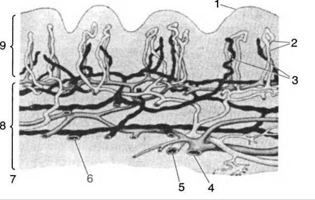
Fig. 180. Blood and Lymphatic vessels and capillaries of the skin:
1 — epidermis; 2 — blood capillaries; 3 — lymphatic capillaries; 4 — vein; 5 — artery; 6 — lymphatic vessel; 7 — subcutaneous layer (adipose tissue); 8 — deep (basal) layer of the skin; 9 — papillary layer of the skin
There is no sharp boundary between the skin and the subcutaneous layer. The subcutaneous layer consists predominantly of loose connective tissue, within the meshes of which lie accumulations of fat cells varying in shape and mass. In some areas, fat is entirely absent from the subcutaneous connective tissue, such as in the eyelids, the auricle of the ear, and the scrotum. The largest amounts of fat are deposited in the abdomen, Mammary Glands, and pelvic regions. The subcutaneous Adipose tissue is better developed in women than in men. It protects Internal Organs from trauma and hypothermia, and also serves as a nutrient reserve that can be utilized by the Organism when necessary.
Skin glands. The skin contains A large number of glands: Sweat Glands, Sebaceous Glands, and mammary glands.
Sweat glands are long, straight tubes terminating in a secretory coil. The opening of the duct leads directly onto the skin surface. Located in the deep layers of the skin, their total number reaches up to 2 million. They are most abundant on the palms and soles (up to 1,000 per 1 sq. cm). In children, sweat glands are not yet fully developed. Sweat glands secrete sweat, which is composed of Water, nitrogenous Metabolic waste products, and mineral salts. The evaporation of water helps regulate body heat dissipation. During muscular exertion, sweat also contains lactic acid, a product of muscle METABOLISM. Sweating is regulated by the Central Nervous system, which contains specialized sweat centers.
Sebaceous (alveolar) glands are located within the dermis. The majority of them are concentrated around hair roots inside the hair follicles. The sebum secreted by these glands lubricates the hair and the entire surface of human skin. Sebaceous glands are absent on the palms and soles.
Modified sebaceous glands include the meibomian glands found in the eyelids, as well as the ceruminous Glands of the external auditory canal, which produce earwax.
Mammary glands are paired organs, phylogenetically related to sweat glands and functionally linked to the reproductive organs. The mammary glands in women have a complex structure. Each gland consists of 15–20 radially arranged lobes surrounded by adipose tissue. The ducts of the glands open into lactiferous sinuses located around the nipple. The nipple is composed of Smooth Muscle tissue, with an opening at its center through which milk is secreted during Lactation, i.e., during infant feeding.
Skin derivatives are keratinized appendages: Nails and hair. All of them share an epidermal origin.
Hair covers the entire body, with the exception of the red part of the Lips, palms, soles, and certain parts of the external genitalia. Hair is most densely concentrated on the scalp, eyebrows, axillae, pubic region, and in men, on the face (mustache, beard) and frequently on the chest and legs. The hair root is located within the dermis; its expanded base is called the hair bulb, which rests inside the hair follicle. The Free part of the hair projecting above the skin surface is called the shaft. Hair grows at an oblique angle to the skin surface. Under a Microscope, the microscopic structure of a hair can be observed: it consists of a central medulla (pith), surrounded by a somewhat denser cortex, and covered externally by a hard protective layer, or cuticle. An arrector pili muscle is attached to the hair follicle, and a sebaceous gland opens into it. Upon the simultaneous contraction of many of these Muscles, the hairs are raised, producing what is commonly known as "goosebumps" in humans. This phenomenon occurs during hypothermia, intoxications, or severe pain. Muscle contraction also causes blood vessels to constrict, reducing blood flow and heat loss. In a living person, hair grows continuously, though its lifespan is relatively short. For instance, scalp hair is replaced every 2–4 years, and eyelashes every 4–5 months. Hair grows at a rate of about 1 cm per month. Hair color varies widely—from pale yellow to black. This coloration depends on the presence of the pigment melanin in the cortical layer, and its intensity depends on The amount of pigment. When pigment is lost, hair turns gray. Graying is associated with the infiltration of air between the cells of the hair's cortical layer.
Nails are located on the dorsal surfaces of the terminal Phalanges of the fingers and toes. They are thin, keratinized plates of the epidermis. The largest part of the nail—its body—terminates in a free edge known as the distal edge, while its proximal thin end, the root, is embedded in a skin fold known as the nail bed. At the junction where the nail body transitions into the root lies a crescent-shaped, whitish germinative zone called the lunula. Nails grow continuously, reaching up to 4 mm per month in growth. The cornified layer of the nail contains no blood vessels or nerves. Nails appear pink due to the underlying blood vessels situated beneath the nail plate, which show through it. During vascular stenosis and venous congestion, nails acquire a bluish tint. The nail is surrounded by a low skin fold known as the nail fold (cuticle/perionychium).
Skin Receptors. The organism interacts with the external environment through receptors. Receptors are divided into two main types: external receptors, or exteroceptors, which are localized on the body surface—in the skin, the mucous membranes of the Mouth AND Nose, and in the organs of Vision, Hearing, etc.; and internal receptors, or interoceptors, which lie within the walls and linings of internal organs. Both external and internal receptors serve as the initial link of the central nervous system, which, upon receiving a signal, responds to it accordingly.
Exteroceptors are located On the surface of the body within the skin and are divided into two groups. The first group includes contact receptors: pain, temperature, touch, pressure, warmth, and cold receptors. Stimuli make direct contact with these receptors. The second group comprises distance receptors: the organs of vision, hearing, and Olfaction, through which stimuli originating at a distance from the body are perceived.
Interoceptors are also divided into two groups: proprioceptors and visceroceptors. Proprioceptors include receptors of muscles, tendons, fasciae, and joint capsules, as well as receptors of the vestibulocochlear apparatus. Visceroceptors are located within internal organs. Skin receptors are nerve endings that form dense networks beneath the epidermis. It is believed that unmyelinated nerve endings function as pain receptors. Touch receptors include tactile menisci (Meissner's corpuscles), which are found in the dermal papillae in the form of spiral fibers. Hair follicles also serve as tactile receptors. Pressure is perceived by lamellar corpuscles (Pacinian corpuscles), which consist of outer and inner capsules enclosing terminal nerve branches. Cold is perceived by Krause end bulbs, which feature a capsule containing a basket-like nerve plexus. Warmth is perceived by Ruffini corpuscles, which resemble spindle-shaped plexuses lying beneath the epidermis.
Muscle receptors (Fig. 181) are linked to numerous reflex reactions that ensure the maintenance of a specific body posture in space. Muscle receptors include Pacinian corpuscles, bare nerve endings, Golgi tendon organs, and muscle spindles. Pacinian corpuscles are found in tendons, joint capsules, fasciae, and the periosteum. Golgi tendon organs (bulbous corpuscles) consist of a Lymph-filled capsule entered by tendon fibers surrounded by bare nerve fibers. When a tendon is stretched, the receptor is excited. Muscle spindles are striated fibers 1–4 mm in length, surrounded by a lymph-filled capsule containing between 3 and 13 intrafusal fibers. Muscle spindles respond to both muscle stretch and contraction because they possess dual innervation: efferent and afferent.
The density of receptors varies across different Regions of the skin. For example, there is an average of one receptor per 10 sq. mm of skin on the shin, about 30 in the HEAD region, whereas on the fingertips, their number reaches 230.

Fig. 181. Muscle receptors:
a — free endings of an afferent fiber enveloping the end of a muscle fiber; b — muscle spindle; 1, 4 — endings of an efferent fiber; 2 — ending of an afferent fiber; 3 — capsule; 5 — intrafusal muscle fibers; 6, 8 — efferent fiber; 7 — afferent fiber
External stimuli, mediated through the Cerebral Cortex, govern Human and Animal behavior and also influence the Functions of internal organs.
Human skin performs A number of vital functions, including body thermoregulation, protection against harmful environmental factors, excretion of metabolic waste, and blood pooling. The skin must be protected from overheating and excessive sun exposure, which can cause persistent vascular changes. Unclean skin leads to blocked sweat pores and sebaceous gland ducts, potentially triggering hair follicle inflammation. The skin is extremely sensitive to chemicals—a fact that must be kept in mind by those working in relevant industries. Frequent skin irritation by chemical agents can become a cause of skin Cancer. Various toxic substances can be absorbed through the skin, such as nicotinic acid, aniline Dyes, chromium compounds, mercury preparations, and others, which may lead to systemic poisoning of the body.
Burns. Damage to body tissues resulting from thermal factors, chemicals, electric current, or ionizing radiation is referred to as burns. Depending on the depth of tissue damage, burns are classified into four degrees.
First-degree burns are characterized by skin redness (erythema), mild Swelling, and a slight rise in local temperature. This condition lasts for 2–5 days and heals without a trace.
Second-degree burns also cause skin redness and swelling, along with The formation of blisters filled with a yellowish fluid. These burns are accompanied by pain and an elevated body temperature.
Third-degree A burns are accompanied by partial skin necrosis, third-degree B burns involve necrosis of all skin layers, and fourth-degree burns involve Necrosis of the skin and underlying deep tissues.
Emergency care involves the immediate removal and neutralization of the harmful agent. First of all, the affected skin and mucous membranes should be rinsed with plenty of cold running water for at least 15 minutes. In the case of skin burns caused by sulfuric acid or quicklime, washing the affected area with water is strictly prohibited, as it will only intensify their effect; instead, vegetable oil or animal fat should be used.
Patients with severe injuries are hospitalized.
Frostbite is a localized tissue injury caused by exposure to low temperatures. There are four degrees of frostbite.
First-degree frostbite is characterized by pallor of the skin in the affected area. Upon rewarming, the skin becomes edematous and cyanotic, accompanied by a burning pain.
Second-degree frostbite involves the formation of blisters containing clear, yellowish fluid.
Third-degree frostbite is characterized by the death of deep skin layers. The blisters are filled with a blood-tinged exudate.
Fourth-degree frostbite results in necrosis of all layers of soft tissue and sometimes even the bones.
For mild second-degree frostbite, gentle massage of the affected areas is required; if blisters are present, an aseptic cotton-gauze bandage should be applied. The victim should be given hot, strong tea or coffee and wrapped in warm blankets. The skin should never be rubbed with snow, as this causes further trauma to the tissues. In cases of third- and fourth-degree frostbite, the victim must be hospitalized urgently.
Last update: 08/08/2026
Editorial and Educational Adaptation: This material has been compiled based on the primary/original source text. The project team performed an editorial review, corrected technical inaccuracies, structured sections, and adapted the content for an educational format.
What was processed:
- elimination of formatting defects (OCR errors, structural breaks, corrupted characters);
- editorial organization of content;
- standardization of terminology in accordance with academic sources;
- verification of factual statements against the original source text.
All mentions of the author, publication year, and origin of the primary text have been preserved in accordance with the source.