Military Surgery with Emergency Surgery - V.Ya. Bilyi 2004
Thermal Injury
Introduction. Thermal injury (TI) is tissue Damage caused by thermal agents or exposure to low temperatures. However, thermal injuries encompass not only thermal Burns, but also chemical burns, Electrical injuries, and localized radiation damage (radiation burns).
Burn injury. Burn trauma is one of the most severe types of injuries in both peacetime and wartime. It occurs more frequently in peacetime, accounting for 5 to 12% of domestic injuries. Proper and timely medical care significantly improves Treatment outcomes for burn patients.
In cases of extensive and deep burns, the clinical course of burn trauma evolves into a systemic disease affecting the entire body and involving vital Organs.
Etiology of burns. Based on the etiological factor, burns are classified into thermal, electrothermal, chemical, radiation, combined injuries, and burns caused by military incendiary mixtures.
Thermal factors include flames, steam, hot liquids, molten metal, and thermal radiation.
Causes of electrothermal burns include electric arcs and contact electrical burns; chemical burns are caused by acids and alkalis; radiation burns result from ionizing radiation; and incendiary mixture burns are caused by metal-based substances (thermite, electron), white phosphorus, viscous incendiary mixtures (Napalm), metallized viscous incendiary mixtures (Pyrogel), and incendiary liquids. Combined injuries involve thermomechanical, radiation-thermal, and radiation-thermomechanical factors.
The severity of thermal impact depends on The Nature of the thermal agent, its Temperature, exposure time, and the duration of tissue hyperthermia.
The most severe burns are caused by flames (in fires, bonfires, clothing ignition, natural gas explosions, and other incidents).
Scald burns from boiling Water are most commonly superficial. Hot steam burns can be extensive, but are rarely deep. Hot sticky substances, such as tar, molten bitumen, and asphalt, exert a more prolonged effect on the Skin.
Burns can also occur through contact with hot objects (contact burns) and molten metal, as well as from electric current and electric arcs.
The damaging effect of an electric current passing through Tissues manifests as thermal, electrochemical, and mechanical phenomena. Due to tissue resistance, electrical energy is converted into heat, leading to cellular overheating and death. Skin damage at the entry and exit points of the current varies in shape and size depending on the nature of contact with the conductors, ranging from punctate marks to complete charring of an entire limb. Notably, the extent of skin necrosis is usually smaller than that of the deeper underlying tissues. Electrical burns may be combined with thermal injuries from the electric arc generated at the moment of electrocution. The combined thermal and mechanical action of high-voltage current sometimes causes tissue delamination and traumatic amputation of body parts.
Chemical burns are caused by aggressive substances capable of inducing tissue necrosis within a short period. Strong inorganic acids, alkalis, and heavy metal salts possess such properties.
The action of acids results in protein coagulation. The dissolution of certain acids in tissue fluid releases heat and causes tissue overheating, leading to Cell death. The damaging effect of aggressive liquids begins upon contact with tissues and continues until the chemical reaction is complete. Afterward, organic and Inorganic Compounds remain in the burn wound, exerting an adverse effect on tissue regeneration processes.
Based on the depth of pathological changes and treatment requirements, burns are divided into two groups. The first group comprises superficial burns (degrees I, II, and IIIA). They undergo spontaneous epithelialization during conservative treatment thanks to the preserved papillary layer or skin epithelial appendages. Degree IIIB to IV burns constitute the second group—deep burns that require surgical restoration of the skin via autodermoplasty (Fig. 21).
Class="center">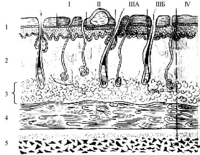
Fig. 21. Classification of burns by depth of injury (Roman numerals indicate burn degrees): 1 - epidermis, 2 - dermis, 3 - subcutaneous adipose tissue, 4 - fascia, 5 - Muscles.
Pathomorphological changes in first- and second-degree burns present as aseptic inflammation, leading to dilation and increased permeability of dermal capillaries.
First-degree burns manifest as erythema and edema of the skin (sustained arterial hyperemia and inflammatory exudation) accompanied by localized hyperthermia. The edema resolves quickly, and the process concludes with epithelial desquamation.
Second-degree burns are characterized by The formation of blisters of various sizes resulting from the accumulation of fluid in the epidermis. These blisters appear immediately after the burn, within a few hours, or by the next day. They are typically filled with clear, yellowish fluid. The content of large blisters gradually thickens and becomes gelatinous due to fibrin precipitation and the reabsorption of water. The basal layer of the epidermis remains exposed beneath the detached epidermal layers.
When complicated by secondary infection, the blister fluid becomes purulent. Edema and hyperemia of the burn wound increase. In second-degree burns, barring purulent complications, complete re-epithelialization of the skin and recovery occur within 14 days.
Degree IIIA burns (dermal burns) involve partial-thickness skin damage that does not extend through the entire dermis. Often, the damage is limited to the germinal layer of the epidermis only at the tips of the papillae. In other cases, Necrosis of the epithelium and superficial dermis occurs while the deeper layers and Skin Appendages (Hair follicles, sebaceous and sweat gland ducts) remain preserved.
Local changes in degree IIIA burns vary. Depending on the causative agent, a superficial, light brown or whitish-gray moist eschar forms. Because exudation and necrosis coexist, the aforementioned blisters appear in some areas. On the 14th day, eschar Separation begins, concluding within 2 to 3 weeks. In degree IIIA burns, island and marginal epithelialization of the wound surface occurs via remnants of the germinal epidermal layer and skin appendages. Island epithelialization is a crucial clinical sign for assessing the depth of tissue necrosis and diagnosing a degree IIIA burn.
Degree IIIB burns involve partial or total destruction of the subcutaneous fat layer accompanied by full-thickness skin necrosis and the formation of a necrotic eschar (see insert, Fig. 22).
Fourth-degree burns typically occur with prolonged thermal exposure in areas lacking a significant subcutaneous fat layer. Muscles and tendons are usually affected first, followed by bones, large and small joints, major nerve trunks, and Cartilage.
Diagnosis of burn depth and area. It is worth noting that the depth of skin damage in burns can only be estimated approximately During the first days following trauma. Full-thickness skin injuries are characterized by pale coloration or charring, tissue compaction, and a loss of pain and tactile sensitivity. Patients typically present with a combination of burns of varying degrees.
Based on clinical presentations, history taking, examination of the burn wound, and various diagnostic tests, both the depth and total area of the injury can be determined. It is essential to question the victim or accompanying persons about the circumstances of the trauma: the Nature of the thermal agent, its temperature, duration of exposure, as well as the timing and nature of the first aid provided.
A thorough examination of the burn wound is of utmost importance. The symptoms used to determine the depth of the injury are appropriately divided into three groups:
1) external signs of tissue necrosis;
2) signs of circulatory impairment;
3) level of pain and temperature sensitivity.
Cytology/practical/136.html">Differential diagnosis OF grade IIIA and IIIB burns is more complex. A reliable sign of a deep burn is the presence of thrombosed Veins beneath a necrotic eschar ranging in color from yellowish-brown to dark brown, with a parchment-like thickness.
The diagnosis is most often clarified after the rejection of necrotic tissues and the spontaneous epithelialization of the wound, which begins from islands of preserved epithelium 20-30 days following exposure to the thermal agent.
The simplest and most accessible method for objectifying burn depth is assessing pain sensitivity, which is tested by multiple needle pricks (Billroth's test) on various areas of the burn or by touching it with a cotton pellet soaked in alcohol. In areas of deep injury, Pain Sensation is absent.
Areas of second-degree burns are always acutely painful. The examination should be conducted so that the patient cannot see what the physician is doing, starting in areas where deep necrosis is suspected and gradually moving toward superficial burns and healthy skin. Testing pain sensitivity on intact skin shows how accurately the patient perceives their sensations. The state of sensitivity can also be determined using the epilation test by pulling out individual hairs in the burned area with tweezers. If the patient feels pain and the hair offers some resistance when pulled, the injury is superficial. In deep burns, the hair is removed easily and painlessly.
Temperature sensitivity is tested using two test tubes. One is filled with water at room temperature (20-25 °C), and the second with water at 50-55 °C. The tubes are applied alternately to the burn surface. If the patient perceives the temperature difference, the burn is superficial; if not, the burn is deep.
Determination of Burn Area. An important factor in assessing the severity of a thermal injury is determining the burn area.
The simplest and most convenient method for use during the Initial Stages of evacuation is the "rule of nines" or the "rule of palms" METHOD FOR DETERMINING burn area.
THE PRINCIPLE OF determining the burn area using the "rule of nines" is based on dividing the entire body surface into regions whose areas are multiples of 9% of the body surface. Thus, the HEAD surface equals 9%, the anterior trunk surface 18%, the posterior trunk surface 18%, the thigh surface 9%, the lower leg and FOOT 9%, and the Perineum 1% (Fig. 23).
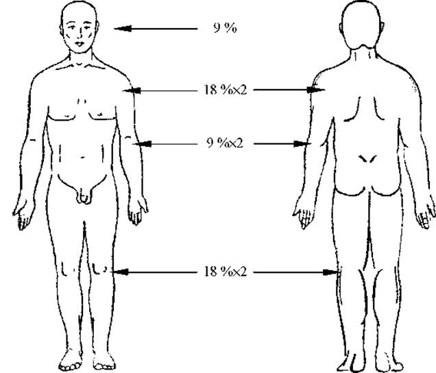
Fig. 23. Determining burn area using the "rule of nines".
In extensive burns, it is simpler to determine the area of uninjured skin and then subtract that figure from 100.
The area of the victim's palm is approximately 1-1.2% of their body surface. Taking this into account, the burn area can be determined with sufficient accuracy.
B.M. Postnikov proposed determining the surface area of burns by tracing the BOUNDARIES OF THE burn surface onto washed X-ray film, transferring the film onto graph paper, and calculating the burn area in square centimeters.
G.D. Vilyavin proposed calculating burn area by sketching the burns onto body silhouettes printed on graph paper, where the total number of squares (17,000) corresponds to the number of square centimeters of an adult's skin surface with a height of 170 cm.
V.O. Dolinin recommended a more modern approach: plotting the burn contours onto a human body silhouette divided into 100 segments, each representing 1% of the body surface. The degree of the burn is indicated by appropriate hatching.
Prognosis of Burn Injury Severity. To predict the severity of a burn and its outcomes, calculations are used that are based on determining the total affected area and establishing the area of deep burns with relative precision. The simplest prognostic method for determining burn severity is the hundred rule.
The prognostic index According to the hundred rule (age and total burn area) has the following values:
up to 60 — favorable prognosis;
61-80 — relatively favorable prognosis;
81-100 — questionable;
101 and above — unfavorable.
The Frank index is used to determine the severity and potential consequences of a burn. To calculate it, one must know the area of deep partial-thickness and full-thickness burns. For calculation purposes, 1% of a superficial burn corresponds to 1 unit, and a deep burn corresponds to 3 units. The Frank index is obtained by adding the area of the superficial burn to the calculated area of the deep burn (Table 7).
Table 7 Express Diagnostics of Burn Shock
|
No. item |
Degree of shock |
Frank index (units) |
|
|
Without inhalation injury |
With inhalation injury |
||
|
1 |
Mild |
30-70 |
20-55 |
|
2 |
Severe shock |
71-130 |
65-100 |
|
3 |
Extremely severe |
More than 130 |
More than 100 |
For example, the total burn area is 55% of the body surface, with deep burns accounting for 30% and superficial burns accounting for 25%. The sum of the superficial burn indicator (25 units) and the deep burn indicator multiplied by three (90 units) yields a Frank index of 115 units.
To account for The impact of inhalation injury (IHI), 15 units are added to the Frank index for mild IHI, 30 units for moderate IHI, and 45 units for severe IHI.
The prognosis of burn disease depending on the Frank index is established as follows:
up to 30 units — favorable prognosis;
31-60 units — questionable prognosis;
61-90 units — unfavorable prognosis.
Inhalation injury (IHI). IHI is typically caused by the combined multifactorial impact of flame, hot air, hot particles, and toxic chemical compounds—products of combustion. Mortality among casualties with inhalation injuries remains high, ranging from 22.3% to 82%.
As a rule, IHI occurs in victims who at the time of injury were in a confined space: a vehicle, a residential or office room, basements, cellars, tanks, ship compartments, an airplane cockpit, etc. (see insert, Fig. 24).
IHI can also occur in open spaces if a person is briefly exposed to an atmosphere with a high concentration of smoke, for example, in napalm strike zones or during large fires.
The Pathogenesis of disorders developing in the respiratory organs upon their burn is complex and involves A number of factors. Thus, victims experience rapid breathing that takes on the character of dyspnea. These changes can progress to severe respiratory failure, caused by mechanical airway obstruction, bronchospasm, and alterations in the pulmonary tissue.
Hemoconcentration, metabolic acidosis, and increased Blood viscosity combined with sluggish blood flow promote the activation of intravascular coagulation. Diffuse intravascular coagulation in the lung tissue causes severe circulatory and Metabolic Disorders. These disorders impair external Respiration Functions, worsening blood oxygenation and carbon dioxide elimination.
In burns of the face, neck, and chest, the possibility of IHI must always be kept in mind. Its presence is confirmed by charred nasal hairs, burns of the Lips, Tongue, Hard and Soft palate, and the posterior pharyngeal wall. The diagnosis is supported by a hoarse voice, cough, dyspnea, cyanosis, sputum production, and sloughing of the affected mucous membrane. Mucosal edema and membrane shedding can lead to mechanical asphyxia.
Electrical injuries. According to B.S. Vikhriev, Electrical Injury victims account for 1–2.5% of all trauma cases in peacetime, with approximately 10% resulting in fatalities.
In combat conditions, electrical injuries may occur when overcoming electrified barriers or operating autonomous electric power equipment units.
A specific type of electrical injury is lightning strike.
Contact electrical injury should be differentiated from electric arc injury. The latter is of a thermal nature: As a result of a flash caused by a short circuit, ionization of the air occurs between the current source and The surface of The Human Body.
Contact action of electric current causes both systemic changes and local tissue damage.
Systemic disorders depend on the path of the current through the body—the "current loop." Life-threatening current loops are those passing from one hand to the other, from the head to the hands or feet, or from the hands to the feet. In these cases, vital organs such as the Brain and Heart lie within the current pathway.
Clinically, this manifests as Central Nervous system and cardiovascular disorders, as well as paralysis of the Respiratory Muscles. This can lead to the victim's death at the time of the injury, or hours or days later.
The local effect of electric current in tissues manifests as thermal, mechanical, and electrochemical effects.
At the points of current entry and exit on the skin, various lesions are observed, ranging from punctate electrical marks to carbonization of an entire limb.
It should be noted that electrical burns are virtually always deep. A characteristic feature is the combination of moist and dry necrosis. Furthermore, secondary necrosis may develop due to compromised Blood supply to the tissues.
At the accident scene, the victim must be immediately disconnected from the electrical circuit while the rescuer takes strict safety precautions. For this purpose, an insulated object or tool may be used (such as a shovel, an axe, or a dry tree branch). Prior to this, the rescuer must insulate themselves from the ground using a rubber mat, a cardboard box, or a dry board.
Since the victim is frequently unable to open their hands due to Muscle spasms, they should be pulled away by the hem of their dry clothing, after first wrapping the rescuer's hands in insulating fabric.
Combined radiation-thermal injuries (CRTIs). Combined injuries occur when a person is exposed to two or more damaging factors simultaneously. Their clinical course is uniquely characterized by The phenomenon of mutual aggravation.
The impact of radiation injury on the course of burns and burn disease manifests in the following ways:
- reparative and regenerative processes in the burn wound are significantly slowed down;
- the incidence of infectious complications increases, with anaerobic infection and Sepsis being particularly common;
- burn disease develops even with a smaller total burn surface area;
- the mortality rate increases.
At the same time, the presence of burns aggravates the clinical course of radiation sickness:
- the latent period of radiation sickness is shortened;
- the severity of the radiation injury may increase by one degree (for example, grade II radiation injuries combined with burns correspond in severity and clinical outcome to grade III radiation injuries);
- the threshold for The Development of radiation sickness in the presence of extensive and deep burns decreases to 0.5-0.75 Gy (instead of 1 Gy in isolated injuries).
These manifestations of the mutual aggravation phenomenon in combined thermal and radiation injuries occur due to the impairment of protective and adaptive mechanisms caused by the suppression of immunological and nonspecific host defense factors, pancytopenia, and hemorrhagic syndrome (reduced blood clotting, increased capillary fragility and permeability).
Along with the aggravated course of radiation sickness, combined thermal and radiation injuries (CTRI) are characterized by local complications in the healing process of the burn wound.
During the latent period of radiation sickness, no significant deviations from the normal course of the burn wound are observed, since the Main Components of the wound repair process—inflammation and regeneration—are not yet suppressed at this stage.
At the peak of radiation sickness, the inflammatory response in the burn area is suppressed, the rejection of necrotic tissues, the formation of granulation tissue, and epithelialization are delayed. Hemorrhages appear beneath the burn escar, along with suppuration of the burn wound and surrounding tissues. In severe cases, regressive tissue changes within the wound and necrosis of newly formed granulations occur.
At the height of radiation sickness, the most serious complications are sepsis and the so-called necrotizing leukopenic pneumonias, which are characterized by an extremely severe clinical course.
During the recovery period of radiation sickness, reparative and regenerative processes begin to gradually resume. The burn wound is cleared of necrotic tissues, healthy granulation tissue develops, and the processes of epidermal and Connective Tissue regeneration are activated.
It is now established that preventive measures come to the forefront in the management of CTRI. The latent period of radiation sickness must be utilized for effective Surgical treatment, which includes both local interventions on the burn wound (necrotomy, necrectomy, autodermoplasty) and comprehensive treatment for radiation sickness.
Features of combined thermal and mechanical injuries (CTMI). Due to industrial and transportation disasters, accidents, and local wars, the issue of organizing care and treatment for casualties with CTMIs is acquiring critical importance.
According to I.G. Leshchenko and M.M. Zyryanov, in peacetime, up to 35% of all hospitalized patients with combined thermal injuries present with moderate severity injuries. Given that patients with mild CTMIs are mostly treated on an outpatient basis, while those with severe and extremely severe injuries require prolonged treatment, the socio-economic significance of treating and rapidly restoring the working capacity of patients with moderate CTMIs becomes evident. In wartime, this category of casualties can serve as one of the primary reserves for army replenishment.
Characteristics of napalm-induced burns. Today, incendiary agents, including napalm, are in service with the armies of many countries.
Napalm is a viscous mixture consisting of a liquid fuel and a thickening agent. The fuel is typically thickened (gelatinized) gasoline. The thickener most frequently consists of organic acid salts, making up 8 to 12% of the napalm composition.
The combustion temperature of napalm reaches 800 to 1200 °С. Its burning releases a large amount of carbon monoxide, which can cause severe poisoning in the victims. Upon ignition, napalm spatters, adheres to skin and clothing, and burns for 4-7 minutes. Napalm is lighter than water, allowing it to float on the water surface while continuing to burn.
Napalm burns are localized predominantly on exposed areas of the body, with the face affected in 75% of cases. These burns are distinguished by their depth of injury. A multifactorial nature of the trauma is typically observed: frequently, skin burns are combined with Upper Respiratory Tract burns, Carbon monoxide poisoning, overheating, and psychiatric disorders. A high mortality rate has been documented both at the scene of the injury and in medical facilities, with only 15-20% of the wounded surviving. Many victims perish directly at the site of napalm deployment due to carbon monoxide poisoning and hyperthermia. Subsequently, high mortality is driven by the severe course of burn shock and the unfavorable progression of burn disease.
The wound heals in approximately 2.5-3 months. Scars emerging at the sites of burn wounds are typically keloid in nature and frequently ulcerate.
Burn disease. When the area of deep burns reaches 10% or more, and superficial burns exceed 25-30%, victims invariably develop severe systemic disorders manifested as burn disease. Burn disease is a pathological process caused by widespread and deep skin burns, manifested by impaired functions of all Internal Organs and physiological systems, and characterized by a specific periodicity in its clinical course.
The probability of burn disease sharply increases in combat-related thermal trauma due to the adverse complex of factors that invariably accompany military operations. Consequently, the clinical course of burn disease typically becomes more severe.
The course of burn disease is divided into several periods. According to B.M. Postnikov's classification, there are four distinct periods in the clinical progression of burn disease:
Period I – burn shock with erectile and torpid phases, lasting 24-72 hours;
Period II – acute burn toxemia, lasting up to 15 days;
Period III – septicotoxemia, lasting on average 2-3 months, determined by the persistence of burn wounds;
Period IV – convalescence, lasting 1-2 months.
The periods of the disease are characterized by general syndromes and a specific set of organopathological changes and visceral complications.
Burn shock (BS). BS is the primary systemic response of the body to a burn injury. Unlike Traumatic shock, in burn shock the erectile phase is always pronounced and more prolonged. Depending on the severity of the clinical course, BS is classified as mild, severe, or extremely severe. When assessing the severity of shock, clinicians rely On the surface area of deep burns: 10% to 20% of the body surface indicates mild shock, 20% to 40% severe, and greater than 40% extremely severe. It should be borne in mind, however, that the final assessment of BS severity must be based on clinical criteria of severity (ACUTE RENAL FAILURE, tachycardia, etc.).
The erectile phase of shock is characterized by generalized agitation, euphoria, elevated blood pressure, increased respiratory rate and heart rate, and thirst, with fluid intake frequently accompanied by vomiting.
The torpid phase of shock usually sets in several hours after the burn injury and is characterized by the depression of all vital body functions. The cardinal feature of this phase is the development of acute renal failure (ARF), the severity of which serves as an important criterion for BS severity. Blood pressure exhibits a downward trend, primarily involving a decrease in maximum and pulse pressure. Minimum blood pressure remains within normal or subnormal ranges. In BS, a significant drop in blood pressure (maximum systolic pressure below 90 mmHg) is observed only in cases of extreme severity.
The pathogenesis of BS is determined by two main factors—neuro-reflex and plasma loss—which drive the development of neuroendocrine, humoral, and hemodynamic disorders, acute renal failure, metabolic disturbances, and impaired external respiration. These listed disorders constitute the core links in the pathogenesis of burn shock.
Psychological trauma and a massive barrage of pain impulses reaching the central nervous system from the lesion site and surrounding tissues lead to generalized excitation across all Divisions of the CNS. From the subcortical structures (the primary collector of autonomic and endocrine functions), excitation is transmitted to the pituitary-adrenal system (PAS). Increased amounts of catecholamines and glucocorticoids enter the bloodstream. As a result of the activation of the vasomotor and respiratory centers, the PAS, and the sympathoadrenal system, tachycardia develops, cardiac contractions intensify, stroke volume and Cardiac Output increase, blood pressure rises, and Blood Circulation becomes centralized (spasm of peripheral vessels, primarily in the skin, subcutaneous tissue, and parenchymatous organs). Breathing becomes faster and deeper. These changes correspond to the erectile phase of shock and represent a specific form of the body's nonspecific reaction to burn injury (stress response). Subsequently, depression of the CNS, PAS, and sympathoadrenal system is observed, which explains the onset of the torpid phase of shock.
Plasma shifts primarily into the tissues surrounding the burn wound (burn edema). The primary role in plasma loss belongs to increased vascular wall permeability. This occurs due to the release into the bloodstream of BIOLOGICALLY ACTIVE SUBSTANCES—histamine, serotonin, prostaglandin E2, changes in plasma globulin fractions, and kinins. As a result of reduced levels of Lipoproteins responsible for retaining water in intact skin, a large portion of the edema fluid constantly evaporates, creating a hyperosmotic zone in the destroyed tissue, which in turn extracts water from underlying areas, thereby establishing a peculiar "vicious circle".
In extensive injuries, this process is not limited to the edges of the burn wound and its surrounding tissues, but spreads systemically throughout the entire body. The magnitude of plasma loss corresponds to the severity of the injury and can reach 10% or more of body weight.
As a result of plasma loss, circulating blood volume (CBV) decreases, the concentration of formed elements increases—leading to an elevated hematocrit—blood rheological properties are altered, and blood viscosity rises. Concurrently, the number of circulating erythrocytes and their absolute count decline. The causes of this phenomenon are thermal hemolysis of erythrocytes and their pathological sequestration. In severe thermal injuries, the reduction in erythrocyte count can reach 30% or more.
Due to neuroendocrine, humoral, and hemodynamic disorders, acute renal failure (ARF) arises, serving as the leading syndrome of burn shock. The development of ARF involves extrarenal and intrarenal factors. Extrarenal factors include impaired neuroendocrine regulation, as well as humoral and hemodynamic disturbances. The centralization of blood circulation is accompanied by spasms of peripheral vessels, primarily in parenchymatous organs, including renal vessels, which leads to ischemia of the renal tissue. In response to renal ischemia, the juxtaglomerular apparatus (JGA) floods the bloodstream with increased amounts of renin. The renin-angiotensin-aldosterone system is activated, intensifying the spasm of renal vessels, which leads to a reduction in the filtration capacity of the Kidneys and a decrease in primary filtrate production. Humoral and hemodynamic disorders deepen renal tissue ischemia and further depress the volume of primary filtrate. Hypoxia and the resulting acidosis further promote renal vasoconstriction and diminish Glomerular Filtration. Elevated aldosterone secretion enhances sodium reabsorption, and an antidiuretic hormone surge is triggered, further suppressing renal filtration. Thus, extrarenal factors play the primary role in the development of ARF.
Intrarenal factors are secondary in nature. They encompass degenerative-destructive Changes in the epithelium of the renal tubular apparatus, arising as a result of profound microcirculatory disturbances within the renal tissue and the toxic impact of nephrotoxic substances and free Hemoglobin present in the primary filtrate. The appearance of free hemoglobin is driven by high concentrations of erythrocytes in peripheral blood resulting from massive thermal hemolysis.
The severity of ARF (determined by hourly diuresis volume) serves as the primary criterion for the severity of burn shock. If Kidney function cannot be restored within the first 72 hours, the patient typically succumbs to uremia.
Existing microcirculatory disorders and the closely associated hypoxia of all organs and tissues lead to the onset and progression of metabolic acidosis. The victim's body constantly experiences an energy deficit, which forces a shift toward anaerobic oxidation and Lipid METABOLISM for energy production, which in turn exacerbates metabolic acidosis. The development of ARF also contributes to the accumulation of acidosis. Metabolic acidosis disrupts cellular membrane permeability, promotes potassium efflux from Cells, and fosters the development of hyperkalemia. Protein metabolism is severely impaired. The causes of this disruption include plasma loss, intensified Protein Catabolism, suppression of Protein Synthesis, and the development of ARF. The loss of the skin's thermoregulatory function in the presence of an extensive burn wound leads to continuous heat loss, thereby worsening the body's energy debt and deepening all types of metabolic disorders.
Pronounced impairment of external respiratory function occurs when the respiratory mucosa is burned—a condition known as inhalation injury syndrome (IIS). The potential presence of IIS is indicated by the anatomical Location of burns (face, neck, anterior chest wall). The resulting mucosal edema, hypersecretion, and bronchospasm lead to airway obstruction, the development of areas of Atelectasis and pulmonary emphysema, which subsequently impairs gas exchange, reduces blood oxygen saturation, and sets the stage for the development of Pneumonia. Thus, impaired external respiration contributes to heightened hypoxia across all organs and tissues in victims. In patients with IIS, the course of burn shock is significantly more severe. The presence of isolated IIS is considered equivalent in severity to a deep burn covering 10% of the body surface area.
Thus, the most characteristic signs of burn shock are:
- persistent oliguria (hourly diuresis less than 30 mL);
- uncontrollable vomiting;
- macrohemoglobinuria;
- subnormal body temperature;
- rectocutaneous temperature gradient greater than 9 °C;
- azotemia (above 40-50 mmol/l);
- pronounced acidosis (blood pH 7.3 and below).
Below is a table that allows differentiating burn shock by the severity of clinical manifestations into mild, severe, and extremely severe (Table 8).
A formidable complication of burn shock is myocardial infarction. The cause of its development is coronary artery thrombosis. It should be emphasized that the Clinical presentation of myocardial infarction is veiled, lacking prominent pain syndrome. The course of burn shock may be complicated by the development of acute cardiovascular failure with a corresponding clinical picture (dyspnea, cyanosis, acute weakness, cold sweat, tachycardia, stagnant rales in the lower PARTS OF THE Lungs, etc.).
The development of acute erosive and hemorrhagic gastritis is possible, as a rule, in the severe course of burn shock. Its signs include pain in the epigastric region, nausea, vomiting, sometimes resembling "coffee grounds", and the development of flatulence.
A frequent complication of severe burn shock is acute gastrointestinal ulcers (Curling's ulcers). The clinical course of these ulcers is generally asymptomatic. Corresponding clinical manifestations occur upon their complications (perforation, penetration, ulcer bleeding). It must be remembered that the development of an acute abdomen presentation in this context may be masked due to the patient's overall critical condition.
Characteristic blood changes during the period of burn shock include hemoconcentration (increased hematocrit), high neutrophilic leukocytosis with a shift to the left, slowed ESR, hypoproteinemia, and hyperglycemia.
Acute burn toxemia. The patient's exit from burn shock and the Development of the second period of the disease—acute burn toxemia—are indicated by the following clinical and laboratory signs: normalization of hourly diuresis; normalization of blood pressure; resolution of burn edema; onset of fever; disappearance of hemoconcentration; renewed increase in neutrophilic leukocytosis with a shift to the left, increased ESR, anemia, hypo- and dysproteinemia, decrease in blood bilirubin, protein metabolites, and sugar levels; disappearance of signs of coronary insufficiency on the ECG.
Fever is the primary and mandatory symptom of the second period of burn disease. Its severity is directly dependent on the severity of the disease. The fever is remittent in character, with small differences between evening and morning temperatures. It is not accompanied by chills. Sweating is absent. Symptomatic therapy yields no effect.
The leading pathogenetic mechanisms of this period are non-microbial and microbial toxemia, infection, as well as generalized dystrophic processes in all organs and tissues, disruptions in all Types of Metabolism, especially protein metabolism. Catabolism (enhanced protein breakdown) is a characteristic feature of the pathogenesis of the second period of burn disease. It is caused by increased activity of the adrenal cortex under The Influence of pituitary ACTH. In the later stages of this period, insufficiency of the sympathoadrenal and hypothalamic-pituitary-adrenal systems, as well as anemia, play a role. Non-microbial toxemia (histiogenic) develops concurrently with the normalization of blood circulation, when rapid absorption of burn edema fluid with corresponding hemodilution begins. This creates conditions for the resorption of biologically active substances from the burn zone, and increased capillary permeability leads to the rapid spread of toxic substances throughout the body.
Subsequently, as infection develops in the burn wound, microbial toxemia acquires a leading role in the pathogenesis of acute burn toxemia.
Organopathological changes during the period of burn toxemia are universal in nature. Numerous dystrophic changes and infectious processes are detected in parenchymatous organs.
Changes in The Cardiovascular system manifest as the development of myocardial dystrophy, the most frequent myocardial pathology in this period. Patients may complain of dyspnea that worsens with physical exertion, palpitations, and pain in The Heart region. The development of toxic myocarditis is possible. An uncommon but severe complication of this period is myocardial infarction.
Regarding the respiratory organs, the development of pneumonia is highly characteristic, representing one of the most frequent internal organ disorders in victims.
Pneumonia In the second period of burn disease is of bronchogenic origin: bronchopneumonia, most often large-focal, confluent, and also lobar. The diagnosis of pneumonia in victims is often difficult, especially when localized in the chest area (hindered physical examination). Furthermore, symptoms such as fever, neutrophilic leukocytosis with a shift to the left, and increased ESR are characteristic of patients with burn disease even without the development of pneumonia. In such cases, decisive importance is given to X-ray Examination, which allows confirming or ruling out this condition.
Regarding the kidneys, a worsening of ARF that originated during the shock period may be observed.
Pronounced dystrophic changes closely associated with Carbohydrate Metabolism disorders are observed in the Liver. In severe burns, the development of early acute parenchymatous toxic hepatitis is possible (within the first 20 days of the disease). Its clinical picture resembles acute viral (serum) hepatitis. The development of hepatorenal syndrome (hepatorenal failure) is prognostically extremely unfavorable.
Anemia in patients with burn disease differs from all Other types of anemia and is therefore termed burn anemia. It consistently complicates the course of the disease and is regarded not as a complication of burn disease, but as one of its leading syndromes.
Burn septicotoxemia. This period of the disease is most frequently observed in patients with extensive deep lesions exceeding 20% of the body surface. Burn septicotoxemia is associated with the suppuration of the burn wound. Clinically, it manifests from the 4th to the 10th day. This timeframe marks the beginning of the third period of burn disease. The clinical picture of the septicotoxemia period is characterized by the sequential development of three general syndromes: purulent-resorptive fever, burn sepsis, and burn exhaustion. These syndromes may overlap one another.
The main clinical signs of burn septicotoxemia are: hectic fever, burn exhaustion, persistent bacteremia, secondary necrosis of granulations, significant purulent discharge from the burn wound, appearance of necrosis foci on the affected skin, metastatic purulent foci, bedsores, Osteoporosis, alopecia, multiple joint contractures, and infectious-allergic and septic lesions of internal organs.
The generalization of infection leads to the development of a significantly more severe syndrome—burn sepsis.
The development of sepsis is heavily driven by secondary immunodeficiency, which is typically caused by a decline in the proliferation rate and the destruction of granulocytes and lymphocytes. Lymphocyte destruction and plasmacytosis can already be observed during the burn shock stage. Furthermore, other anti-infective mechanisms become suppressed, affecting both non-specific defense factors and multiple links of The Immune System. Consequently, patients' susceptibility to infectious agents increases dramatically.
As with other types of sepsis, burn sepsis is most commonly caused by pathogenic staphylococci, gram-negative bacilli (predominantly Pseudomonas aeruginosa), mixed microflora (staphylococcus combined with Pseudomonas aeruginosa), and less frequently, Fungi. Sepsis induced by poly-resistant coagulase-positive staphylococci typically features pronounced wound suppuration, purulent microthrombophlebitis, subcutaneous infiltrates, multiple purulent vasculitides, and metastatic abscesses in internal organs. This specific type of sepsis is also more frequently associated with septic endocarditis, Arthritis, and ulcerative lesions of The Stomach and intestines.
Prolonged purulent-resorptive fever facilitates the development of burn exhaustion (burn cachexia) and persists against its Background.
This stage of the disease is characterized by profound dystrophic changes across all tissues and internal organs.
Cardiovascular alterations manifest primarily as myocardial dystrophy. A less common yet severe complication of burn sepsis is the development of septic endocarditis.
The most frequent respiratory complications during the 3rd stage of burn disease include hypostatic and toxic-septic pneumonias. These are frequently complicated by abscess formation, pyopneumothorax, and Pleural Empyema. Pneumonia can also be the direct cause of death.
Liver changes are characterized by pronounced dystrophic processes. Among hepatic disorders, the development of serum hepatitis is typical during this period.
Gastrointestinal complications are also possible. Gastrointestinal paresis is a frequent occurrence, alongside erosive gastritis, gangrenous or ulcerative cholecystitis, and chronic colitis. Acute ulcerative lesions of the gastrointestinal tract are likewise not uncommon.
Pronounced intestinal dysbiosis, triggered by prolonged broad-spectrum antibiotic therapy, combined with mucosal damage, can lead to severe mycotic sepsis.
Renal disorders most frequently include acute diffuse Glomerulonephritis and purulent (apostematous) nephritis.
A prolonged course of burn septicotoxemia may lead to systemic amyloidosis affecting the kidneys, liver, and gastrointestinal tract.
A characteristic hematological pathology is burn anemia. During the 3rd stage of the disease, it becomes most pronounced and is notoriously difficult to correct.
Death in the 3rd stage of burn disease may result from sepsis, severe pneumonia, amyloidosis, or profound dystrophy of internal organs. This period lasts anywhere from 1–1.5 months to a year or more. The persistence of large granulating wounds renders exhaustion irreversible and leads to inevitable death.
The convalescence period. The healing of burn wounds, which marks the beginning of the convalescence period, does not yet signal the patient's full recovery. The inertia of the pathological process continues to drive numerous dysfunctions involving internal organs, systems, and The Musculoskeletal System. The newly formed skin cover also undergoes a specific evolution before acquiring The properties of normal skin. A paradoxical situation arises: the wound, which was the initial cause of the burn disease, is gone, yet the disease persists. A considerable amount of time (from 1–1.5 months to a year or more) is required for the gradual restoration of all bodily functions impaired during the burn disease.
During this stage, the patients' general condition begins to improve markedly. Body temperature drops and gradually normalizes. The psycho-emotional status of the patients improves; their mood lifts, they willingly engage in communication, and become more active. Nevertheless, throughout the entire recovery period, about two-thirds of patients exhibit astheno-neurotic manifestations, such as poor Sleep, increased irritability, and rapid physical fatigue.
Internal organ disorders during the convalescence period may include pneumonia, acute parenchymatous hepatitis, hemorrhagic vasculitis, diffuse glomerulonephritis, urolithiasis, Pyelonephritis, and visceral amyloidosis. These complications represent both residual pathology from earlier Stages of the burn disease and new complications, predominantly of an infectious-allergic nature, which develop against the backdrop of lowered bodily defenses resulting from the debilitating illness.
General Principles of burn disease treatment. Treatment must be comprehensive and pathogenetic. Recovery can only occur following spontaneous epithelialization or successful autodermoplasty of the burn wounds. Today, the combined open treatment method is recognized as the best approach; it integrates care in general and local avacterial isolators with early necrotomy performed within the first 4–5 days post-injury, followed by autodermoplasty.
General treatment for burn disease is multicomponent, aiming to achieve the maximum possible compensation for impaired or lost functions, provide the most favorable background for the pre- and postoperative periods, and prevent and treat emerging complications.
General treatment consists of specific therapeutic complexes, the primary ones being: shock-preventive therapy, Transfusion Therapy during the periods of acute burn toxemia and septicotoxemia, management of infectious complications, treatment of internal organ disorders, medical Nutrition, and hormone therapy. Physiotherapy and therapeutic exercise also play a vital role. The Scope of general therapy depends on the specific stage of the burn disease.
Modern principles of burn wound management. Conservative treatment is applied for superficial burns, whereas deep burns typically require surgical restoration of the skin cover.
When applying local treatment to a burn wound, one should differentiate between The concepts of "initial burn toilet" and "surgical debridement of a burn".
The initial burn toilet involves cleaning the skin surrounding the burn using swabs moistened with gasoline, a 0.25% ammonia solution, or warm water. Detached epidermis, foreign bodies, and soil fragments are removed from the burn surface. The burn surface is then cleaned with gauze Sponges moistened with antiseptics (such as furacilin or boric acid solutions) or a 3% hydrogen peroxide solution. Small and medium blisters may be left intact, whereas large ones are merely incised. The Procedure concludes by drying the burn wounds with sterile wipes.
Subsequent burn treatment is carried out using either the open (dressing-free) method or the closed method utilizing dressings.
With the open method, exposure to air accelerates the formation of a dry eschar and allows for continuous monitoring. The disadvantages of this method include greater plasma loss from the burn surface compared to the closed method, The Need for special measures to protect against dust and flies, constant cooling of the burn surface, and high linen consumption.
The dressing-free method is used for burns of the face, perineum, and genitalia. In these cases, antiseptic solutions and water-soluble ointments are used to treat the burns. For facial burns, good results are achieved by applying a 1:5000 potassium permanganate solution 3–4 times daily during the first 2–3 days. This forms a thin, dark-colored eschar beneath which epithelialization of superficial burns takes place.
Today, the open method is experiencing a "second birth" as treatment is carried out within a controlled antibacterial environment. However, currently only specialized medical facilities can utilize this technique, as it requires dedicated equipment.
The closed method is the most widely used approach. Its advantage lies in facilitating patient care, while the bandage protects the burn wound from contamination and trauma—a consideration of utmost importance in wartime. Disadvantages of this method include the time-consuming and painful dressing changes, as well as the high consumption of dressing Materials.
Deep burns are characterized by the development of purulent-demarcation inflammation. In the early stages, the primary management goal in cases of wet necrosis is to promote the formation of a dry escarp, whereas in dry necrosis, it is to prevent its purulent liquefaction. This is achieved by applying moist-drying dressings soaked in antiseptic solutions (Decasan, 0.2% furacilin, 3% boric acid, 0.5% silver nitrate, etc.). The Use of ointments is inappropriate, as they promote the moistening of the escarp. To prepare for surgery, burn wounds are cleansed of dead tissues through bloodless necrectomy. During this process, necrotic tissues are removed step-by-step during dressing changes performed every other day.
For the same purpose, Proteolytic Enzymes (Trypsin, Chymotrypsin, chymopsin) or chemical keratolytic agents (40% salicylic acid ointment on a petrolatum base) can be utilized. Keratolytic therapy is applied starting from the 6th–8th day post-burn. Following the rejection of dead tissues, it is necessary to alternate antiseptic solutions or antibacterial agents with emulsions and ointments.
The treatment of deep burns is fundamentally a surgical challenge. It is conducted in two stages: preventive and reconstructive-restorative. The objective of preventive surgery is to restore the lost skin cover as rapidly as possible, thereby eliminating the source and ROOT cause of burn disease. The objective of reconstructive-restorative surgery is to address the long-term consequences of thermal trauma.
Preventive burn surgery is essentially urgent surgery. The time factor—during which burn wounds are prepared for closure and the skin deficit is restored—significantly impacts treatment outcomes. Preventive burn surgery consists of averting the development of irreversible systemic changes in a patient suffering from burn disease.
Surgical interventions for deep burns are divided into two groups.
Operations in the first category are necessary to prepare the burn wound for plastic closure. These include necrotomy—the incision of the burn escarp down to bleeding tissues to relieve its tourniquet-like constricting effect on limb segments and the chest. Auxiliary operations include necrectomy, which involves the removal of non-viable tissues (see insert, Fig. 25).
Management of thermal burns during medical evacuation. Providing care to casualties in combat conditions has a number of specific features.
Burns will occur against a backdrop of severe physical and psychological stress. Under these conditions, medical sorting at the stages of medical evacuation acquires critical importance, allowing for the identification of groups of casualties who require urgent interventions.
First aid and emergency medical care are delivered directly at the site of the lesion through self-aid and buddy aid. Urgent measures include evacuating the casualty from the fire zone and extinguishing burning clothing. Flames are smothered using improvised materials (overcoats, tent-shelters, etc.) by tightly covering the burning area of the body. Attempting to extinguish flames with bare hands can result in additional burns to both the victim and the rescuer. Melted or burnt clothing remnants that have adhered to the skin should not be removed; instead, an aseptic dressing must be applied over the entire burn surface, followed by transport immobilization. It is not recommended to apply vegetable oils, animal fats, or petroleum jelly to burned areas.
For extensive burns, analgesics are administered using a syrette, and the patient is given hot tea to drink. During the cold season, casualties must be wrapped warmly and evacuated for the delivery of primary medical care.
Primary medical care. The scope of primary medical care primarily entails the Prevention of burn shock using Methods available in the given situation.
Relieving pain is one of the MAIN OBJECTIVES OF primary medical care. Therefore, the mandatory intravenous administration of one of the analgesics (promedol 2% 1.0; analgin 50% 2.0) supplemented with antihistamines (dimedrol 1% 1.0; pipolphen 2.5% 2.0) is required.
Patients exhibiting psychomotor agitation require the administration of aminazine or its analogues.
Casualties are fitted with contour or dry aseptic dressings, or improvised dressings if standard ones are unavailable. One should not attempt to remove remnants of charred clothing from the burn surface; the applied primary dressing will prevent secondary infection of the burn wound and reduce the influx of pain impulses.
Transfusion therapy plays a vital role, aimed at restoring circulating blood volume (CBV) and supporting cardiovascular function. Patients are intravenously administered 400–800 ml of Rheosorbilact (Sorbilact) or Neomodez, 400 ml of Ringer's solution, and 200 ml of a 0.1% novocaine solution. If these are unavailable, the administration of normal saline or a 5% glucose solution is advisable. In the absence of vomiting, casualties are provided with oral Hydration. A solution of the following composition may be recommended for oral use: 3.5 g of table salt and 1.3 g of sodium bicarbonate per 0.5 L of water, or table salt, citric acid, and baking soda per 0.5 L of water.
Cardiovascular agents are administered according to clinical indications. All casualties are given tetanus toxoid as well as antibacterial drugs (lefloxin, ciprofloxacin, fluconazole, ofloxacin).
Thus, the complex of primary medical care measures includes local or general anesthesia, the prevention of secondary microbial contamination of wounds, the initiation of CBV restoration, and the evacuation of casualties.
Burn patients are quite sensitive to changes in ambient temperature; therefore, during evacuation, additional measures must be taken to prevent hypothermia.
Evacuation transport for casualties should be as rapid as possible. Air transport may be utilized for this purpose. Patients in a state of shock are to be evacuated on a priority basis.
Upon the admission of A large number of casualties from a mass casualty zone, the scope of care is streamlined. Every effort should be made to identify a group of lightly burned patients at the very First stage. In doing so, one must be guided not only by the expected duration of treatment, but also by the general condition and the preservation of The ability to move independently and perform self-care. Under these conditions, primary medical care is provided to casualties exclusively for vital indications (severe shock; asphyxia; burns combined with massive trauma requiring urgent intervention).
All other casualties, following the administration of analgesics, should be evacuated urgently.
Casualties whose clothing and skin are found to be contaminated with radioactive substances in quantities exceeding permissible limits are subject to partial sanitation.
Qualified medical care. At this stage of medical evacuation, two primary tasks are resolved: organizing the rapid transport of burn victims to specialized medical institutions, and providing care according to emergency indications.
Therefore, burn casualties are divided into four groups:
1) those requiring immediate (life-saving) Emergency care at The current stage;
2) those to be evacuated to specialized facilities (hospitals);
3) those with minor burns;
4) those to be managed within the convalescent group at the current stage.
The first group comprises victims showing signs of asphyxia and those in a state of shock. Tracheostomy should be performed strictly on indication: in severe inhalation injuries when conservative therapy proves insufficient to manage asphyxia and progressive respiratory failure resulting from a depressed cough reflex and impaired bronchial drainage.
Burn victims in shock must receive anti-shock therapy.
Comprehensive anti-shock management for burns involves the prevention and relief of pain and respiratory failure, cardiovascular support, along with adequate infusion and Oxygen therapy.
For oxygen delivery, a nasal catheter is inserted; for infusion therapy, catheterization of a major vein is performed; and to monitor hourly diuresis, the Urinary Bladder is catheterized.
During the burn shock period, novocaine blockade and the initiation of infusion therapy should be performed as early as possible.
Anti-shock therapy is based on the intravenous administration of colloidal and crystalloid solutions, as well as amino acid-containing preparations.
When using crystalloid solutions, the fluid requirement for the first 24 hours in ml is calculated as: 3 ml × % burn × 1 kg of body weight. Overloading of the systemic circulation can be compensated for by using hypertonic crystalloid solutions. In this case, the calculation should be based on the sodium content in the solutions: 0.5–0.7 mEq of sodium, 200 mEq of lactate, and 10 mEq of chlorine are required per kg of body weight. In subsequent days, the required volume of solutions depends on the patient's condition. Initially, patients are administered intravenous analgesics combined with neuroleptics, antihistamines, and cardiac drugs: 2 ml of 50% analgin solution, 1–2 ml of 1% diphenhydramine solution, 1–2% promedol solution 3–4 times a day; 0.25% droperidol solution at a dose of 0.1 ml per kg of body weight twice a day; 0.05% strophanthin 0.5–1 ml or 0.06% corglycon 1 ml in 20 ml of 40% glucose solution 2–3 times a day; ATP 2 ml, cocarboxylase 100 mg 2–3 times daily.
The daily dose of transfused fluids and other medications includes:
|
rheosorbilact (sorbilact) |
800 ml |
|
non-hemodez |
400 ml |
|
aminol |
400-800 ml |
|
glucose 5-10 % |
1000-1500 ml |
|
ascorbic acid 5 % |
5 ml |
|
vit B1 6 % |
1 ml |
|
vit B6 2.5 % |
1 ml |
|
vit B12 |
1 ml |
|
Ringer's solution |
800-1200 ml |
|
mannitol |
30.0 1-2 times daily |
|
novocaine 0.1 % |
400-500 ml |
|
prednisolone or hydrocortisone |
30-60 mg each, respectively, 2-3 times daily |
|
glucose 40 % |
20-40 ml 2-3 times daily |
|
euphylline 2.4 % |
10 ml |
This regimen can be used at the stage of qualified medical care and in any specialized medical facility for patient treatment.
It is advisable to add heparin to this regimen. Initially, 20,000 IU (4 ml) of heparin in 10 ml of Ringer's solution is administered as a bolus, followed by half the dose of these drugs every 6 hours. Depending on the blood rheological properties, the heparin dose may be adjusted. To normalize blood pressure, rheosorbilact (sorbilact) is preferred. By the end of the first 24 hours, transfusion of 400 ml of aminol, 500 ml of plasma, 500 ml of protein, or 100 ml of 20% albumin solution is anticipated. Other similar preparations may also be used.
In extensive superficial burns ranging from 30 to 70%, as well as in deep burns, compensated metabolic acidosis develops, requiring the transfusion of 5% sodium bicarbonate solution.
For inhalation injuries, antispasmodic agents are used: 2% papaverine solution 2 ml, 5% ephedrine solution 0.5–1 ml, 2.4% euphylline solution 5–10 ml 2–3 times a day.
The criteria for therapeutic efficacy are a urine output of 0.5 mg/kg of body weight per hour or more, normalization of blood pressure, and a decrease in hemoconcentration. It should be kept in mind that in burn victims in a state of shock, absorption from subcutaneous tissue and muscles is significantly reduced; therefore, all medications should preferably be administered intravenously only (except those contraindicated for intravenous administration).
Attention should be paid to the careful handling of veins, and venesection should be used less frequently, as it precludes repeated infusions when the need for infusion therapy remains high. In cases where the victim is delayed at the current medical evacuation stage, catheterization of the subclavian or another vein of similar caliber is advisable for long-term intravenous infusion therapy. In oliguria and anuria, it is advisable to start with a bolus infusion of 500–1000 ml of fluid, followed by a drip infusion.
In cases of shock, burn wound toilet is not performed. During this period, one can limit interventions to covering the burned surface with contour or sterile dressings.
For circumferential burns of the chest and limbs, necrotomy in the form of 2–3 longitudinal relieving incisions is advisable, as it prevents compression of underlying tissues, improves microcirculatory perfusion in the burn zone, and reduces necrosis.
Patients with extensive burns frequently experience gastric and intestinalparesis, which worsens their condition. To prevent and eliminate gastric dilation and improve gastrointestinal motility, leaving a nasogastric tube in place is indicated. This tube can be used for gastric lavage and continuous drip administration of an alkaline-salt solution or nutrient fluids, provided that gastric absorption is preserved.
Since extensive and deep burns are frequently accompanied by shock, Setting up an anti-shock tent for victims is necessary.
Casualties with limited superficial burns who are capable of self-care may be retained within convalescent groups for up to 10 days.
Burn patients scheduled for evacuation to various hospitals are given analgesics, bandaged, and provided with other necessary care that was not completed at the previous stage to ensure a safe transfer.
Casualties with superficial burns—regardless of localization and area—as well as those with localized deep burns of the torso and limb segments (excluding joint areas) exceeding 5–6% of the body surface, showing no signs of burn disease and capable of self-care within the first days following trauma or hours after recovery, are sent to hospitals for slightly wounded personnel for subsequent military service.
Specialized burn hospitals treat casualties exhibiting pronounced symptoms of burn disease, particularly those requiring early skin grafting of the face, bones, and joints, as well as patients with eye and respiratory tract burns.
Specialized medical care. Specialized hospitals primarily admit casualties with deep burns who will subsequently require skin grafting, as well as those with severe injuries to the visual organ.
In specialized burn centers, special attention is paid to combating toxemia and wound exhaustion, closing skin defects, treating early secondary complications, and preventing the formation of contractures, scar deformities, and cosmetic flaws.
Upon admission of casualties in a state of shock, the previously described anti-shock treatment protocol is administered.
The primary objectives in treating burn patients during the toxemia period are combating intoxication, infection, anemia, and general hypoxia.
For detoxification purposes, the intravenous administration of low-molecular-weight dextrans is advisable.
Antibiotic therapy is administered according to the type of microflora and its sensitivity; however, antimicrobial therapy is not always sufficiently effective. The USE OF ANTIMICROBIAL sera and hemoglobulin is recommended.
A balanced diet is of great importance, containing 200–250.0 g of protein and a caloric value of at least 4000 kcal per day.
One of the determining factors in treatment outcomes is the rapid healing of the burn wound.
The local management of burn wounds depends on their depth and localization. Second-degree burns of the face or perineum are treated using the open method. After wound cleansing, the burn surfaces must be irrigated 2-3 times daily with antiseptic solutions and aerosols.
Second-degree facial burns with proper treatment typically heal within 8–12 days, while perineal burns heal within 12–16 days.
The main objective of local treatment for IIIA-degree burns is to create favorable conditions for islet and marginal epithelialization utilizing preserved skin derivatives. This can be achieved through the timely removal of necrotic tissues from the wound and successful control of wound infection.
As a rule, for IIIA-degree burns, starting from the 9th to 10th day, a staged removal of the thin, moist necrotic scab is performed. A dry scab can be left on the wounds, as it may detach spontaneously by the 17th–20th day, leaving an epithelialized surface underneath.
During dressing changes for infected IIIA-degree burns, it is advisable to use dressings soaked in ANTIBIOTICS AND CHEMOTHERAPEUTIC antibacterial agents (such as iodobac or a 1% iodopyrone solution).
For IIIA-degree burns, especially covering more than 15% of the body surface, casualties must be transferred to specialized departments no later than the 2nd day post-trauma. There, sequential necrectomy and wound closure using lyophilized xenogenic skin grafts should be performed promptly. This prevents the development of burn disease and associated complications.
In outpatient and inpatient settings, on days 10–15 for superficial injuries—when wound exudate significantly decreases and there are no signs of an active inflammatory response—ointment dressings may be used. In this case, preference should be given to non-greasy formulations.
Third-A-degree burns of the face or perineum, as well as similar burns in other locations, are managed using the closed method. When the area of thermal injury exceeds 5–6% of the body surface, daily dressings are recommended during the first 10 days after trauma. Antiseptic-soaked dressings are applied to the burn wound. Ointments are applied to a 4-layer gauze pad, which is then covered with 6–10 layers of dry sterile gauze and secured with bandages.
Dressings for extensive burns require special anesthetic support, and if necessary, multiple general anesthetics. Bandages must be removed gradually, layer by layer, to avoid traumatizing the epithelium.
For patients with extensive burns, general baths are recommended once every 4–5 days, whereas patients with localized burns should receive local baths.
The treatment of casualties with deep burns (exceeding 2–3% of the body surface), particularly those complicated by burn disease, must be specialized.
The ultimate goal of local treatment for deep burns is the surgical restoration of the lost skin cover. Early surgical interventions performed within the first 10 days post-trauma align best with the principles of preventive surgery, during which necrotic tissues covering up to 10–15% of the body surface are removed in a single stage. The resulting wounds are closed with autografts or, temporarily, with lyophilized xenografts. Repeat surgeries are performed after 2–3 days.
Combating infection occupies a crucial place in the management of deep burn wounds. Antibiotic therapy is combined with the topical application of antimicrobial agents on the wounds. It is advisable to use combinations of several antimicrobial agents (such as streptocide and nitazole) or antiseptics (such as dioxidine, miramistin).
Following the cleansing of burn wounds in cases of stage III and IV injuries, all available physical methods should be employed to combat infection (wound drying; daily dressing changes; controlled abacterial environment; wound oxygen therapy; various types of wound irrigation; hydrotherapy; and dressing changes performed in detergent baths).
Management of burn wounds during the first two periods of the wound healing process should be as active as possible. Patients who do not undergo early surgical intervention must necessarily undergo gentle bloodless or relatively bloodless necrotomies and necrectomies.
If there are contraindications to early surgery, in order to accelerate the preparation of wounds for skin grafting, necrolytic therapy should be performed starting from days 7-9 using necrochemical agents (30-40% salicylic acid, benzoic acid) or animal, plant, and bacterial protease enzymes (travase-sutilains ointment, alprin, Papain, debrican, protelin, Iruxol, etc.). Eschar detachment occurs within 2-3 days, and over the subsequent 5-6 days, the wound becomes ready for autodermatoplasty.
Of great importance is the temporary closure of burn wounds (granulating wounds after necrectomies, residual wounds after autodermatoplasty, donor sites) using lyophilized xenodermografts, which engraft for 2-3 weeks. Thus, the wounds are protected against infection, the patient loses fewer Proteins and salts through wound exudate, and intoxication is reduced.
Implementing this local wound treatment tactic in combination with comprehensive general therapy for burn disease makes it possible to prepare the patient and their burn wounds for autodermatoplasty on days 18-22 after trauma, ensuring the engraftment of at least 95% of the transplanted skin grafts.
Frostbite and freezing. Functional and morphological changes in body tissues resulting from exposure to low temperatures are termed frostbite (freezing). Frostbite in warfare is observed not only in winter but also in spring.
Contributing factors to frostbite include: wind, wet and tight footwear, damp clothing, forced immobility, wounds and blood loss, fatigue and malnutrition, and previous history of frostbite.
In almost all past wars, frostbite accounted for a significant percentage of non-battle casualties (casualties due to disease and non-combat injuries). In World War I, the German army had 10,000 men suffer from cold injuries in a single night. In the European theater of operations within a week in February 1945, the U.S. Army hospitalized 35,424 casualties of cold-related trauma.
During the Great Patriotic War, extremity frostbite was combined with wounds in 32.2% of cases. Such a high percentage is explained by the wounded person's blood loss and helplessness, causing them to remain lying in the snow or on frozen ground for a long time.
Thus, frostbite is considered a frequent companion of wars; sometimes it assumes epidemic proportions and accounts for a significant percentage of casualties. Frostbite predominantly affects the fingers of the upper and lower extremities, ears, Nose, and occasionally external genitalia. According to wartime and peacetime statistics, lower extremity frostbite predominates. This is explained by the fact that the lower extremities are exposed to cold more frequently than the upper ones, and furthermore, the feet and toes are constricted by tight footwear, leading to Circulatory Disorders in the distal parts of the extremities.
Functional and morphological changes in body tissues under the influence of low temperatures. The Mechanism of cold injury and the causes of tissue death during local exposure to low temperatures are complex. Currently, two theories exist to explain this process: the direct damaging effect of cold on cells, and tissue death resulting from circulatory and innervation disorders in the segments.
The direct damaging effect of low temperatures consists of ice crystal formation, initially in the intercellular fluid and subsequently intracellularly. This causes a sharp increase in electrolyte concentration within the cells and triggers osmotic disturbances, leading to cell death. A leading role in the pathogenesis of frostbite is played by functional impairment due to local circulatory disorders. During body temperature reduction, hyper-excitation of the sympathoadrenal system occurs with the release of large amounts of catecholamines, blockade of myoneural synapses, arteriolar and postcapillary venular spasm, enhanced arteriovenous shunting, and blood hemoconcentration in the form of erythrocyte aggregation into "rouleaux," which promotes slowed blood flow, stasis, and capillary thrombosis. Ultimately, oxyhemoglobin dissociation ceases, leading to tissue hypoxia and metabolic acidosis. Oxygen starvation, metabolic acidosis, and the accumulation of metabolic products in tissues lead to cell death.
Following tissue rewarming, the permeability of cold-injured endothelium sharply increases, internal plasma loss occurs, hemodilution develops, and blood cell aggregation leads to thrombosis of arterioles and larger vessels within the first 2-3 days post-rewarming. The uneven resistance of tissues to low temperatures leads to necrosis, a delayed Formation of the demarcation line and sloughing of necrotic tissues, and a decrease in tissue regenerative properties.
In the genesis of frostbite, three periods are distinguished: latent (prereactive); the period of tissue hypoxia; and reactive (following tissue rewarming).
Classification and clinical signs of frostbite. In the prereactive period, damaged areas of the body (auricles, tip of the nose, fingers, toes) are white, cold to the Touch, and pain and tactile sensations are sharply reduced or absent.
Upon rewarming, the white color of the skin changes to bright hyperemia or cyanosis. Sensory disturbances are noted—anesthesia, hyperesthesia, and various paresthesias.
Signs indicating the onset of the reactive period, alongside rising tissue temperature, include the appearance and progression of edema in the frostbitten areas.
Local signs indicative of tissue death appear only after 3-5-10 days.
Depending on the depth of tissue damage, four degrees of frostbite are distinguished:
First-degree frostbite. Characterized by the reversibility of functional disorders and morphological changes. In the prereactive period, a sensation of tingling, numbness, and loss of skin elasticity is noted. After rewarming, the skin acquires a red or cyanotic color, accompanied by itching, pain, paresthesia, and edema. All these phenomena resolve by the end of the week, though cyanosis, edema, and pain persist for a prolonged period—up to 10 days. Cold hypersensitivity increases and may persist for 2-3 months or longer.
Second-degree frostbite. The prereactive period is similar to first-degree frostbite. In the reactive period, blisters form, filled with clear, straw-colored fluid. The blisters have a tendency to merge. The bottom of the blister forms a pink, sharply painful wound surface (the germinative layer of the epidermis). Healing of such a wound is completed in 2-3 weeks, but skin cyanosis, joint stiffness, and increased cold sensitivity may persist for up to 2-3 months and longer.
Third-degree frostbite. In the prereactive period, skin pallor, numbness, and loss of pain and tactile sensation are observed. In extremity frostbite, There is a sharp functional impairment. The reactive period is characterized by the formation of blisters with hemorrhagic content, which become suppurated after 5-6 days. At this degree, the skin and subcutaneous tissue undergo necrosis, and nail rejection occurs on the fingers. Following the sloughing of necrotic fingers, granulations form. In extensive third-degree frostbites, wounds heal over a long period by secondary intention with the formation of coarse scars. Residual phenomena in the form of skin cyanosis, increased cold sensitivity, pain, and joint stiffness last longer than after second-degree frostbite. Specific consequences of third-degree frostbite include the development of obliterating endarteritis, impaired venous outflow, and extremity elephantiasis.
Fourth-degree frostbite. Necrosis involves all layers of tissue in the distal parts of the extremities. Subjective signs depend on the extent of the injury. The clinical picture in the reactive period manifests in two variants:
- dry necrosis type, with blister formation, where dark cyanotic areas of the skin and damaged fingers begin to turn black and dry out by the end of the 7th-8th day;
- wet necrosis type in frostbite of larger extremity segments, accompanied by suppuration and pronounced intoxication, sepsis, and exhaustion. The demarcation line is noted by the end of the 7th-10th day post-trauma. Spontaneous sloughing of fingers may occur in 4-6 weeks.
The consequence of fourth-degree frostbite is invariably extremity gangrene.
Diagnosis of the degree of tissue damage is usually possible only on the 7th–10th day. The diagnosis is refined through subsequent dynamic observation of the local process during dressing changes (Fig. 26).
Trench foot is a form of frostbite that develops as a result of prolonged cooling of a limb in a damp environment for at least 3–4 days. Its direct signs include pain in the JOINTS OF THE feet, paresthesias, pronounced impairment of all types of sensation, and loss of limb function. When walking, the patient steps on the heels. The skin of the feet is pale and waxy. Later, edema develops, spreading up the leg to the knee joint, and blisters containing hemorrhagic fluid form. In more severe cases, wet Gangrene of the foot develops.
The diagnosis of trench foot is based on anamnestic data, the localization of the lesion, and the moist character of the necrosis.
Contact frostbite is the most acute form of cold injury.

Fig. 26. Zones of the pathological process in grade III–IV frostbite: 1 – zone of total necrosis, 2 – zone of irreversible changes, 3 – zone of reversible changes, 4 – zone of ascending pathological processes.
Injuries occur as a result of contact between bare skin and metal objects, as well as certain liquids and gases cooled to 40 °C and below. Such frostbite occurs during repair work on tanks, artillery, and other equipment outdoors in winter conditions. The latent period is practically absent. In terms of severity, grade I, II, and III frostbite may occur. In grade III frostbite, healing is prolonged and accompanied by scarring.
Immersion foot is frostbite caused by exposure to cold water, observed mainly at sea during the cold season. Pathological tissue changes develop due to intensive cooling of the limbs in a highly thermally conductive medium, the temperature of which ranges from -21,9 °C in winter to +8 °C in the spring and autumn periods. The severity of the injury depends on the water temperature and the duration of the victim's stay in cold water. In the first minutes of being in the water, the victim experiences a feeling of numbness, difficulty and pain when trying to move the fingers, fibrillations, shivering and cramps in the calf muscles, edema in the distal parts of the limbs, and blisters with clear fluid may form. General signs of hypothermia often appear very quickly: chills, depression, general weakness, drowsiness, and sometimes increased excitability. The patient cannot walk because they "cannot feel the floor." The reactive phase begins 2–5 hours after removal from the water. The border of the hyperemic zone corresponds to the level of immersion of the limb in water, and increasing pain appears in the limb. In grade I–II frostbite, skin hyperemia and pronounced edema are noted, and multiple blisters form—so-called cold neurovasculations, which persist for 2–3 months. In grade III–IV frostbite, skin hyperemia and blisters appear much later, and a moist escarp forms. Sharply pronounced purulent-resorptive fever, frequent lymphangitis, lymphadenitis, and thrombophlebitis invariably develop. In later periods, obliterating endarteritis is observed.
Freezing (General Hypothermia). Freezing is a general pathological hypothermia of the body caused by a progressive drop in body temperature under the Influence of the cooling environment, when the protective thermoregulatory Properties of the body prove insufficient. The basis of freezing is a disruption of the body's thermothermoregulation. General hyperthermia causes a decrease in all types of metabolism, resulting in conditions where heat loss significantly exceeds heat production.
Three phases are distinguished in the clinical course of freezing.
Adynamic phase – a mild injury. The victim is sluggish, apathetic, and has a desire to sleep. The skin is pale, the limbs are cyanotic or marbled, and there is "goosebumps." The pulse is slowed, blood pressure is elevated, respiration is within normal limits, and body temperature is reduced to 34–35 °C.
Stuporous phase – a moderately severe injury. Body temperature is reduced to 31–32 °C, adynamia and chills occur, limbs are pale and cold to the touch, and acrocyanosis may be present. The pulse is thready and difficult to palpate, blood pressure is slightly elevated or lowered, and respiration is shallow, at 8–10 breaths per minute.
Convulsive phase – a severe injury. Body temperature is below 30 °C, consciousness is lost, convulsions and vomiting are noted. The pulse is detected only on the carotid artery, blood pressure is lowered, and respiration is shallow.
Complications life-threatening to the patient join in (cerebral and pulmonary edema, hemorrhages into tissues and organs). A fatal outcome is possible when the rectal temperature drops to 25 °C.
Prevention and treatment of frostbite and freezing in field medical evacuation (FME). A rational set of preventive measures can prevent frostbite or significantly reduce its frequency. This requires coordinated efforts by the command, military medical services, and other rear services. The Role of junior commanders—platoon, squad, and company commanders, senior sergeants, etc.—is particularly significant.
Proper Selection of clothing and footwear is key in preventing frostbite. Units and ships must be equipped with special dryers for foot wraps, socks, damp clothing, and footwear (footwear must not be dried near an open fire, as it becomes stiff). Regular consumption of hot food (at least twice a day) is important to prevent cold injury.
In freezing weather on the march, soldiers at the front of the Column or on its flanks must be rotated periodically. When transporting personnel in unequipped vehicles, they must sit with their backs to the direction of travel and be covered with tarpaulins or tent canvas. When billeting personnel in field conditions, dry and wind-protected areas must be selected, and snow barriers constructed, etc. When troops are airdropped, servicemen must be provided with waterproof clothing and footwear, and arrangements must be made to change and dry wet underwear and footwear.
The only pathogenetically substantiated and rational pre-medical aid is the rapid normalization of the temperature of tissues exposed to cold and the restoration of Blood Circulation in them.
To achieve this, first of all, the cooling effect of the environment must be stopped by any available means: warmly wrap the victim, give hot drinks, change wet clothing and footwear, and transport the victim to a warm room. In case of frostbite of the auricles, tip of the nose, and Cheeks, they should be treated with alcohol and smeared with sterile petroleum jelly or any fat-based antiseptic ointment.
In recent years, in providing first and pre-medical aid in the pre-reactive period for limb frostbite, the method of thermal insulation of frostfrozen limbs by applying a grey cotton wool bandage has become widespread. The limb in the bandage must be kept until it warms up and the victim is brought into a warm room. Warming the limb indoors can be achieved by immersing it in warm water (37–38 °C).
It is strictly forbidden to warm frostbitten areas with snow, near a stove or a campfire, as these yield the worst long-term outcomes, increasing the severity of the injury (causing abrasions from ice crystals, localized skin overheating, etc.).
Primary medical aid. When providing PMA in the reactive period, it is necessary to perform a conduction or case novocaine block on the injured limbs, administer analgesics, intravenous sorbilact, rheosorbilact, antispasmodics (No-shpa, papaverine, nicotinic acid, etc.). Antihistamines (diphenhydramine, pipolfen, suprastin, diazolin), 10,000 IU of heparin, and aspirin per os are administered. Warming the limb in warm water or general warming in a warm bath (if possible) is applied.
After warming, the limbs are treated with alcohol, a gauze-cotton warming bandage is applied, and a ladder splint is placed. Tetanus toxoid is administered. Blisters formed during grade II–III–IV frostbite must not be opened.
Qualified surgical care. Victims with grade I–II frostbite are treated on an outpatient basis among convalescents. An ointment dressing is applied to the frostbitten areas. Frostbitten patients undergo outpatient treatment. For extensive grade III–IV frostbite covering a large area, patients undergo the full complex of qualified surgical care measures:
- intravenous administration of a medicinal mixture (euphylline 2.4% 10 ml; novocaine 0.5% 10 ml; nicotinic acid 1% 5 ml; papaverine hydrochloride 2% 2 ml; diphenhydramine 2% 2 ml; heparin 8,000 IU; trypsin 5 mg);
- fibrinolysin 20,000 IU;
- fascial novocain block of the Base of the frostbitten limb;
- intravenous administration of rheosorbilact (sorbilact) or rheopolyglucin;
- glucose-novocain mixture (novocain 0.25% 250 ml; glucose 5% 700 ml);
- gentle massage of the frostbitten limb in a warm bath during the pre-reactive period;
- oral administration of 1.0 aspirin.
The frostbitten areas are treated with alcohol, blisters are removed, and an aseptic dressing is applied. Before evacuation to a specialized hospital, the limb is wrapped in a cotton or fur cover. When providing full-scale qualified care, if the patient is not evacuated by the second day of the reactive period, the toilet of the frostbitten skin area is performed. After alcohol treatment, blisters are removed in cases of second-to-third-degree frostbite. In cases of third-to-fourth-degree frostbite, deep necrosis is usually revealed after blister removal. The toilet concludes with the application of a moist-drying dressing, which helps reduce edema and minimizes the risk of wound suppuration. Limb immobilization is performed using a plaster cast.
In cases of general hypothermia with a body temperature below 35 °C, and especially rectally below 30 °C, the victim must be undressed, undergarments removed, given hot tea and warmed wine, and their limbs and torso rubbed with clean hands until the skin turns red. If possible, immerse the patient in a bath with a water temperature of 37-38 °C (gradually adding hot water). Continue rubbing in the bath until skin redness appears, pulse normalizes, and consciousness returns (if absent). Simultaneously, a medicinal mixture is administered intravenously, or preferably intra-arterially (euphyllin 2.4% 10 ml; novocain 0.5% 10 ml; nicotinic acid 1% 5 ml; papaverine hydrochloride 2% 2 ml; diphenhydramine 2% 2 ml; heparin 8,000 IU; trypsin 5 mg);
- fibrinolysin 20,000 IU;
- intravenous administration of rheosorbilact (sorbilact) or rheopolyglucin;
- glucose-novocain mixture (novocain 0.25% 250 ml; glucose 5% 700 ml);
- oral administration of 1.0 aspirin;
- cardiovascular and respiratory analeptics are administered according to indications, and oxygen is supplied.
If general freezing is diagnosed along with frostbite of the distal limbs, a fascial novocain block is performed at the base of the frostbite.
After warming up and improvement of the general condition, affected individuals are evacuated to specialized burn hospitals. Before evacuation, the patient should be given hot tea and warmed wine (vodka), wrapped up warmly, and fitted with padded trousers for the limbs. Air transport is the preferred mode of transportation in these cases.
Specialized surgical care. Specialized surgical care and treatment of frostbite and freezing injuries are carried out in a specially equipped burn hospital, specialized departments, a trauma hospital, or a hospital for mildly wounded patients.
Under these specified conditions, the average recovery time for patients with large-area second-degree frostbite is reduced by 2-3 weeks, and for third-degree frostbite by 1-2 months.
Patients with extensive second-to-fourth-degree frostbite, mainly of the distal limbs, require specialized surgical care. Casualties will be admitted to these hospitals during the reactive period of frostbite.
The best therapeutic effect in frostbite treatment is achieved if it is initiated in the pre-reactive period. The main condition is the application of Pathogenetic Therapy—the use of antispasmodics, anticoagulants (heparin, aspirin), as well as agents that improve blood rheology and microcirculation. Treatment initiated in the second half of the day, and especially on the second day of the reactive period, while improving the course of the local process (manifested by reduced pain, limb edema, and reactive inflammation, and accelerated tissue mummification), does not reduce the spread of necrosis in the proximal part of the limb.
Long-term practice in inpatient thermal injury treatment centers shows that pathogenetic therapy initiated within the first two to three days consists of infusion-transfusion therapy according to a developed regimen:
- heparin 10,000-15,000 IU 4 times a day intravenously (intra-arterially);
- rheosorbilact (sorbilact) 400 ml intravenously twice a day;
- 0.25% novocain solution 100 ml twice a day;
- nicotinic acid 1% solution 1 ml twice a day;
- papaverine 2% 2 ml or no-spa;
- fibrinolysin 20,000 IU (not administered without heparin);
- 5-10% glucose solution 200-400 ml intravenously twice daily;
- trental (pentoxifylline) intravenously or 0.15 - 0.3 three times daily;
- aspirin 0.25 three times daily every 8 hours;
- cardiac Glycosides are administered as indicated by clinical conditions.
Anticoagulation therapy should be monitored by blood clotting time and fibrinogen levels, with the latter maintained at no less than 1 g/L and the prothrombin index at no less than 40%. Anticoagulation therapy is contraindicated in gastric ulcer disease and on the day of necrotomy.
In such cases, infusion therapy is administered without heparin and fibrinolysin. Concurrently, detoxification therapy is indicated (sorbilact, rheosorbilact, Ringer's solution, hemodez, normal saline, plasma, aminol).
All victims with grade II-IV frostbite are given antibiotics and tetanus toxoid to prevent infectious complications. Concurrently, local treatment is administered. For grade I-II frostbite, antiseptic ointment dressings are applied until complete epithelialization of the damaged skin segments occurs. For grade II frostbite, ointment dressings are used to accelerate the epithelialization process.
Local treatment of grade III frostbite is generally conservative during the first 2-5 days. Ointment dressings are applied during this period. During the phase when the demarcation line of deep necrosis begins to form, dressings with enzymatic preparations (lipase, Pepsin, trypsin, chymotrypsin) are applied to accelerate wound cleansing from necrotic tissue.
In grade IV frostbite, the main objective of local treatment is to convert wet necrosis into dry necrosis to prevent infectious complications. Two methods of local treatment are used in these cases: open and closed. In the open method, the extremities are placed in a dry-air bath, antibacterial isolators, or chambers. Mummification occurs within 3-5 days, as opposed to 2-3 weeks when dressings are applied. The application of solcoseryl promotes the desiccation of necrotic tissue and improves blood circulation in the area of reversible degenerative changes.
During the reactive period in a hospital setting, surgical debridement of wounds is performed for grade III-IV frostbite. Blisters are removed on the day of admission, necrotomy is performed on the 3-5th day, and necrectomy on the 10-12th day. Following the removal of blisters and necrotomy, a moist-drying alcohol-furacilin dressing is applied to the wound to reduce edema, pain, and the risk of suppuration. Ointment dressings are contraindicated as they impede the transition of wet necrosis to dry necrosis.
In cases of grade IV frostbite of the distal extremities where mummification of the fingers, toes, foot, or lower leg and the formation of a demarcation line are absent, amputation (exarticulation) of the extremities is performed. During amputation, skin incisions are made within healthy tissue limits. Bones are resected at the level of the skin incision or 2-3 cm proximally to prevent reamputation when preparing the stump for prosthetics.
Skin plasty is indicated for the closure of large skin defects on the stump. Non-free skin grafting yields the best results. To prevent infectious complications, antibiotics, antitetanus plasma, and toxoid are prescribed, plasma is transfused, and hemodez, physical therapy, ultraviolet blood irradiation, and other symptomatic treatments are administered. Therapeutic Exercises are of great importance for functional restoration (indoors during the winter season).
When extreme conditions occur in winter, accompanied by various types of trauma alongside cold injuries (primarily frostbite), rapid response rescue teams will concurrently provide first medical aid to the victims. For grade I-II frostbite of the tip of the nose, auricles, cheeks, and fingers, the damaged areas are rubbed with a clean hand until the skin reddens. For grade II-III frostbite, once blood circulation is restored, an aseptic dressing is applied to the affected areas using woolen scarves, handkerchiefs, blankets, or similar items; painkillers are administered; and if possible, hot tea, warmed wine (50-100 ml), or vodka is given. Medical sorting is carried out, and decisions are made regarding the evacuation of victims to the surgical departments of central district hospitals or the burn units of regional hospitals. Victims with grade I frostbite of the tip of the nose, cheeks, or auricles do not require evacuation after receiving first aid (skin reddening and smearing with sterile petroleum jelly) and, in the absence of other injuries, may participate in rescue operations.
In cases of grade II-IV frostbite of the distal segments of the extremities, following first aid and initial medical care, victims are evacuated to central district hospitals, burn units, trauma departments, or specially deployed units for specialized care and treatment of frostbite patients.
Upon admission of patients with grade III-IV frostbite in the reactive period, complex infusion-transfusion therapy for frostbite is administered. Spasmolytics and anticoagulants that improve blood rheological properties and microcirculation are administered intravenously, or intra-arterially in cases of severe freezing. In cases of hypothermia and freezing, the patient is placed in a bath with a water temperature of 38-39 °C. Gentle massage of the extremities and torso is performed in the bath, hot tea is given, cardiac glycosides are administered, respiratory analeptics are given in case of respiratory failure, and oxygen inhalation is performed.
One of the Specific features of the clinical pathology of cold injury is the absence of clear recovery criteria. While the inevitable consequence of grade IV frostbite is the loss of distal extremity segments ranging from finger Phalanges to entire fingers, toes, and feet, and grade III frostbite invariably results in scarring with facial disfigurement and functional Impairment of the extremities, grade I-II frostbite is very frequently followed by chills and endarteritis as direct consequences of the sustained injury. Therefore, a certain degree of disability following tissue rejection can be considered a consequence of frostbite.
Unsatisfactory treatment outcomes for frostbite are often explained by physician errors during patient management: refusal to timely perform primary surgical debridement of frostbitten extremity areas (necrotomy and necrectomy) in grade III-IV frostbite, incorrect amputation of distal extremity segments—primarily errors in the level of bone amputation requiring reamputation to prepare the limb stump for prosthetics—along with other mistakes. This leads to prolonged average recovery times and increased disability rates. As a result of various infectious complications, mainly sepsis and pneumonia, frostbite cases have sometimes resulted in fatalities.
Conclusion. Sustaining frostbite in a combat environment is classified as a combat injury, and such injuries can constitute a high percentage of non-battle casualties.
To reduce the number of non-battle casualties from cold injuries, frostbite prevention is of paramount importance. The necessity of conducting health education and awareness campaigns among troops must be emphasized.
Thus, the prerequisite for achieving positive outcomes in the treatment of extremity frostbite is early infusion-transfusion therapy adhering to the specified administration methods and timely prevention of infectious complications.
Last update: 08/08/2026
Editorial and Educational Adaptation: This material has been compiled based on the primary/original source text. The project team performed an editorial review, corrected technical inaccuracies, structured sections, and adapted the content for an educational format.
What was processed:
- elimination of formatting defects (OCR errors, structural breaks, corrupted characters);
- editorial organization of content;
- standardization of terminology in accordance with academic sources;
- verification of factual statements against the original source text.
All mentions of the author, publication year, and origin of the primary text have been preserved in accordance with the source.