Review of Medical Physiology - William F. Ganong 2002
Functions of the Nervous System
Cutaneous, Deep, and Visceral Sensation
Pain Sensation
Receptors and Pathways
Pain receptors are free nerve endings found in almost all body Tissues. Pain impulses are transmitted to the Central Nervous system via two fiber systems. One nociceptive system consists of thin, myelinated Aδ fibers with a diameter of 2–5 µm, transmitting impulses at a velocity of 12–30 m/s. The other system comprises unmyelinated C fibers with a diameter of 0.4–1.2 µm. These fibers are located in the lateral Regions of the dorsal roots and are frequently referred to as dorsal ROOT C fibers; they conduct impulses at a low velocity of 0.5–2 m/s. Both fiber groups terminate in the dorsal horns: Aδ fibers predominantly on Neurons in laminae I and V, whereas dorsal root C fibers terminate on neurons in laminae I and II. Evidence suggests that the synaptic transmitter released by primary afferent fibers mediating fast, moderately intense pain (see below) is glutamate, while the transmitter conveying slow, severe pain is substance P.
The synapses between peripheral nociceptive fibers and dorsal horn Cells in the Spinal Cord are sites of considerable plasticity. Consequently, the dorsal horn is often described as a gate where pain impulses can be modulated. For example, stimulating large-diameter afferent fibers originating from the painful region reduces the sensation of pain. Collateral branches of dorsal Column Touch fibers enter the substantia gelatinosa; it has been suggested that impulses from these collaterals, or the interneurons on which they terminate, inhibit transmission from dorsal root pain fibers to spinothalamic neurons. The underlying mechanism may involve presynaptic inhibition (see Chapter 4) at the terminals of primary afferent neurons that convey pain impulses.
Some axons of dorsal horn neurons terminate within the spinal cord and Brainstem. Others ascend into the anterolateral system, including the lateral spinothalamic tract. A few ascend in the dorsolateral funiculus of the spinal cord. Some of the ascending fibers project to the ventral posterolateral nuclei, which are specific sensory Relay nuclei of the thalamus, and subsequently reach the Cerebral Cortex. Studies in normal human subjects using PET and functional MRI have revealed that pain activates the SI and SII cortical areas, as well as the contralateral cingulate cortex. In addition, the medial frontal gyrus, the insular cortex, and the Cerebellum become activated.
Sherrington described pain as 'the psychic adjunct to an imperative protective reflex.' The standard response to a noxious stimulus is a rapid withdrawal followed by avoidance behavior. Furthermore, pain is unique among sensory modalities because it inherently carries an unpleasant affective load. In experiments designed to isolate the anticipation of pain from pain itself, the anticipation of pain activated the medial frontal gyrus, insular cortex, and cerebellum, recruiting frontal cortical areas even faster than actual pain did.
Many pain-activated fibers terminate in the reticular formation, which projects to the midline nuclei and non-specific projection intralaminar nuclei of the thalamus, and from there to various cortical regions. Other fibers project to the Hypothalamus, while some terminate in the periaqueductal Gray matter, known as the pain-modulatory area (see below).
Fast and Slow Pain
The existence of two distinct pain pathways—one slow and one fast—accounts for the physiological observation of Two Types of pain. A noxious stimulus elicits a distinct, sharp, localized sensation that transitions into a dull, intense, diffuse, and unpleasant ache. These two sensory modalities are termed fast and slow pain, or First and Second pain, respectively. The greater the distance of the noxious stimulus from the Brain, the wider the time gap between these two components. This and other observations demonstrate that fast pain is mediated by Aδ pain fibers, whereas slow pain is mediated by C fibers.
Adequate Stimulus
Pain receptors are specific; pain does not arise from the excessive stimulation of other receptor types. On the other hand, the adequate stimuli for pain receptors are less specific than those for other Senses, allowing them to be activated by a variety of intense stimuli. For instance, nociceptors respond to high temperatures, although their thermal threshold is estimated to be 100 times higher than that of dedicated thermoreceptors. Nociceptors also respond to electrical, mechanical, and, notably, chemical energy.
Scientists have hypothesized that pain signaling is chemically mediated and that, regardless of the form of energy applied, noxious stimuli release a chemical factor that irritates nerve endings. This chemical factor could be ATP, which acts via P2X receptors to open Ligand-gated channels on sensory neurons (see Chapter 4). Moreover, ATP is present in pain-producing cytoplasmic extracts and elicits pain when injected intradermally. Another candidate is a yet-unidentified endogenous ligand for the capsaicin receptor. Capsaicin is the compound responsible for the burning sensation of red hot chili peppers. The capsaicin receptor, which Functions as a non-selective ion channel, permits the influx of Na+ and Ca2+ into activated nociceptive neurons, leading to their depolarization. This receptor—also called the vanilloid receptor due to the structural similarity between capsaicin and vanilla—is likewise activated by heat (see above) and may function as a thermal receptor. In addition, it is activated by protons. Mice with blocked capsaicin receptors show a diminished response to thermal and chemical stimuli, while maintaining normal responses to mechanical stimulation.
Subcortical Perception and Affect
There is compelling evidence that sensory perception occurs even in the absence of the cerebral cortex, particularly regarding pain. Cortical sensory areas are apparently involved in the discrimination, refinement, and interpretation of pain and its emotional components, but the conscious perception of pain itself does not require the cortex.
Post-Traumatic and Neuropathic Pain
Wound healing is typically accompanied by post-traumatic pain. In this state, stimulation of the injured area that would normally cause only mild discomfort evokes an exaggerated pain response (hyperalgesia), and normally non-painful stimuli, such as a light touch, also trigger pain (allodynia). If the nerves innervating the injured area are damaged, pain may persist and become excruciating even after the wound has healed (neuropathic pain). Neuropathic pain can be modeled experimentally by nerve injury. Clinically, it is characterized by insensitivity to standard analgesics and can be remarkably refractory to Treatment.
Post-traumatic and neuropathic pain are accompanied by hypersensitization of peripheral pain receptors due to the local release of inflammatory mediators. Furthermore, the intensity of synaptic transmission between first- and second-order neurons in the dorsal horns of the spinal cord is elevated. Multiple mechanisms may contribute to the onset of this pain. One involves enhanced activity of presynaptic NMDA receptors on primary afferent terminals coupled with increased production of substance P. Another mechanism involves a genetic switch whereby a subpopulation of Aβ mechanoreceptive fibers begins to synthesize substance P. Additionally, studies show that neuropathic pain is attenuated or abolished in mice lacking protein kinase C gamma, in prostacyclin receptor knockout mice, and in rats with depleted substance P receptors on lamina I neurons. While the specific contribution of each mechanism is still being elucidated, a common feature of these observations is the augmented release of substance P by primary afferents and its production by convergent nerve fibers that normally process only touch and pressure.
Deep Pain
The principal difference between superficial and deep sensibility stems from the distinct nature of pain elicited by noxious stimuli. Unlike superficial pain, deep pain is poorly localized, induces nausea, and is frequently accompanied by sweating and Blood pressure fluctuations. It can be experimentally modeled by injecting a hypertonic saline solution into the periosteum or ligaments. Such experimental pain triggers reflex contractions of adjacent skeletal Muscles, mimicking the Muscle spasms associated with injuries to bones, tendons, and joints. These persistently contracted muscles become ischemic, which in turn stimulates their nociceptors (see below). The resulting pain provokes further spasm, creating a vicious cycle.
Muscle Pain
During rhythmic Muscle contraction under conditions of adequate blood supply, pain generally does not occur. When Circulation is impaired, however, muscle contractions rapidly induce pain that persists even after contraction ceases, lasting until normal blood flow is fully restored.
These observations are difficult to explain without the concept that pain is caused by the local accumulation of a specific chemical factor within the muscle (Lewis's P-factor). Upon restoration of blood flow, this substance is washed away by the blood or metabolized. Although the exact identity of this factor and substance P has not been proven, K+ ions are also potential candidates.
The clinical development of retrosternal pain during myocardial ischemia induced by physical exertion (known as angina pectoris) is a classic example of P-factor accumulation in muscle. Anginal symptoms subside with rest because myocardial oxygen demand decreases and blood flow clears the P-factor. Intermittent claudication is associated with pain in the lower extremities in individuals with occlusive vascular disease, classically characterized by pain during walking that resolves with rest.
Visceral Pain
Pain originating from Internal Organs is poorly localized, unpleasant, and accompanied by nausea and autonomic symptoms. It frequently refers to or is associated with other PARTS OF THE body.
Like the somatic nervous system, the Autonomic nervous system comprises afferent components, integrative centers, and effector pathways. Receptors for pain and other sensory modalities in internal organs resemble those in the Skin, but their distribution pattern differs markedly. Proprioceptors are absent in visceral structures, and thermoreceptors and touch receptors are sparse. Nociceptors are present, though much more sparsely distributed than in somatic structures.
Afferent fibers from visceral structures reach the central nervous system via sympathetic or parasympathetic pathways. The Cell bodies of these neurons are located in the spinal ganglia and the homologous ganglia of the Cranial Nerves. Specifically, visceral afferents are part of the facial, glossopharyngeal, and vagus nerves, as well as the dorsal roots of the thoracic, upper lumbar, and sacral nerves (Fig. 7-7). Visceral afferents from the eyeballs belong to the Trigeminal nerve. At least some afferent fibers containing substance P form connections via collaterals with postganglionic sympathetic neurons in collateral sympathetic ganglia, such as the inferior mesenteric ganglion. These connections may play a relevant role in the reflex control of internal organs, independent of the central nervous system.
Within the central nervous system, visceral sensation travels along the same pathways as somatic sensation, namely the spinothalamic tracts and thalamic projections; the cortical receptive areas for visceral sensation alternate with somatic areas.
Irritation of Pain Fibers
Due to the relatively low number of pain receptors in internal organs, visceral pain is poorly localized. Everyone knows from personal experience that visceral pain can be exceptionally intense. Receptors in the walls of hollow internal organs are particularly sensitive to stretch. Such stretching can be simulated experimentally by inflating a swallowed balloon connected by a tube to the external environment. Balloon inflation is accompanied by bouts of pain that periodically subside (known as intestinal colic), corresponding to the contraction and relaxation of the intestine around the balloon. Similar colic occurs in intestinal obstruction due to contractions of the dilated bowel proximal to the site of the occlusion. When internal organs are inflamed or hyperemic, even a relatively mild stimulus can trigger severe pain. This is likely a form of primary hyperalgesia (see above). Mesenteric traction also causes pain, although its exact role in the onset of visceral pain remains unclear. Visceral pain is particularly unpleasant because many afferents from internal organs—stimulated by the same process that causes pain—form reflex connections that trigger nausea, vomiting, and other autonomic Reflexes.
Muscle Spasm and Rigidity
Visceral pain, much like deep somatic pain, induces reflex contraction of adjacent skeletal muscles. This spasm typically involves the abdominal wall, causing it to become rigid. Such rigidity is especially characteristic when the peritoneal cavity is involved in an inflammatory process, though it can also occur without it. The anatomical details of the reflex pathways underlying the spasmogenic effect of affected internal organs on skeletal muscles are not fully understood. Reflex spasm of the abdominal wall muscles protects inflamed intra-abdominal structures from accidental trauma, which is why it is referred to as defensive rigidity.
Class="center">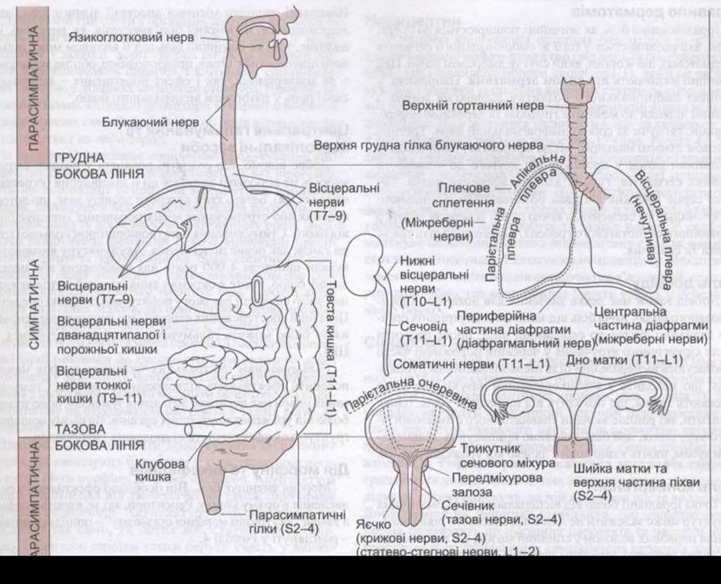
Fig. 7-7. Pain innervation of internal organs. Ascending impulses from structures located above the thoracic pain line and below the pelvic pain line cross the parasympathetic pathways (after White JC. Reproduced with permission from Ruch TC: Physiology and Biophysics, 19th ed. Ruch TC, Patton HD [editors]. Saunders, 1965).
The classic signs of intra-abdominal inflammation include pain, tenderness, autonomic changes such as hypotension and sweating, and abdominal wall spasm. THE ORIGIN OF all these symptoms is clear: tenderness is caused by sensitized visceral pain receptors, autonomic changes result from the activation of visceral reflexes, and the spasm arises from reflex contraction of the abdominal wall muscles.
Referred Pain
Irritation of internal organs frequently causes pain sensations not in the viscera themselves, but in somatic structures that may be located at a considerable distance. This is known as referred pain to a somatic Structure. Deep somatic pain can also be referred, whereas superficial pain typically is not. When visceral pain is both localized and referred, it produces a diffuse sensation of pain (irradiation) that encompasses both the site of the lesion and distant areas.
Clearly, knowledge of the referred pain phenomenon, as well as the somatic areas most commonly associated with pathology in specific internal organs, is crucial for a clinician. Perhaps the best-known example is the referral of cardiac pain down the medial aspect of the left upper extremity. Another example is shoulder pain resulting from irritation of the central Diaphragm, and testicular pain caused by ureteral stretching. Such cases are widely encountered in the practice of general medicine, surgery, and dentistry. As a rule, the localization of referred pain is not entirely stereotypical, and unusual sites of referral are quite common. For instance, cardiac pain may radiate to the Abdominal cavity, the right arm, or even the neck. Referred pain can be modeled experimentally by stimulating the severed end of a visceral nerve.
The Dermatomal Rule
Referred pain typically projects to a structure that develops from the same embryonic segment (dermatome) as the organ serving as the source of the pain. This principle is known as the dermatomal rule. For instance, during embryonic development, the diaphragm migrates from the cervical region to the border between the thoracic and abdominal cavities, dragging the phrenic nerve along with it. One-third of the phrenic nerve fibers are afferent, entering the spinal cord between the second and fourth cervical segments—the exact same region where afferents from the shoulder enter. Similarly, The Heart and upper extremity develop from a shared embryonic segment, while the Testis and its nerve migrate from the primitive urogenital ridge, which also gives rise to the Kidney and Ureter.
The Role of Experience
Prior experience also plays a role in the localization of referred pain. Although pain from inflamed internal organs typically radiates to the midline, post-operative abdominal surgery patients frequently attribute their visceral pain to their surgical scars. Pain originating from the maxillary sinus is usually perceived as originating from adjacent Teeth; however, patients with a history of traumatic dental Procedures typically attribute this pain to the traumatized teeth, even if they are located far from the affected sinus.
The Role of Convergence
The site of referred pain from visceral or somatic structures may depend not only on the shared spinal cord entry level of the nerve fibers, but also on the fact that peripheral sensory nerve endings outnumber the fibers in the spinothalamic tracts. Consequently, there is evidently significant convergence of peripheral sensory nerve fibers onto spinothalamic neurons. One prominent theory regarding the origin of referred pain is based on this concept. According to this theory, somatic and visceral afferents converge on the exact same spinothalamic neurons (Fig. 7-8). Because somatic pain occurs much more frequently than visceral pain, the brain is "trained" to interpret activity in a given pain pathway as originating from a specific somatic area. When signals from visceral afferents reach the brain via this same pathway, the pain is projected onto the corresponding somatic area.
Facilitation Effects
Facilitation may also play a role in the origin of referred pain. Collateral connections between visceral afferents and dorsal horn neurons in the spinal cord—which normally process pain impulses from somatic structures—can provide pathways through which enhanced activity of visceral afferents produces EPSPs, thereby increasing neuronal excitability. As a result, minimal activity in somatic afferents can trigger persistent pain.
If convergence were the sole explanation for referred pain, local anesthesia applied to the somatic referral zones would have no effect on the pain, whereas under the facilitation model, the anesthesia should abolish it.
In reality, the effects of local anesthesia on referral zones vary: severe pain is usually unaffected, whereas moderate pain may be completely abolished. Therefore, it is likely that both mechanisms—convergence and facilitation—play a role in the Pathogenesis of visceral pain.
Central Inhibition and Distraction Methods
It is well known that soldiers wounded in the heat of battle may feel no pain until the engagement is over (stress-induced analgesia). Many people know from personal experience that touching or shaking the site of an injury reduces the resulting pain. Electrical vibratory stimulation of a pain point also provides a degree of relief. Acupuncture has been used for 4,000 years for analgesia or pain relief, and in rare cases, this method alone serves as sufficient anesthesia to perform major surgeries. These and other observations indicate that pain transmission and perception can be inhibited, just as they can be facilitated, within the central nervous system.
Inhibition of central sensory pathways explains the effectiveness of counter-irritants and distraction methods. Stimulating the skin overlying an area of inflammation provides relief from pain originating in the affected internal organs. The effectiveness of traditional mustard plasters is based on this very principle.
Action of morphine and enkephalins
Morphine relieves pain and is particularly effective when administered rectally. Receptors that bind morphine, as well as the body's "internal morphines"—opioid Peptides—are discussed in Chapter 4.

Fig. 7-8. Illustrations of convergence and facilitation theories in The Mechanism of referred pain.
There are at least three sites in the body where opioids can act to produce analgesia: peripherally, at the site of injury; at the "gate" area of the dorsal horns, where nociceptive fibers synapse with dorsal root ganglion cells; and in brainstem regions closer to the cerebrum. Opioid receptors are formed by dorsal root ganglion cells and migrate along their processes both centrally and peripherally.
In the periphery, inflammation induces immune system cells to produce opioid peptides, which likely act on corresponding receptors on afferent nerve fibers to reduce pain. In the dorsal horn region, opioid receptors may act at the presynaptic level to decrease substance P release, although the relevant presynaptic nerve terminals have not been identified. Finally, injection of morphine into the periaqueductal gray matter of the Midbrain reduces pain by activating descending pathways that inhibit primary afferent transmission in the dorsal horns. There is evidence that this activation occurs via projections from the periaqueductal gray to the neighboring Nucleus raphe magnus, and that descending serotonergic fibers from this nucleus exert inhibitory activity. The precise mechanism of serotonin's inhibitory effect on impulse transmission in the dorsal horns remains unclear.
Placebos are capable of triggering The production of endogenous opioids that reduce pain. Their effect is partially blocked by morphine antagonists, notably naloxone. Acupuncture of areas remote from the pain source also induces the release of endorphins. Acupuncture of a trigger point acts much like touch or shaking (see above). Endogenous opioids are also involved in stress-induced analgesia, as animal experiments have demonstrated that naloxone can block some forms of it. Naloxone does not affect the occurrence of Other forms of stress-induced analgesia, suggesting the involvement of other components.
Acetylcholine
Epibatidine, a cholinergic agonist first isolated from frog skin, is a potent non-opioid analgesic whose synthetic derivatives exhibit even higher pain-relieving activity. Their action is blocked by cholinergic antagonists; to date, no evidence has been obtained that they cause addiction. Conversely, the analgesic effect of nicotine is less pronounced in mice deficient in the a4 and ß2 subunits of neuronal nicotinic acetylcholine receptors. These observations prove that the nicotinic-cholinergic mechanism plays an active role in The regulation of pain perception, although the precise mechanism of this regulation still requires investigation.
Cannabinoids
As noted in Chapter 4, the cannabinoids anandamide and PEA are produced endogenously and bind to CB1 and CB2 receptors, respectively. Anandamide exhibits pronounced analgesic activity, and anandamide-containing neurons have been found in the periaqueductal gray and other brain regions associated with pain sensation. When administered, PEA acts peripherally, potentiating the analgesic effect of anandamide.
Nociceptin
During the cloning of μ, k, and δ opiate receptors, an orphan receptor, ORL1 (opioid receptor-like 1), was identified, which did not bind any known opioids with high affinity. Recently, the endogenous ligand for this receptor was identified. It turned out to be a 17-amino-acid polypeptide resembling dynorphin-17 (see Chapter 4). When injected intracerebrally into experimental animals, it induced hyperalgesia. For this reason, it was named nociceptin. Nociceptin and its receptors are present in many brain areas, including the hypothalamus, brainstem, and dorsal horns. The precursor protein molecule of nociceptin also contains nocistatin, a polypeptide antagonist of nociceptin. The mechanism of interaction between these Polypeptides is not yet fully elucidated, but it is likely that both participate in pain transmission.
Chronic pain syndromes
Neuropathic pain in humans takes various forms. One of these is pain accompanying Other Sensations from an amputated limb (so-called phantom limb; see Chapter 5). In causalgia, spontaneous burning pain persists for a long time after relatively minor injuries and is frequently accompanied by hyperalgesia and allodynia. Reflex sympathetic dystrophy is also quite common. In these cases, the skin of the affected area is thin and shiny, with increased Hair growth. Animal studies have shown that nerve damage leads to sprouting and subsequent ingrowth of noradrenergic sympathetic nerve fibers into the dorsal root ganglia providing innervation to the affected areas. In this case, sympathetic impulses mediate the pain sensation. Thus, the periphery is short-circuited, and the affected fibers are stimulated by noradrenaline at the dorsal root ganglion level. Alpha-adrenergic blockade alleviates causalgia-type pain in humans, although for unknown reasons, a1-adrenergic blockers are more effective than a2-agents.
Spontaneous pain sensations can be generated at the thalamic level. In thalamic syndrome, damage to the posterior thalamic nuclei is characteristic, most often caused by occlusion of the thalamogeniculate branch of the posterior cerebral artery. Patients with this syndrome experience attacks of prolonged, severe, and extremely unpleasant pain that occur spontaneously or in response to ordinary stimuli.
Pain can often, though not always, be relieved by appropriate doses of analgesics. To reduce intractable pain, chronic stimulation of the spinal dorsal horns using implanted electrodes has been proposed. The electrodes can be connected to a portable stimulator, allowing patients to independently control their pain. Self-stimulation of the periaqueductal gray matter also helps reduce intractable pain, presumably due to increased release of ß-endorphins.
In some cases, the unpleasant symptoms of severe pain can be relieved by severing the deep connections of the frontal lobes with the rest of the brain (prefrontal lobotomy). Following this surgery, patients still feel pain, but it no longer bothers them. However, such an operation causes significant personality changes (see Chapter 16). Pain reduction can also be achieved by removing the cingulate gyrus. These operations, as well as other options for surgical interruption of pain pathways, are illustrated in Fig. 7-9. One such surgical intervention is anterolateral cordotomy. This Procedure involves inserting a scalpel into the lateral part of the spinal cord to encompass the anterolateral areas. When properly performed, this intervention severs the lateral spinothalamic and anterolateral pain-conducting fiber systems while leaving most ventral spinothalamic touch sensitivity fibers intact. Occasionally, pain may recur months or even years later, reflecting the potential for short-circuiting of its pathways.
Last update: 10/08/2026
Editorial and Educational Adaptation: This material has been compiled based on the primary/original source text. The project team performed an editorial review, corrected technical inaccuracies, structured sections, and adapted the content for an educational format.
What was processed:
- elimination of formatting defects (OCR errors, structural breaks, corrupted characters);
- editorial organization of content;
- standardization of terminology in accordance with academic sources;
- verification of factual statements against the original source text.
All mentions of the author, publication year, and origin of the primary text have been preserved in accordance with the source.