Military Surgery with Emergency Surgery - V.Ya. Bilyi 2004
Infectious Complications of Wounds and Injuries
Introduction. Purulent wound infection of various origins is one of the most complex problems in both peacetime and wartime surgery.
The following types of wound infection are distinguished: purulent, putrefactive, anaerobic, and tetanus. The development and clinical course of a wound infection are determined by the body's defense mechanisms, on the one hand, and the virulence of the microorganisms that have entered the wound, along with favorable conditions for their vital activity, on the other.
A core principle of military surgery is that any gunshot wound is considered microbially contaminated. It invariably contains a mixed microbial flora, as well as necrotic tissue foci, which provide a fertile ground for The Development of the infectious process.
METABOLISM/2.html">THE CONCEPT OF surgical infection refers to a pathological state of the wound in which the presence of microflora impairs reparative processes and causes infectious complications both directly within the wound area and far beyond its boundaries.
Infectious complications of gunshot wounds have been common across all wars, significantly worsening injury outcomes. For instance, the incidence of purulent complications in gunshot wounds ranged between 45–60% during World War I, and 45–50% during the military operations near Lake Khasan; during the Great Patriotic War, soft tissue gunshot wounds were complicated by severe purulent infection in more than 18% of the wounded. The frequency of infectious complications is also rising in peacetime surgery: suppuration of "clean" surgical wounds reaches 5%, and contaminated ones 20–30%; as a cause of death in the postoperative period, infectious complications are observed in 25% of patients.
Factors contributing to the development of wound infection include: the extent of tissue damage, which in turn depends on The Nature of the wounding projectile (bullet or shrapnel), supersonic projectile velocity, or a wound inflicted by a bullet (shrapnel) at the end of its trajectory; the degree of soil contamination in the wound; the presence of Foreign bodies in the wound; the degree of Blood supply impairment caused by tight wound packing or hematoma; the anatomical Location OF THE wound—such as the HEAD, chest, gluteal region, or FOOT, where wound infection develops more frequently; inadequate first aid, delayed evacuation of casualties from the battlefield, and late or technically imperfect primary surgical debridement of the wound; Shock, blood loss, hypothermia, fatigue, malnutrition, and other factors causing general weakening of the body.
Etiology AND Pathogenesis of purulent wound infection. Among the aforementioned factors, three have a decisive influence on the development of wound infection: 1) the morphological and functional state of the traumatized Tissues; 2) microbial contamination of the tissues—the number of microbes per 1 g of tissue must exceed a critical threshold (105–106 Bacteria per 1 g of tissue sampled from the depth of the wound); 3) the presence of foreign bodies in the wound.
From the perspective of their impact on The Human Body, all microbes are divided into 3 groups: pathogenic, opportunistic, and non-pathogenic. The differences between pathogenic and opportunistic species of microbes are due to the varying intensity of their invasive properties.
Invasiveness is a key criterion of pathogenicity, reflecting the ability of microorganisms to multiply within the host Organism while overcoming its diverse defense and immunological reactions aimed at preventing The entry of genetically foreign agents (bacteria, Viruses, somatic Cells, etc.).
Thus, ultimately, wound infection develops when the equilibrium between the microbes contaminating the wound and the host's defense mechanisms is disrupted.
Microbiology of wartime wounds. Even during the Great Patriotic War, it was established that the microflora of muscular wounds, particularly of the extremities, shares similarities with intestinal microflora. Such wounds are predominantly colonized by *Escherichia coli*, various intestinal and soil anaerobes, the tetanus bacillus, *Pseudomonas aeruginosa*, *Proteus*, enterococci, anaerobic intestinal streptococci, *Sarcina*, and others. This is evidently explained by the similarity of living conditions for microbes in the gut and in necrotic tissues of a muscular wound. Microorganisms with robust enzymatic properties capable of inducing fermentative, putrefactive, and other organic degradation processes adapt and survive best in a muscular wound.
Recent studies have established that gram-negative microflora predominates in gunshot wounds.
The causative agents of wound infection are opportunistic aerobic or anaerobic microorganisms that constantly coexist with the human body—staphylococci, streptococci, *Escherichia coli*, *Proteus*, klebsiellae, *Pseudomonas aeruginosa*, clostridia, bacteroides, and fusobacteria. The etiology of most wound infectious complications is mixed (aerobic-anaerobic). However, microbial associations under various conditions can trigger either aerobic (purulent) or anaerobic wound infections.
Pathogenesis of the purulent wound process. Any infected wound heals by secondary intention. Healing by secondary intention is viewed as a unified purulent-granulation process that encompasses suppuration and granulation.
Suppuration is the biological process of wound cleansing that involves the participation of microorganisms (proteolysis). The formation and development of granulation tissue are inextricably linked with suppuration.
The course of the wound process comprises 3 phases (Girgolav S.S., 1956): the first is the preparatory or inflammatory period, during which complex biochemical and pathophysiological processes take place, although morphological signs of regeneration are not yet detectable; the second phase is the regeneration period, which ends with the Filling of the wound cavity by newly formed tissue; the third phase is the scar formation period.
The Classification of the wound process proposed by M.I. Kuzin (1977) is somewhat more detailed.
The first (inflammatory) phase is divided into 2 periods:
1) vascular changes; 2) cleansing of the wound from necrotic (dead) tissues.
The second phase is the regeneration phase, characterized by the formation and maturation of granulation tissue.
The third phase is the scar Organization and epithelialization phase.
The first period of the inflammatory phase involves sequential vascular reactions characteristic of acute inflammation—vasoconstriction followed by vasodilation, elevated capillary pressure, increased permeability, edema, leukocyte migration, etc. During this period, substances that catalyze the vascular reaction play a crucial role:
1) proteases—plasmin, kallikrein; 2) Polypeptides—leukotaxine, bradykinin, kallidin; 3) amines—histamine, serotonin.
The period of wound cleansing from dead tissues is clinically significant as it determines the normal progression of regeneration and the entire healing process.
The primary criterion for evaluating wound healing is the clinical dynamics of the wound process, supplemented by two Laboratory Research Methods: cytological and bacteriological.
Classification of surgical infection.
A. Local forms of surgical infection.
1. By etiological factor: aerobic microflora (indicating the causative agent: staphylococcus, streptococcus, etc.); anaerobic non-clostridial microflora (indicating the causative agent: peptostreptococcus, etc.); aerobic clostridial microflora (indicating the causative agent: gas-producing bacillus, etc.); rare forms (scarlet fever, wound diphtheria).
2. By the nature of microflora: monoinfection; polyinfection (caused by several aerobes or several anaerobes); mixed (aerobic-anaerobic).
3. By clinical and morphological manifestations: abscess; Phlegmon; fistula; Osteomyelitis; thrombophlebitis; lymphangitis; regional lymphadenitis.
1) By localization: head, neck; spine; chest; abdomen; pelvis; extremities (soft tissues, bones, joints).
2) By source of infection: endogenous; exogenous.
3) By causes of occurrence: spontaneous; traumatic; iatrogenic.
4) By origin: community-acquired; nosocomial.
B. General form of surgical infection.
1. Severe Sepsis (sepsis syndrome) is sepsis accompanied by the development of multiple organ dysfunction (failure of two or more body Organs or systems).
2. Septic shock is sepsis accompanied by hypotension refractory to ongoing therapy (systolic BP of 80 mmHg and below).
Clinical forms of sepsis are determined by the characteristics of toxin production by the pathogens and can simultaneously represent Phases of the septic process:
1) The toxemic phase of sepsis is characterized by the development of toxicosis syndrome As a result of the systemic influx of microbial toxins (primarily exotoxins) and inflammatory mediators from the primary infectious focus. Bacteremia may be absent in this phase.
2) Septicemia — this phase involves the dissemination of pathogens with The formation of multiple secondary microbial foci in the form of infectious vasculitis and septic microthrombi within the microcirculation system. Bacteremia acquires a persistent character.
3) Septicopyemia — this phase is characterized by the Formation of secondary purulent foci (abscesses) in the Lungs, Kidneys, bones, Spleen, Skin, etc.
Clinical picture of a purulent wound. Classic signs of inflammation — edema, hyperemia, pain — characterize the stage of vascular changes. They are accompanied by hyperthermia and impaired function. Within 2–5 days, inflammatory demarcation of non-viable tissue takes place, leading to the stage of rejection of dead tissue, which completes the inflammatory phase.
Against the Background of demarcation and gradual rejection of non-viable tissue, islands of granulation appear in certain areas of the wound, usually no earlier than 5–6 days after injury. The appearance of granulations signifies the beginning of the wound process transition from the inflammatory phase to the regeneration phase.
Granulation tissue is represented predominantly by newly formed capillaries. Concentrated around them are mast cells (labrocytes), which secrete BIOLOGICALLY ACTIVE SUBSTANCES that facilitate the Formation of the capillary network, essentially serving as the construction site for the healing process. Being rich in Blood Vessels, normal granulation tissue is pinkish-red, granular, and easily bleeds.
In a complicated course of The first phase of the wound process, pronounced inflammatory changes of the wound edges and walls are observed — edema, progressive skin hyperemia, tissue infiltration, and tenderness upon Palpation. As a result, the wound process proceeds via secondary intention healing. This is characteristic of wounds with a large volume of dead tissue, which serves as a substrate for pus formation.
Varieties and signs of local purulent infection.
Purulent pockets (extensions) are pus-filled cavities communicating with a purulent wound. The cause of their formation is impaired outflow and prolonged retention and spread of pus along fascial spaces and cellular tissues with the formation of infiltrates and abscesses at the periphery of the wound. This form of wound infection spread is characterized by a discrepancy between the severe General condition of the wounded patient and the minor local manifestations of the infectious process.
The surface and walls of the wound have a gray tint, becoming covered with continuous fibrinous-purulent deposits with areas of necrosis. Externally, the wound appears dry, but The amount of pus within it progressively increases. Sometimes pus appears only upon pressing the wound edges, which is an unmistakable sign of purulent pockets. In other cases, there is no pus discharge, but the wound walls are heavily infiltrated with seropurulent exudate.
Frequently, the purulent process spreads into the subcutaneous tissue surrounding the wound or affects the fascia, developing into periwound phlegmon. In this case, only tissue edema with minor pus drainage may be observed against the background of high fever. The Clinical presentation of periwound phlegmons is difficult to diagnose and requires immediate surgical revision and debridement of the wound.
In some cases, isolated accumulations of pus — wound tract abscesses — form near the wound canal.
A Connective Tissue capsule, impregnated with pus, forms around abscesses. Pus from long-undiagnosed abscesses discharges externally, as well as into cavities through developing fistulae, or causes deep fascial spaces infection (sapremia/pockets).
When foreign bodies that promote suppuration (metal fragments or free-lying bone fragments) remain deep within the wound canal, long-term functioning fistulae develop.
As a rule, the character of the pus discharged from a wound (type, quantity, odor, consistency) is determined by the pathogen of the purulent process. Staphylococci almost always produce abundant yellowish pus, whereas streptococci produce thin, yellowish-green pus or fluid resembling serous discharge tinged with blood. Pseudomonas aeruginosa produces a characteristic coloration and a distinct sweetish odor.
The type of pathogen causing a local purulent infection largely determines its clinical course. For instance, Staphylococcal infections are characterized by an acute local process, whereas streptococcal infections tend to spread diffusely in the form of phlegmon with relatively mild local and general manifestations. Underestimating the threat of developing complications can lead to severe consequences. Pseudomonas aeruginosa and Proteus typically exhibit a slow, protracted local course accompanied by pronounced systemic intoxication, which frequently leads to wound exhaustion.
Young connective tissue protects the wound from environmental factors, prevents infection invasion, and inhibits the absorption of toxins and tissue breakdown products. It is well established that normal granulation tissue typically secretes a small amount of blood elements, Proteins, and electrolytes. However, when wound infection develops, the Functions of granulations change. First, they lose their protective role, and the absorption of tissue breakdown products and microbes from the wound surface increases. Second, the loss of protein and electrolytes from the wound surface sharply rises. Third, microbes overcome the granulation tissue and begin to penetrate the surrounding tissues.
Toxico-resorptive fever and sepsis. The body responds to the development of a severe local purulent infection with a systemic reaction proportional in scale and character to the local process. This systemic response is intensified by the formation of deep fascial extensions and phlegmon. The body's overall reaction to the development of a purulent wound process was termed purulent-resorptive or toxico-resorptive fever by I.V. Davydovsky, who regarded it not as a complication, but as a typical systemic syndrome accompanying purulent-necrotic processes in the body.
Purulent-resorptive fever manifests as deterioration of the patient's well-being, the onset and progression of pain in the wound area, persistent fever, and hematological changes (leukocytosis, left shift in the leukocyte formula, elevated ESR). Protein appears in the urine, leukocyte counts increase, and hyaline casts emerge. Hypoproteinemia and a decreased albumin-globulin ratio are consistently observed.
An important feature of toxico-resorptive fever is its dependence on the primary purulent focus; upon the elimination of this focus, the toxico-resorptive fever subsides.
The most serious complication of purulent wound infection is sepsis. The Diagnosis of sepsis is based on the detection of systemic inflammatory response syndrome combined with a clinically identified infectious focus or bacteremia. The criteria for systemic inflammatory response syndrome are: tachycardia exceeding 90 beats per minute; tachypnea exceeding 20 breaths per minute; Temperature above 38 °C or below 36 °C; peripheral blood leukocyte count greater than 12,000/mm3 or less than 4,000/mm3, or immature leukocytes exceeding 10%.
Sepsis occurs particularly frequently in severe gunshot fractures of Bones and joints, chest injuries complicated by Pleural Empyema, and pelvic injuries, including those involving damage to pelvic organs. The incubation period for sepsis varies widely—from a few hours to several weeks or even months. Acute, subacute, and chronic sepsis are distinguished. In an unfavorable clinical course, acute sepsis leads to patient death within 2 to 15 days, subacute within 16 days to 2 months, and chronic within 2 to 3–4 months.
Acute sepsis is characterized by a high temperature of a continuous fever type with very rare remissions. The patient's condition is severe. The skin is pale with an earthy tint, and hectic flushes appear on the Cheeks. In the final days of life, 25% of wounded patients exhibit varying degrees of jaundice. Euphoria, insomnia, irritability, and tachycardia are possible. The pulse is of low volume and tension. Blood pressure drops. In 35% of wounded patients, bedsores develop at the peak of the disease. Anemia progressively worsens. The leukocyte count increases, although sepsis can also occur with a normal leukocyte count. The course of the wound process changes. The wound becomes dry, granulations appear pale and bleed easily, and a white coating appears on the wound surface. Marginal epithelization ceases.
As an independent infectious disease, sepsis loses its connection with the primary focus of purulent infection, develops according to its own pathophysiological laws, and proceeds against the background of suppressed immunological defense mechanisms.
General Principles of wound infection Prevention. At all stages of medical evacuation, the prevention of wound infection includes the following measures.
During first aid administration: 1) apply a primary aseptic dressing correctly; 2) perform transport immobilization; 3) administer oral Antibiotics.
Pre-medical care involves: 1) adjusting dressings; 2) improving transport immobilization; 3) administering antibiotics (orally or parenterally).
First medical care involves: 1) administering high doses of antibiotics; 2) improving the application of aseptic dressings; 3) transport immobilization using standard-issue means; 4) administration of tetanus toxoid; 5) procaine blockades; 6) anti-shock measures.
During the provision of qualified surgical care, it is necessary to perform: 1) early and radical primary surgical debridement (PSD) of the wound; 2) adequate antibiotic therapy and timely correction of blood loss.
Cautious evacuation of the wounded from one medical evacuation stage to another.
Treatment of purulent infection in gunshot wounds at medical evacuation stages.
The first fundamental tenet is comprehensive treatment of purulent wound infection; surgical and medical methods Complement each other, yet the primary role belongs to surgery. In most cases, only surgery provides the necessary conditions for optimal healing, as it eliminates the infection focus and creates favorable conditions for draining wound contents. Adequate surgical intervention creates the best conditions for the action of antibacterial agents.
THE PRINCIPLE OF the paramount role of surgical intervention is fundamental in the treatment of any purulent wound.
The elimination of the purulent focus, which a purulent wound represents, can be achieved by only one method: radical surgical debridement. According to its purpose, surgical debridement of a purulent wound is classified as secondary surgical debridement, as it aims to eliminate an already developed wound infection and prevent more severe purulent-septic complications.
Technique of secondary radical surgical debridement (SRSD)
The technique of SRSD for a purulent wound includes wide incision of the wound, purulent pockets, and fascial extensions, along with the excision of all necrotic, non-viable, and pus-impregnated tissues.
The excision of necrotic tissues helps prevent the generalization of infection, reduces the resorption of necrolysis products from the wound surface, and thereby prevents endotoxicosis.
Preparation and management of the operative field follow the standard rules established in general surgery. The incision must be sufficient for a thorough exploration of the wound, ensuring the most complete removal of necrotic tissue and subsequent drainage of wound exudate. The skin edges of the wound should be excised via two wide incisions, taking into account the topography of major anatomical structures in the area and the direction of skin Langer's lines. To ensure adequate access to the deep layers of the wound, a wide incision of the aponeurosis is mandatory. Pus, detritus, foreign bodies, and blood clots are first evacuated from the wound, and the extent of tissue damage is assessed. Next, the main stage of the operation is performed—the removal of non-viable tissue. Due to the lack of objective criteria for determining tissue viability, the surgeon must rely on standard clinical signs during the Procedure: the degree of tissue bleeding, the presence of visible destruction, and tissue infiltration by purulent or serous exudate. Although tissue excision should generally be conservative, leaving questionable tissue in the wound can cause more harm to the body than creating a larger wound surface.
The Outcomes of surgical management for purulent wounds are significantly improved by treating them with surfactant solutions (detergents), a pulsating stream of antibacterial fluid, vacuum therapy, ultrasound, laser, etc.
Purulent wound drainage. Effective wound drainage must ensure adequate outflow of wound exudate, creating optimal conditions for the rapid sloughing of dead tissue and the transition of the healing process into the regeneration phase.
Active drainage methods, which combine drainage with wound irrigation using various antibacterial agents and surfactant solutions, allow for a targeted approach in combating wound infection.
Types of drains and drainage methods. There are three mechanisms of drain action. The first is the outflow of purulent contents that separate and drain through the drainage tube due to gravity, provided the drain is placed at the lowest point of the purulent cavity (Fig. 27).
The second is passive drainage of pus from the wound.
The third mechanism of drainage action is active drainage of a purulent wound (Fig. 28).
The principle of active antibacterial drainage involves continuous wound irrigation with antibacterial solutions (antiseptics) or detergent solutions. This provides mechanical removal of purulent exudate and detritus, creating conditions for the direct action of the antibacterial solution on the wound's microbial flora (Fig. 29).
Class="center">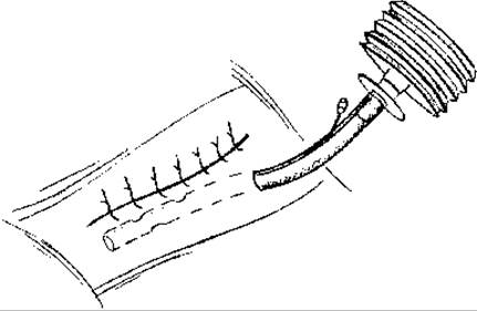
Fig. 27. Passive drainage of a blind wound tract using a PVC tube.
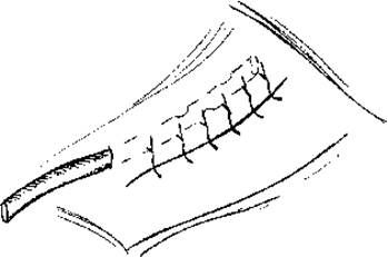
Fig. 28. Active drainage of a blind wound tract using a PVC tube.
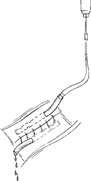
Fig. 29. Active continuous-flow drainage of a blind wound tract using a PVC tube.
A variation of active drainage is aspiration drainage, performed using devices that create negative pressure within the drainage system.
The most effective drainage, achieved through continuous wound irrigation, is constant vacuum aspiration.
When treating any wounds, including those complicated by wound infection, surgical efforts are always aimed at accelerating their healing after secondary debridement. Early secondary sutures are applied to a wound covered with healthy granulation tissue and having mobile edges, prior to the development of scar tissue. Late secondary sutures are applied to a granulating wound in which scar tissue has already formed and the wound edges are fixed. In this case, granulations and scar tissue are excised beforehand, and the wound edges are mobilized.
Early closure of a purulent wound after surgical debridement—using secondary sutures or skin grafting—significantly shortens the treatment period for wounded patients and yields favorable functional and cosmetic results.
Postoperative care. Immediately after surgery, wound irrigation through the drain is initiated using antibacterial agents (0.1% dioxidine solution, 0.1% furagin solution, furacilin, 3% boric acid solution). Irrigation is performed 3 to 6 times daily, or continuously in severe cases. The drain is removed on the 8th–14th postoperative day. Antibacterial therapy is mandatory. When selecting an antibiotic, the pathogen's sensitivity, its distribution in organs and tissues, and compatibility with other drugs are taken into account. The duration of antibiotic therapy depends on the severity of the infection: 12–21 days for localized purulent processes, and 1.5–2 months or more for sepsis. Infusion-Transfusion Therapy is administered for detoxification, restoration of lost plasma, and management of anemia (parenteral administration of plasma, blood products, aminol, sorbilact, reosorbilact, hemodesis, albumin, hydrolysates, Ringer's solution, glucose, etc.). To restore immune function, staphylococcal toxoid is administered subcutaneously following a schedule of 0.1, 0.5, 1.0, 1.5, and 2.0 ml with an interval of 2–3 days between doses.
During the acute phase of purulent wound infection, anticoagulants, fibrinolytics, Proteolytic Enzymes, and protease inhibitors may be used as indicated, under laboratory monitoring. Anabolic processes are stimulated using Hormones such as retabolil or nerabol, administered intramuscularly at 1 ml once a week. A high-calorie, vitamin-rich diet contributes to the rapid recovery of patients.
Anaerobic infection. This type of wound infection is one of the most severe complications of combat trauma, including wounds, frostbite, Burns, etc.
During World War II, anaerobic infection occurred in approximately 0.5–2% of the wounded.
Anaerobic infection must be considered generalized from the very beginning, as anaerobic toxins possess an extraordinary ability to penetrate protective barriers, subsequently exerting a necrotic effect on living tissues.
Practically only clostridia and anaerobic Gram-positive cocci can cause a monoinfection. Much more frequently, the anaerobic process involves multiple species and genera of bacteria—both anaerobic (bacteroides, fusobacteria, etc.) and aerobic—and is referred to as "synergistic." The following forms of anaerobic wound infection are distinguished:
1. Anaerobic monoinfections (clostridial cellulitis, clostridial myonecrosis; anaerobic streptococcal myositis, anaerobic streptococcal cellulitis).
2. Polymicrobial synergistic (aerobic-anaerobic) infections (synergistic necrotizing fasciitis; synergistic necrotizing cellulitis; progressive synergistic bacterial gangrene; chronic burrowing ulcer).
Regardless of the form of anaerobic infection, the wound develops distinct zones: a zone of purulent melting, a zone of necrosis and phlegmon, and a vast zone of serous edema comprising living tissues heavily saturated with toxins and anaerobic enzymes, lacking clear boundaries.
Anaerobic wound infections are caused by microbes of the genus Clostridium (Cl. perfringens, Cl. septicum, Cl. oedematiens, Cl. histolyticum).
Pathogens of anaerobic infection exhibit several distinctive features.
Cl. perfringens is the most common CAUSATIVE AGENT OF gas gangrene in humans. This microbe is widespread in nature and found in large quantities in the intestines of humans and animals, as well as in the soil. It is non-motile, spore-forming, and produces a toxin composed of hemolysin, myotoxin, and neurotoxin. The action of this toxin on living tissues leads to the formation of bloody exudate and gas, tissue Swelling and necrosis—particularly of the Muscles. Affected muscles turn pale, resembling boiled meat, and contain numerous gas bubbles. Large doses of the toxin are fatal.
Cl. oedematiens is a motile, spore-forming microbe that produces hemolysin and an exotoxin. Its toxins are characterized by high activity and The ability to rapidly induce edema in the subcutaneous tissue, intermuscular spaces, and muscles. The toxin also exerts a constant and specific hemolytic effect. Bacterial spores survive boiling for up to 60 minutes.
Cl. septicum is a motile, spore-forming microbe discovered by Pasteur in 1861. Its toxin has a hemolytic effect, causing rapidly spreading blood-serum edema, serous-hemorrhagic infiltration of the subcutaneous tissue and Muscle, and, in rare cases, muscle destruction. Entering the bloodstream, the toxin causes a rapid drop in blood pressure, vascular paralysis, and damage to the myocardium. The microbe resides in the soil and the intestines of humans and animals. Its spores withstand boiling for 8 to 20 minutes.
Cl. histolyticum is a spore-forming, motile microbe discovered in 1916. Its toxin contains a proteolytic enzyme—fibrinolysin—which rapidly liquefies muscles, subcutaneous tissue, connective tissue, and skin. The liquefied tissues turn into an amorphous mass resembling raspberry jelly. The toxins of gas gangrene pathogens are complexes of various protein-based enzymes (lecithinase, hyaluronidase, deoxyribonuclease, hemolysins, etc.).
The primary sources of wound contamination by anaerobic pathogens are soil and soil-contaminated clothing. In cultures from fresh wounds, Cl. perfringens is found in 60-80% of cases; Cl. oedematiens in 37-64%; Cl. septicum in 10-20%; and Cl. histolyticum in 1-9%. Along with these microbes, fresh gunshot wounds reveal other anaerobic and aerobic microorganisms (anaerobic and aerobic streptococci, staphylococci, Escherichia coli, Proteus, etc.). Proliferating aerobic Microorganisms in the wound—especially streptococci and staphylococci—can act as activators for the "group of four" anaerobes, enhancing their reproduction, pathogenicity, and their hemolytic and necrolytic properties. Thus, the flora of gas gangrene can be associative, though anaerobic microbes remain the primary pathogenic factor.
Despite the high rate of gunshot wound contamination by anaerobic microorganisms, anaerobic infection develops relatively rarely (0.5-2%) upon the convergence of specific local and general factors. Local factors primarily include extensive tissue damage, most frequently observed in fragment wounds, especially those involving bone fractures.
Experience from the Great Patriotic War confirmed that in gunshot fractures of the extremities—which are typically accompanied by significant soft tissue damage—anaerobic infection occurs 3.5 times more frequently than in extremity injuries without bone involvement. The type of wound also affects the incidence of anaerobic infection: complications were observed 1.5 times more frequently in fragment wounds than in bullet wounds, and twice as often in penetrating (blind) wounds as in through-and-through wounds.
Wound localization plays a significant role in the onset of anaerobic infection. In the majority of cases (75%), the anaerobic process developed in INJURIES OF THE lower extremity, which is presumably due to large muscle masses enclosed within tight fascial sheaths. Post-injury traumatic edema leads to the compression of Muscles and their blood vessels within these fascial sheaths, resulting in muscle ischemia—a condition known to favor the development of anaerobic infection. The fact that lower extremities are more easily contaminated with soil also plays a contributory role.
Factors predisposing to the development of anaerobic infection include local Circulatory Disorders caused by major vessel damage, tourniquet use, and tissue compression by hematomas.
Shock, blood loss, and general weakening of the body caused by fatigue, hypothermia, or malnutrition further facilitate the onset of anaerobic infection.
Anaerobic infection occurs more frequently when the evacuation of the wounded from the battlefield (or incident site) is delayed, or when first aid is inadequate and untimely.
Nevertheless, the primary role in the development of anaerobic infection is played by delayed and technically inadequate primary surgical debridement of the wound, or the omission of this procedure when indicated. The risk of developing anaerobic infection increases significantly if the wound is tightly sutured following primary debridement.
Clinical presentation of anaerobic infection. The most critical period for the development of anaerobic infection is the first 6 days post-injury. It is precisely during this window that optimal conditions are created within the wound for the proliferation and activity of pathogenic anaerobes.
Occasionally, the course of anaerobic infection becomes fulminant. Tissue necrosis and edema develop before one's eyes. Proteolysis of muscles and erythrocytes leads to the Formation of tissue gases—hydrogen, hydrogen sulfide, ammonia, and carbonic acid. A hemorrhagic exudate appears in the subcutaneous tissue, accompanied by hemolytic skin discoloration. Rapid proliferation of anaerobes in the wound, combined with abundant bacterial tissue toxins, triggers severe intoxication.
Anaerobic infection is characterized by diverse and dynamic clinical manifestations. As pathological processes progress, the symptomatology of the infection changes; however, from a practical standpoint, early symptoms are of the utmost importance.
1. Acute, excruciating pain unresponsive to analgesics. Following injury, pain follows a specific trajectory: initial injury-related pain subsides, but as the anaerobic infection develops, the pain sharply intensifies and quickly becomes unbearable. With the formation of extensive soft tissue necrosis and deepening intoxication, the pain subsides or disappears once again. In a state of severe toxicoinfection, wounded patients make no Complaints whatsoever (late stage!).
2. Rapidly progressive tissue edema of the extremities, causing a sensation of tightness or bursting in the limb. To gauge The rate of edema progression, A.V. Melnikov proposed applying a ligature around the limb 8-10 cm above the wound (the "ligature sign"). The sign is considered positive if a tightly applied ligature above the wound begins to cut into the soft tissues. According to Melnikov, if the ligature cuts to a depth of 1-2 mm within 2-3 hours of application, amputation is required.
Upon the appearance of these two symptoms, the wound dressing must be removed immediately to thoroughly examine both the wound and the entire affected extremity.
3. Wound changes. Dryness with minimal bloody discharge ("varnish blood"). The muscles appear gray, resembling boiled meat. Due to edema and tissue gas impregnation, Muscle tissue may protrude from the wound opening above the skin level; muscle fibers neither contract nor bleed and tear easily. In cases of delayed diagnosis of anaerobic infection, devitalized muscles turn dark gray or brown. Characteristic blisters filled with bloody, clear, or turbid fluid frequently form on the skin of the affected segment. The skin acquires a "bronze," "saffron," brown, or bluish discoloration. This is caused by the diapedesis of erythrocytes, which are rapidly destroyed by bacterial enzymes; Hemoglobin breaks down to form a dirty-brown pigment that imparts this specific coloration to the tissues.
Wounds complicated by anaerobic infection frequently emit an unpleasant, putrid odor resembling that of mice, moldy hay, or sauerkraut.
4. Gas in the soft Tissues of the affected segment is a definitive sign of developing anaerobic infection. Gas production generally follows the onset of edema and indicates tissue destruction resulting from The activity of anaerobic microbes, primarily Cl. perfringens. The presence of gas is determined by Percussion, revealing a tympanic sound over the affected area. In subcutaneous tissue, gas can be detected via palpation as a "dry snow crunch" (the crepitus sign of gas bubbles). Shaving Hair around the wound produces a faint crackling sound—Resonance over the gas-saturated tissue area (the "razor sign"). Tapping with the branches of tweezers yields a characteristic box sound. French surgeon Lemaire recommended diagnostic percussion around the wound perimeter to elicit a characteristic resonant sound.
5. Loss of sensitivity and motor function in the distal extremities is an early and formidable symptom of anaerobic infection. These disturbances appear even when there are outwardly minor Changes in the wound and the limb, and they are critically important: they help detect anaerobic infection when, at first glance, other symptoms are still absent. Therefore, triage officers should always have a needle on hand to test the sensitivity of the distal extremities and fingers.
6. X-ray examinations serve as an auxiliary method for detecting gas in tissues. When gas spreads through muscle tissue, radiographs reveal "feathery clouds" or "fir trees," while the presence of gas in the subcutaneous tissue resembles a honeycomb; occasionally, distinct isolated gas blisters or gas streaks spreading along interfascial spaces can be observed. The toxins of anaerobic infection affect multiple organs and all systems of the wounded person, leading to A number of systemic symptoms.
7. The temperature most frequently ranges between 38–38.9 °С.
8. In the majority of wounded patients, The Heart rate exceeds 120 bpm. A formidable sign is the dissociation between pulse and temperature, the so-called "scissors" phenomenon: the pulse rate increases while the temperature curve drops.
9. Blood pressure progressively decreases as the anaerobic infection advances.
10. Blood abnormalities: marked neutrophilic leukocytosis, a shift to the left, lymphopenia, and eosinopenia.
11. Icteric sclerae due to erythrocyte hemolysis.
12. Gastrointestinal status: the Tongue is dry and coated. Wounded patients experience unquenchable thirst and dry Mouth. The onset of nausea and vomiting undoubtedly indicates significant systemic intoxication.
13. Facial expression. Anaerobic infection alters the appearance of the wounded person. The skin becomes pale with an earthy tint, facial features become sharp, and the eyes sink. A characteristic appearance and facial expression develop—facies Hippocratica.
14. Neuropsychiatric status varies from mild euphoria to acute agitation, and from apathy and lethargy to severe depression. Incorrect orientation and misjudgment of one's own sensations and condition are frequently noted. However, consciousness remains intact until the very end.
Depending on the specific clinical course, the following forms of anaerobic infection are distinguished: 1) fulminant—developing a few hours after injury; 2) rapidly progressive—developing within 1–2 days after injury; 3) slowly progressive—characterized by a long incubation period.
According to the Nature of the pathological process, anaerobic infection is subdivided into gas, edematous, tissue-necrotic, and mixed forms (see insert, Fig. 30).
Based on the depth of tissue involvement, deep (subfascial) and superficial (epifascial) forms are distinguished.
It is important to remember that anaerobic infection does not always present with an extremely grave general condition from the very beginning. Treating such notions as absolute can lead to delayed diagnosis. Only careful monitoring of the wounded patient allows for the timely identification of what may be the sole symptom characteristic of anaerobic infection against a relatively favorable background. Examples include changes in the wound and surrounding skin: muscle bulging, edema, tissue tension, tenderness along neurovascular bundles, skin pallor, the appearance of hemorrhagic spots, etc. In other cases, patients complain of constriction of the limbs by a bandage, anxiety, thirst, or fever. Knowledge of the clinical presentation in all its manifestations and a careful examination of every wounded patient are the guarantees for early detection of anaerobic infection.
Non-clostridial anaerobic infection is a fundamentally new form of surgical infection caused by spore-free anaerobes.
In 1861–1863, Pasteur was the first to describe the possibility of Bacterial growth in the absence of air. The discovery of anaerobiosis and The Study of various anaerobic bacteria—pathogens of surgical diseases and purulent complications—already at the beginning of the 20th century made it possible to distinguish three groups of anaerobic infections. The first includes botulism and tetanus; the second comprises clostridial myonecrosis of soft tissues. Due to their characteristic clinical picture and The properties of the causative agents, they were considered specific infections accounting for a minor fraction of all human anaerobic infections. The third group consists of "ordinary" purulent-putrefactive processes that typically occur with the participation of obligate anaerobes and constitute the largest category of purulent-Inflammatory Diseases.
Etiology and pathogenesis. It has now been established that non-spore-forming anaerobes are the causative agents in 40 to 95% of surgical infection cases.
Currently, representatives of non-spore-forming anaerobic microorganisms from the following genera are known:
- Gram-positive anaerobic cocci (Ruminicoccus, Peptococcus, Peptostreptococcus);
- Gram-negative anaerobic cocci (Veillonella, Arachnia);
- Gram-positive anaerobic bacteria (Actinomyces, Lactobacillus, Bifidobacterium, Eubacterium);
- Gram-negative anaerobic bacteria (Bacteroides, Fusobacterium, Butyrivibrio, Campylobacter).
It has been established that the microflora of purulent foci is polymicrobial in nature and occurs in associations, with non-clostridial anaerobes predominating in the etiology of surgical infections.
In cases of mixed infection and non-clostridial anaerobic monoinfection, both local and systemic factors contributing to its development should be singled out.
Local factors include crushed wounds with a large mass of necrotic tissue, prolonged tissue ischemia resulting from microcirculatory disorders (edema, prolonged tourniquet application to a limb, etc.), surgical operations, endoscopy, punctures, and other surgical manipulations.
General factors include those that reduce the body's defense mechanisms, such as Traumatic shock, blood loss anemia, hypothermia, malnutrition, hypovitaminosis, and excessive physical and psycho-emotional stress.
The pathogenesis of anaerobic non-clostridial and mixed infections is driven by interdependent factors, including the site of invasion, as well as the pathogen type, pathogenicity, and virulence. The patient's level of immunological and nonspecific reactivity is also of practical significance.
Clinical Presentation. The most typical clinical picture of anaerobic non-clostridial infection manifests when soft tissues are affected by diffuse, boundary-free inflammation known as phlegmon. In this case, skin changes are minimal. The infectious process typically develops in the subcutaneous adipose tissue (cellulitis), fascia (fasciitis), or muscles (myositis). Inflammation may involve all of these anatomical structures simultaneously or predominantly affect just one of them.
Abscesses involving anaerobes are characterized by a deep-seated location. Consequently, local classic signs of manifestation are often subtle and disproportionate to the severity of systemic symptoms. This also explains the discrepancy between mild inflammatory changes on the skin and extensive damage to underlying tissues, such as subcutaneous fat, fascia, and muscles. Occasionally, faint, sharply tender skin patches resembling "flame traces" may appear.
Purulent wounds emit an intense, foul, putrid odor, which most surgeons tend to attribute to *E. coli* overgrowth. However, as early as 1938, W. Altemeier demonstrated that this odor is specific to anaerobes.
Serous exudate discharged from the wound is scanty and typically grey or dark grey in color, containing droplets of fat and fragments of necrotic tissue. If the exudate contains blood, it may appear intensely black.
The presence of gas in soft tissues is characteristic of bacteroides, anaerobic streptococci, and corynebacteria. As a rule, this symptom is not as pronounced as it is around wounds in clostridial infections. Gas production can be confirmed radiographically, as crepitus—a classic sign of gas formation—is rarely detectable.
When a purulent focus is localized in the pelvic region due to invasion by bacteroides or fusobacteria, lower limb thrombophlebitis frequently develops.
A careful inspection of the wound reveals the zone affected by the infection. Non-clostridial necrotic cellulitis is characterized by the softening and splitting of adipose tissue without a tendency to form abscesses. However, in concurrent staphylococcal infections, the subcutaneous tissue resembles a "suppurative honeycomb" upon dissection. In localized processes, the skin overlying the inflammatory focus remains largely unchanged. Conversely, when underlying tissues are severely damaged, skin necrosis occurs due to compromised blood supply.
When the process spreads to the fascial sheath, the fascia darkens due to lysis. Fragments of necrotic fascia then appear within the exudate.
When the infection develops in muscles, they become edematous, flabby, dull, and greyish-red. Upon cross-section, they are bloodless, featuring large, yellowish, easily torn areas soaked in serosanguinous discharge.
Surgeons with extensive experience in treating such patients assert that the presence of even two of the aforementioned clinical signs warrants a diagnosis of non-clostridial infection.
The systemic bodily response to anaerobic non-clostridial infection manifests as toxic-resorptive fever resulting from the absorption of tissue breakdown products, microbes, and their toxins from the wound. Its signs include malaise, headache, chills, fever, and, in severe cases, dizziness or complete loss of consciousness, motor agitation, and delirium. The manifestation of these symptoms depends on the extent of tissue damage, the Specific characteristics of the purulent wound, and the adequacy of surgical debridement.
Treatment of Anaerobic Non-Clostridial and Mixed Infections. Comprehensive treatment of anaerobic non-clostridial and mixed aerobic-anaerobic infections involves targeting the purulent focus, the infectious agents, and the patient's overall condition, alongside combating intoxication.
The mainstay of treatment is timely and adequate surgical intervention, which may be either palliative or radical.
A classic example of surgical wound management for anaerobic or mixed aerobic-anaerobic non-clostridial infection is secondary radical debridement of soft tissue lesions. The procedure is typically performed under general anesthesia. It involves removing previously placed skin sutures and performing a wide excision of the skin. Skin flaps can be unfolded, placed on sterile gauze rolls, and anchored with interrupted sutures to adjacent unaffected areas of skin. Subsequently, the aponeurosis and fascial-muscular compartments are incised to reduce tissue pressure and prevent ischemic complications.
All affected tissues—subcutaneous fat, fascia, and muscles—must be meticulously excised.
During the operation, the wound is periodically irrigated with antiseptic solutions such as dioxidine, chlorhexidine, or hydrogen peroxide. If these are unavailable, furatsilin or saline solution is used.
The operation concludes with the setup of a continuous wound irrigation and drainage system. For this purpose, silicone or polyvinylchloride tubes of various diameters are passed through the wound via counter-incisions. It is often necessary to place two or more drains, arranged in layers depending on the Anatomical Features and size of the wound.
Immobilization of the limb using a plaster cast or an external fixation device is a mandatory requirement.
A frequent and often fatal mistake is closing the wound with tight primary sutures immediately after initial surgical debridement. For most surgical inpatient departments, the protocol must be unequivocal: primary early suturing should be prohibited. During subsequent dressings—provided there are no clinical signs of progressing infection and bacteriological studies are favorable—either a primary delayed suture (after 5–7 days, prior to granulation tissue development) or a secondary early suture (after 10–14 days, following granulation) is applied. Occasionally, a secondary late suture is required. By this time, scar tissue forms along the wound edges, which must be excised prior to suturing to effectively mobilize the margins of the skin wound.
Patients with extensive, flat wounds undergo skin grafting on days 12–16.
In the first phase of the wound healing process, the USE OF ANTIMICROBIAL and analgesic agents is indicated. Modern domestic multi-component Water-soluble ointments meet these requirements, given the broad-spectrum efficacy of levomycetin and dioxidine. However, aqueous solutions of hydrogen peroxide (3%), decasan, potassium permanganate (0.1–0.5%), boric acid (0.1%), dioxidine (0.02%), chlorhexidine, and sodium chloride (10%) remain valuable and can be successfully used in anaerobic non-clostridial and mixed aerobic-anaerobic non-clostridial infections.
For wound infectious complications, sorbents ("SKN", "Dnipro 1-5N", etc.) have gained widespread use. They are applied following surgical debridement either directly to the wound surface or packed as drain-sachets containing sorbent granules.
The application of sorbents helps accelerate the first phase of the wound healing process. They possess high absorption capacity, which leads to the removal of wound exudate, a reduction in microbial load within the tissue, alleviation of wound pain, and normalization of body temperature.
Sorbents are also used as a matrix to immobilize drugs for topical application.
During the second phase of the wound healing process (regeneration, repair), it is essential to use agents that protect granulation tissue from trauma, stimulate its growth, and accelerate epithelialization. They should also possess antimicrobial and anti-inflammatory properties.
Gunshot wounds with extensive crushed and necrotic tissues can provide a breeding ground for putrid infection. Some manifestations of putrid infection are similar to those observed in gas gangrene.
The causative agents of putrid infection include *B. coli*, *B. pyocyanes*, *B. putrificum*, *Streptococcus faecalis*, *B. proteus vulgaris*, *B. emphysematicus*, *Escherichia coli*, and many other anaerobic and aerobic microorganisms. The vital activity of these microbes causes the putrefactive breakdown of dead and non-viable tissues. This is accompanied by the release of hemorrhagic exudate and a large amount of foul-smelling gas. The Absorption of Protein Breakdown Products causes intoxication, fever, and chills, while the presence of gas in the tissues suggests an anaerobic infection. Differential diagnosis from anaerobic infection: in putrid infection, the wounded person's general condition is better than in anaerobic infection.
Local changes in the wound, as well as in the extremity as a whole during putrid infection, have specific characteristics. Wounds with putrid decomposition are characterized by a sharp, unpleasant, sickly-sweet odor. A brownish, foul-smelling pus is found in the wound. The wound edges are edematous and hyperemic. There are always areas of dead tissue within the wound; the connective tissue is infiltrated with seropurulent exudate containing gas bubbles (crepitus symptom), yet upon dissection, healthy, well-perfused muscles are always observed. Although limb edema is pronounced, it develops slowly and is not malignant in nature. There are no sensory disturbances in the distal PARTS OF THE extremity.
Prevention. In a combat zone, it begins with simple yet exceptionally important first-aid measures on the battlefield, including the timely search for the wounded, application of an aseptic dressing to the wound, prompt and correct application of a tourniquet to stop bleeding, transport immobilization of the limb in case of fractures, administration of a pain reliever from a syringe-tube, oral intake of antibiotics, and the extraction and evacuation of the wounded.
At subsequent medical evacuation echelons (MEEs), preventive measures are expanded and supplemented (including parenteral administration of antibiotics) and culminate in primary surgical debridement of the wound, which is the main method for preventing anaerobic infection.
Treatment of wounded individuals with anaerobic infection is carried out in the medical facilities where this complication is detected. It consists of a comprehensive set of measures centered on emergency surgical intervention. Given the contagious nature of anaerobic infection, patients with this condition must be isolated in a dedicated ward or tent set up specifically for this purpose.
At the stage of qualified care, a tent is deployed not only for housing and inpatient treatment of wounded patients with anaerobic infection but also for performing surgical interventions: wide incisions, amputations, and disarticulations of limbs. Accordingly, the tent is divided into two halves using a sheet curtain: one half serves as the dressing (operating) room, while the other serves as a 3–4-bed inpatient ward. The equipment and supplies of this tent must ensure the provision of necessary care to these wounded patients: an operating table, a table for sterile instruments, instrument tables, a table for sterile solutions, dressings and medications, a basin stand, enameled and galvanized basins, a washbasin, a stretcher stand, and an IV pole. On the medication table, In addition to standard supplies, there must be sufficient quantities of potassium permanganate solutions, hydrogen peroxide, hypertonic sodium chloride solution, surfactant solutions, and polyvalent serum. Instruments are selected to allow for wide incisions and debridement, placement of counter-openings, amputations, and disarticulations.
In surgical hospitals for limb-wounded patients, specialized anaerobic units are established: wards for patients with anaerobic infection and operating/dressing rooms equipped with all necessary supplies, instruments, and Materials. Medical and nursing staff are required to strictly adhere to anti-epidemic regulations and personal hygiene rules (thorough handwashing, changing gowns after each dressing or procedure). Surgical interventions and dressings must be performed wearing surgical gloves. Contaminated linen, blankets, and gowns are soaked in a 2% soda solution, boiled in the same solution for an hour, and then laundered. Used dressings, drains, and wooden splints are burned, while metal splints are heat-sterilized. Instruments used during surgeries and dressings, after mechanical cleaning, are sterilized for an hour in a 2% soda solution. Dressing tables, backing oilcloths, stands, and other surfaces are treated with 2–3% carbolic acid solutions, 1–3% lysol solutions, etc.
Surgical intervention for anaerobic infection is performed on an emergency basis at the very first signs of the anaerobic process. It should take minimal time and be as radical as possible. Depending on the localization, nature, and spread of the anaerobic infection, 3 types of operations are used: 1) wide "latticed" (lampas) incisions on the affected segment of the limb and fasciotomies; 2) incisions combined with the excision of affected tissues; 3) amputations (disarticulations).
Before surgery, wounded patients require short (30–40 min) preoperative preparation: administration of cardiac medications, transfusion of blood products and blood substitutes, and intravenous glucose infusions. Drip infusions should also be continued during the operation. These measures increase vascular tone and prevent surgical shock, to which wounded patients with anaerobic infection are prone.
General principles of the surgical technique for tissue excision in anaerobic infection. The wound is widely incised and retracted with hooks. Then, in a longitudinal direction using a Z-shaped incision, the fascial compartments are opened; in deep anaerobic processes, muscle tissue is typically compressed within these compartments due to the accumulation of gas and fluid. Following this, necrotic muscles are widely excised within visually unaffected tissues along the entire course of the wound tract—from the entry to the exit wound. Foreign bodies and freely lying bone fragments are removed, and all blind pockets and recesses extending along the periphery of the wound tract are opened. The wound must be widely gaping and boat-shaped. Suturing is contraindicated. The wound is left widely open. The tissues surrounding the wound are infiltrated with antibiotics. Irrigation tubes are inserted into the wound for subsequent administration of antibiotics, and it is loosely packed with gauze soaked in potassium permanganate solution or a 3% hydrogen peroxide solution.
After surgery, the limb must be properly immobilized with plaster splints or casts.
If Clinical symptoms of rapidly spreading anaerobic infection and severe general intoxication are present, limb amputation is indicated. Amputation for anaerobic infection is the most radical operation if performed promptly and within healthy tissue boundaries. The experience of the Great Patriotic War demonstrated that in cases of anaerobic infection, amputation saves the lives of the majority of wounded patients and yields the best results compared to other interventions (incisions and tissue debridement), provided it is performed in the early stages, before severe intoxication phenomena develop.
Indications for limb amputation in anaerobic infection:
- fulminant forms of anaerobic infection;
- limb gangrene;
- involvement of large muscle masses of the limb in the pathological process, making other surgical interventions unfeasible;
- anaerobic infection spreading from the thigh (arm) to the torso;
- extensive destruction of the limb complicated by an anaerobic process;
- spread of the pathological process with signs of marked toxemia and rapid development of gas phlegmon;
- intra-articular fractures of the Femur or Tibia complicated by gas phlegmon or gonitis;
- gunshot wounds of the hip or shoulder joints complicated by gas gangrene;
- widespread forms of anaerobic infection originating from comminuted, particularly intra-articular, gunshot fractures complicated by damage to major blood vessels;
- prolongation of the anaerobic process following tissue debridement;
- progression of anaerobic infection against the background of radiation sickness or other combined injuries.
The level of amputation in anaerobic infection is of paramount importance for the prognosis: the line of transection must be above the infectious focus—within healthy tissues. It must be borne in mind that amputation through tissues affected by anaerobic infection not only provokes shock phenomena, but invariably exacerbates intoxication, which is the usual cause of the wounded person's death. Sometimes shock and intoxication are so severe that the casualty dies on the operating table or shortly thereafter.
Amputation should be performed without a tourniquet, using a circular or flap technique. Sutures are not applied to the stump. Secondary closure of the amputation stump is permissible only in the absence of signs of anaerobic infection. The stump is covered with moist compresses soaked in a hydrogen peroxide solution. The fashioned skin-fascia flap is placed over the dressings. The stump is immobilized with a plaster U-shaped splint.
Alongside Surgical treatment for anaerobic infection, antitoxic antigangrenous serum must be administered to neutralize specific toxins entering the bloodstream. The therapeutic dose of the serum is 150,000 IU. It can also be administered intravenously as a polyvalent mixture of 50,000 IU each of antiperfringens, antiedematiens, and antisepticum sera.
The serum for intravenous administration is diluted 5–10 times in warm isotonic saline and, following preliminary desensitization according to Besredka's method, is administered by drip infusion. Concurrently with intravenous administration, the antitoxic serum is given intramuscularly to establish a depot. Regardless of the route of administration, careful monitoring of the patient is mandatory. If ARTERIAL BLOOD PRESSURE drops, or if anxiety, chills, or a rash appears—indicating anaphylactic shock—the administration of the serum must be halted, and ephedrine, calcium chloride, concentrated glucose solution, or similar agents should be administered.
In the postoperative period, patients with anaerobic infection must be prescribed antibacterial agents (lefloxacin, ciprofloxacin, ofloxacin, fluconazole).
A wounded person with anaerobic infection requires large fluid intake—up to 3 liters per day. Fluids are necessary to dilute and flush toxins out of the body, reduce hemoconcentration, and compensate for the substantial fluid loss through the wound. Patients are encouraged to drink plentifully. In addition, 5% glucose solution, 0.9% sodium chloride solution, reosorbilact, sorbilact, Ringer's solution, aminol, rheohaemodepz, rheopolyglucukin, and others are administered intravenously by drip.
Hyperbaric Oxygenation—exposure to oxygen under elevated pressure, applied after surgical treatment—significantly improves the treatment outcome of anaerobic infection.
In the postoperative period, a casualty with anaerobic infection requires high-calorie Nutrition and continuous monitoring for the early diagnosis of complications. In severe cases, enteral nutrition via a feeding tube is utilized, through which a nutrient mixture is administered by drip. In the first days after surgery, dressings are changed daily: firstly, the dressing quickly and heavily soaks through and loses its absorptive capacity, and secondly, the wound must be closely monitored to prevent any further progression of the anaerobic process.
Evacuation of patients with anaerobic infection is possible 7–8 days post-surgery, provided the clinical course is favorable.
Tetanus. Since ancient times, physicians have known this formidable wound complication in both peacetime and wartime. In all wars, the incidence of tetanus has consistently been significant. During the first World War, tetanus was recorded in 1.2–1.4% of all wounded personnel. The average mortality rate reached 88.49%. During the Great Patriotic War, all armed forces personnel were immunized with tetanus toxoid, which led to a reduction in tetanus cases: its frequency dropped to 0.6–0.7 per 1,000 wounded.
Etiology. The causative agent of tetanus belongs to the genus Clostridium, specifically Cl. tetani, which includes up to 93 species of bacteria widespread in nature: soil, water, food products, intestines, etc. Cl. tetani is a Gram-positive, spore-forming obligate anaerobe. Tetanus spores exhibit high resistance to environmental factors. Under standard boiling, they are destroyed only after 30–60 minutes; in a dry state, spores withstand heating up to 115 °C for 20 minutes, while in steam they remain viable for 25 minutes. In a 1% mercuric chloride solution or a 5% carbolic acid solution, their destruction occurs within 8–10 hours, and in a 1% formalin solution—within 6 hours.
Pathogenesis of tetanus. Upon the transition of Cl. tetani spores into the vegetative form and their subsequent reproduction, the microbes secrete Two Types of exotoxin: 1) neurotoxin (tetanospasmin), which acts on Nerve Cells and causes the clinical syndrome characteristic of tetanus; 2) tetanolysin, which induces erythrocyte hemolysis.
The tetanus bacillus does not provoke any specific morphological changes within the wound. The clinical manifestations of the disease are driven by the action of tetanospasmin. The latter enters the anterior horn motor Neurons of the Spinal Cord, the Medulla Oblongata, and the reticular formation via the bloodstream and by axonal transport along the motor nerve trunks. The toxin exerts an inhibitory effect from the interneurons of polysynaptic arcs onto motor neurons. Consequently, irritating impulses—arising both spontaneously and under The Influence of nonspecific stimuli (light, noise, etc.)—constantly flow from the motor neurons to the muscles. The muscles enter a state of rigidity, periodically intensified by generalized clonic convulsions. Impairment of neural regulation and the resulting spasms play the primary role in the pathogenesis of tetanus.
Furthermore, tetanus involves intoxication of the medullary centers, leading to depression of respiratory activity, hypotension, cardiac disorders, and hyperthermia. Thus, primary DISORDERS OF EXTERNAL Respiration caused by spasms, laryngobronchospasm, and compromised airway patency due to impaired clearance of sputum and mucus are compounded by central respiratory disturbances. All of this leads to profound Hypoxia, BIOCHEMICAL SHIFTS IN the body, and, above all, acidosis.
Clinical Features of tetanus. The incubation period for tetanus ranges from 1 to 60 days. Most frequently, symptoms appear 7–10 days after wounding.
Clinically, tetanus can manifest as localized (local) and generalized tetanus, both sharing identical etiology and pathogenesis.
Local tetanus is rare. Signs include muscle contractions of the injured limb detected during dressing changes. A repeated spasm attack can be triggered by percussion of the injured limb's muscles. The disease manifests as recurrent tonic and clonic spasms strictly in the affected body part. The prognosis is favorable.
Generalized tetanus is the most frequently encountered form. The clinical picture of generalized descending tetanus comprises 4 distinct periods: incubation, initial, period of full-blown manifestations (acme), and convalescence.
The shorter the incubation period, the more severe the clinical course. Prodromal symptoms of tetanus include headache, pain in the wounded limb, and heightened reactivity to light, noise, dressing changes, transportation, etc.
Initial period. The primary sign of tetanus is contraction of the masticatory muscles (trismus). The wounded person speaks through clenched Teeth and is unable to chew. Palpation reveals tense anterior borders of the masseter muscles. Almost concurrently with trismus, a pulling pain and muscle rigidity develop in the neck, occiput, back, and lumbar region. Early symptoms of tetanus also include swallowing disorders and sore throat during deglutition. Soon, spasms appear, followed by diffuse tension of the facial expression muscles, imparting either a suffering grimace or a sardonic smile (risus sardonicus). The initial period is frequently accompanied by fever and profuse sweating.
The period of full-blown manifestations (acme) is characterized by widespread tonic contraction of the long extensor Muscles of the back (opisthotonos), all muscle groups of the trunk (chest, abdomen), and the limb muscles. In severe cases, the patient's body arches like a bow. Limb muscle spasms can be so intense and prolonged as to cause muscle tears, bone fractures, and joint dislocations. Tonic spasms may alternate with clonic-type seizures resembling an epileptic attack, sometimes severe enough to cause tongue biting. Concurrently, symptoms of Brainstem intoxication progress: hyperthermia, diaphoresis, tachycardia, and vascular hypotonia. With the increasing frequency of clonic-tonic seizures, the patient's condition becomes critical. Associated pharyngolaryngeal spasm and spasms of the Respiratory Muscles (including the Diaphragm) can cause respiratory arrest even prior to overt asphyxia. Consciousness remains clear right up until death.
In favorable cases, the acme period persists until the end of the 2nd to the beginning of the 3rd week, after which the convalescence period begins. The intensity of spasms decreases, they become less frequent, body temperature normalizes, trismus lessens, and swallowing improves. The disappearance of tonic muscle tension, like its onset, proceeds in a craniocaudal direction. Clonic seizures disappear first, followed by the hypertonus of striated muscles. Discharge from the hospital following severe tetanus is possible no earlier than the 5th to 6th week from the onset of the disease, once the rigidity of the back, neck, and abdominal wall muscles, as well as stiffness and slowness of movement, have completely resolved.
Clinicians have noted that the clinical presentation of tetanus is not always uniform. Depending on the clinical course, G.N. Tsybulyak distinguishes 4 grades of tetanus.
Grade I - mild form. Incubation period is 3 weeks. Muscle tone is mildly increased. Clonic seizures are rare or absent. The disease lasts up to 10-12 days. The prognosis is favorable.
Grade II - moderate tetanus. Incubation period exceeds two weeks. All clinical symptoms of tetanus are moderately pronounced. The disease lasts up to 2 weeks. Rare fatal cases are the result of various complications associated with age-related changes.
Grade III - severe form. Incubation period is 9-15 days. The full clinical picture of tetanus develops on days 4-5 from the onset of the first symptoms. The patients' condition is severe. Apneic crises are possible. Symptoms of brainstem intoxication are pronounced. Fatal cases are frequent.
Grade IV - very severe form. Incubation period is 8-9 days. The general condition of the patients is extremely severe. Body temperature reaches 40-41 °С, pulse is 140-160 beats per minute. Fatal cases are associated with asphyxial crises and cardiovascular complications.
Tetanus prophylaxis system in the Armed Forces of Ukraine. In preventing tetanus, the primary role belongs to active immunization of all military personnel with tetanus toxoid. Proper scheduled immunization with tetanus toxoid involves three injections maintaining optimal intervals between them. The first injection is 0,5 ml of toxoid subcutaneously. After 30-40 days, the second injection of 0,5 ml is administered, and after 9-12 months, the third injection of 0,5 ml. Subsequent revaccinations with tetanus toxoid are performed every 5-10 years.
Emergency specific tetanus prophylaxis, regardless of the severity of the injury or wound, is carried out in all previously properly vaccinated military personnel by administering solely 0,5 ml of tetanus toxoid.
In unvaccinated or improperly vaccinated military personnel, emergency tetanus prophylaxis is carried out by administering 1 ml of tetanus toxoid and 3 000 IU of tetanus antitoxin (TAT), followed by continued immunization with 0,5 ml of toxoid after 30-40 days and subsequently after 9-12 months.
Furthermore, a major role in tetanus prevention is played by non-specific measures: early search for the wounded, early and high-quality provision of first medical aid, rapid evacuation, parenteral administration of antibiotics, anti-shock measures, warming of the wounded, and prompt, careful evacuation to the stages of qualified medical care.
When providing qualified surgical care, early and complete primary surgical debridement (PSD) of gunshot wounds is performed, along with the administration of massive antibiotic doses, shock treatment, transfusion and infusion therapy, etc.
Despite all tetanus prevention measures, the possibility of the disease cannot be entirely ruled out. Therefore, a specialized tetanus unit is organized in one of the surgical hospitals, to which wounded patients are sent at the slightest suspicion of developing tetanus or those with a pronounced clinical picture of the disease.
Transportation is not contraindicated even for patients with severe forms of the disease (Grades III-IV), provided that seizures are temporarily eliminated and consciousness is suppressed for the duration of transport. This can be achieved through the combined use of neuroleptic agents and barbiturates. First, administer intramuscularly a neuroleptic mixture (aminazine 2,5 % 2 ml; promedol 2 % 1 ml; dimedrol 1 % 2 ml). After 30 min, administer intramuscularly 10 ml of a 5-10 % hexenal or sodium thiopental solution. This induces deep drug-induced Sleep lasting 4-6 hours. A tetanus patient must be accompanied by a physician and a nurse equipped with everything necessary in case of respiratory disorders due to tongue drop, vomiting, etc.
The only non-transportable patients are those in the terminal stage of tetanus with profound respiratory disorders caused by Pneumonia, pulmonary edema, etc.
The basis of tetanus treatment includes: anticonvulsant therapy, elimination of apneic crises and their consequences in the form of asphyxia; prolonged provision of artificial lung ventilation; prevention of hypoxic damage to the Brain, heart, Liver, and kidneys; hyperbaric Oxygen therapy; prevention and treatment of pulmonary complications; maintenance of cardiovascular function; management of hyperthermia; neutralization of circulating tetanospasmin with tetanus antitoxin; detoxification therapy; correction of blood biochemical imbalances; surgical debridement of the primary infection focus; provision of adequate nutrition for the patient; and careful, continuous monitoring of the patient.
Serotherapy is carried out using TAT and toxoid. TAT is administered for 2 days at 100 000 IU per day (50 000 IU intramuscularly, 50 000 IU intravenously) following a preliminary intradermal skin test. Before intravenous infusion, the serum is diluted 1:10 in warm 0,85 % sodium chloride solution. Toxoid (0,5 ml) is administered intramuscularly three times at 5-day intervals. Homologous tetanus immunoglobulin can be used instead of TAT. The mortality rate from tetanus has decreased significantly in recent years, but still remains quite high: 50-70 % in the best specialized centers (Shaposhnikov Yu.G., 1982). Unfortunately, full recovery after tetanus is rare. Many patients who survive tetanus experience persistent sequelae affecting the central and peripheral nervous systems, respiratory organs, Cardiovascular system, and Musculoskeletal System.
Last update: 08/08/2026
Editorial and Educational Adaptation: This material has been compiled based on the primary/original source text. The project team performed an editorial review, corrected technical inaccuracies, structured sections, and adapted the content for an educational format.
What was processed:
- elimination of formatting defects (OCR errors, structural breaks, corrupted characters);
- editorial organization of content;
- standardization of terminology in accordance with academic sources;
- verification of factual statements against the original source text.
All mentions of the author, publication year, and origin of the primary text have been preserved in accordance with the source.