Military Surgery with Emergency Surgery - V.Ya. Bilyi 2004
Craniocerebral and Brain Trauma
Introduction. Combat traumatic Brain injury (TBI), or craniocerebral trauma, occurs in casualties during military operations and encompasses the full spectrum of injuries and wounds. Combat-related injuries are primarily categorized into gunshot wounds, blast injuries, and physical combat trauma. Gunshot wounds are caused by bullets and various fragments. Blast injuries result from the primary factors of an explosion: thermal effects, Shock waves, and projecting missiles. Combat trauma includes cranial and brain injuries sustained during military operations that are not related to the Direct impact of weapon-related damaging factors. Gunshot wounds to the HEAD accounted for 5.2–7% during the Great Patriotic War. In modern local armed conflicts, the incidence of head injuries reaches up to 19%.
Despite significant advancements in the Diagnosis and Structure/175.html">Implementation of modern TBI management Methods, overall mortality from TBI ranges from 4 to 17%, and in severe TBI, it reaches up to 70% (4). TBI leads to disability in 19% and reduced working capacity in 47% of survivors, predominantly young and middle-aged individuals. Taking into account mortality, disability, and periods of temporary work incapacity, TBI ranks first, outstripping cardiovascular diseases and oncological pathologies.
Pathophysiology of TBI. The application of mechanical energy leads to primary Structural and functional brain damage, both reversible and irreversible, which in turn triggers a cascade of secondary reactions at the molecular, cellular, tissue, organ, and systemic levels.
Primary injuries include focal contusions and lacerations of the brain, diffuse axonal injury, intracerebral hemorrhages, and cranial nerve damage. Secondary injury mechanisms involve both intracranial and extracranial factors. Intracranial factors include brain compression by intracranial hematomas, impaired Blood and CEREBROSPINAL FLUID Circulation, and brain Swelling due to edema, hyperemia, or venous congestion, as well as Hydrocephalus. Extracranial factors include hypoxemia and anemia, arterial hypotension or Hypertension, hypercapnia or hypocapnia, hyperosmolarity or hypo-osmolarity, and hyperthermia.
The pathological process developing in the brain in response to mechanical trauma at THE CELLULAR LEVEL involves membrane destruction resulting from both direct mechanical damage and autolytic processes mediated by intracellular Enzymes and free-radical compounds (oxidative stress). The ultimate outcome of these processes is either necrosis or regeneration and repair. At THE TISSUE LEVEL, it manifests as edema, Circulatory Disorders (including microcirculation), and metabolic disturbances (involving Proteins, CARBOHYDRATES, Lipids, and METABOLISM/26.html">Energy Metabolism).
The uniqueness of brain reactions in TBI lies in the fact that they develop within brain tissue, which is characterized by a high lipid content, high rates of cerebral blood flow, and intensive energy metabolism. Although adaptive (salutary) in nature, reactions such as edema and hyperemia can become pathogenic or even thanatogenic if generalized. A significant intensification of metabolic processes is observed post-trauma. This initial period is characterized as the phase of the "metabolic storm" ("flame of metabolism"), which alters the metabolism of the entire Organism. A crucial aspect of the brain's response to trauma is the alteration of neurotransmitter metabolism—acetylcholine, catecholamines, monoamines, excitatory Neurotransmitters (glutamate, aspartate)—along with the activation of hormonal (adrenaline) and mediator (DOPA, dopamine, noradrenaline) metabolism. Multidirectional disruptions in regulatory neurotransmitter systems lead to altered activity in the brain's regulatory functional systems (reticular formation, Endocrine glands). The acute inflammatory response of the brain to trauma is accompanied by the activation and release of A large number of cytokines (both pro-inflammatory and anti-inflammatory) within both the brain and the body as a whole. These include interleukins, tumor necrosis factor, and interferons, which drive the entire cascade of inflammatory reactions. One of the most critical aspects of the universal inflammatory response of the brain to trauma is The Development of cerebral edema. The primary hallmark of cerebral edema is an increase in fluid content within the brain (in the intracellular and extracellular spaces), corresponding to various types of edema:
1. Vasogenic edema — the Movement of Water from the intravascular space into the extracellular space resulting from a compromised blood-brain barrier (BBB).
2. Cytotoxic edema — develops As a result of cerebral Hypoxia, which disrupts Cell membrane function and promotes the accumulation of osmotically active components in the intracellular space, thereby driving water accumulation within The Cell.
3. Interstitial edema — a form of extracellular edema arising from impaired cerebrospinal fluid (CSF) circulation.
The progression of cerebral edema following TBI spans from several hours to several days, though vasogenic edema due to BBB disruption can occur within minutes.
Pathological anatomy of TBI is characterized by diffuse and focal brain lesions. Focal brain lesions across different periods post-trauma comprise the following main components:
1. Foci of Primary and secondary traumatic necrosis.
2. Hemodynamic disorders.
3. Cerebrospinal fluid circulation disorders.
4. Aseptic or septic inflammatory processes.
5. Organization and scarring processes.
Primary traumatic necrosis (foci of hemorrhagic softening or hemorrhagic infiltration of the Cerebral Cortex) results from the direct action of the traumatic agent on the brain parenchyma, featuring a zone of direct tissue destruction, a zone of irreversible changes, and a zone of reversible changes. Secondary traumatic necrosis develops as a consequence of impaired Blood Circulation, CSF dynamics, and inflammatory processes.
Brain contusion refers to a focus of primary Necrosis of the brain parenchyma combined with Hemorrhage into that area. Hemorrhage typically predominates within the contusion zone, and only rarely does primary necrosis occur without hemorrhage. Contusion foci are classically divided into three zones: the zone of direct tissue necrosis, the zone of irreversible changes, and the zone of reversible changes.
Diffuse brain injuries are characterized by fundamentally different pathomorphological changes. Concussion manifests as damage to the synaptic apparatus and redistribution of tissue fluid (intra- and extracellular fluid), ultrastructural alterations in neuronal and glial Cells, and axonal damage in the form of axonal injury.
Classification of TBI. The classification of TBI (1982) is based on categorization by type, subtype, nature, form, severity, and clinical course periods. According to this classification, closed traumatic brain injury (CTBI) refers to trauma where the integrity of the scalp is unbroken or there are superficial soft-tissue wounds without damage to the aponeurosis. Skull vault fractures without soft-tissue and aponeurosis damage are also classified as closed head injuries.
Open traumatic brain injury (OTBI) refers to injuries where soft-tissue scalp wounds are accompanied by skull fractures and significant aponeurosis damage (extensive scalp avulsions). If the dura mater is breached, the OTBI is classified as penetrating; if the dura mater remains intact, it is non-penetrating. In OTBI, particularly when the dura mater is damaged, favorable conditions are created for infection to penetrate the cranial cavity and brain parenchyma. Basilar skull fractures accompanied by cerebrospinal fluid leakage (otorrhea, rhinorrhea), hemorrhage, and the discharge of brain detritus from the Nose, ears, or nasopharynx indicate a compromise of the cranial vault's seal and are classified as open penetrating injuries.
Based on clinical characteristics, there are seven Clinical forms of TBI:
1. Concussion.
2. Mild brain contusion.
3. Moderate brain contusion.
4. Severe closed head injury.
5. Diffuse axonal injury (DAI).
6. Compression of the brain.
7. Compression of the head.
In turn, brain compression as a form of TBI requiring urgent surgical intervention may be caused by:
1. Hematoma (epidural, subdural, intracerebral, intraventricular).
2. Hygroma (hydroma).
3. Air (pneumocephalus).
4. Acute cerebral edema.
5. Compression by bone fragments or foreign bodies.
Depending on the severity, TBI is classified into mild TBI (concussion, mild closed head injury, soft tissue head wounds, and closed skull fractures without signs of concussion); moderate TBI (moderate closed head injury, subacute and chronic brain compression); and severe TBI (severe closed head injury, acute brain compression, open penetrating cranial wounds with brain contusion).
Gunshot wounds to the skull, in turn, are classified According to the type of wound tract: bullet, shrapnel, and wounds caused by special projectiles such as steel balls, flechettes, and bamboo balls. According to The Nature of the wound tract, they are categorized as blind (penetrating without an exit wound), tangential, through-and-through, and ricochet gunshot wounds. Furthermore, blind tracts resulting from wounds are subdivided into diametral, segmental, and radial.
The most comprehensive classification of TBI and its complications was proposed by A.N. Konovalov, L.B. Likhterman, and A.A. Potapov (1998), which takes into account biomechanics, type, category, nature, form, severity of injuries, clinical phase, period, course, and trauma outcome (Table 8).
Class="center">Table 8 TBI Classification
|
Biomechanics |
Impact-counterimpact |
|
Acceleration-deceleration |
|
|
Combined |
|
|
Type of injury |
Focal, or indirect |
|
Diffuse |
|
|
Associated |
|
|
Primary brain injury |
|
|
Secondary brain injury |
|
|
Category |
Isolated |
|
Associated |
|
|
Combined |
|
|
Nature |
Closed |
|
Open non-penetrating |
|
|
Open penetrating |
|
|
Severity |
Mild |
|
Moderate |
|
|
Severe |
|
|
Clinical form |
Concussion |
|
Contusion of the brain |
|
|
Brain compression |
|
|
Diffuse axonal injury |
|
|
Head compression |
One of the leading clinical manifestations of TBI is impaired consciousness. Adequate and unambiguous Assessment of the level of impaired consciousness is essential for determining the clinical form of TBI, Treatment, and prognosis.
Before describing the grades of consciousness levels, let us examine the scale that allows for the quantitative assessment of impaired consciousness and determines the severity of the patient's condition with TBI. This scale was proposed by British neurosurgeons G. Teasdale and D. Jennett in 1974 and was named the Glasgow Coma Scale (GCS). It was developed and implemented in Glasgow clinics to characterize comas and is based on the evaluation of three parameters: eye opening, motor response, and verbal response to a given stimulus. The severity of the state of consciousness is assessed from 3 to 15 points. Every best response to a stimulus is taken into account. Table 9 presents the parameters and evaluation methodology according to the proposed Glasgow Coma Scale.
Seven grades of consciousness states are distinguished: clear consciousness, obtundation (mild, deep), stupor, coma (moderate, deep, terminal).
Clear consciousness is characterized by the patient's adequate response to any stimulus, full orientation in place, time, and personality. The patient clearly follows instructions and maintains verbal contact. Retro-, antero-, and congrade amnesias are possible. On the Glasgow Coma Scale (GCS), the state of consciousness is rated at 15 points.
Mild obtundation — moderate drowsiness, minor errors in time and spatial orientation, slightly delayed execution of commands and instructions, limited verbal contact against the Background of an elevated threshold for external stimuli: commands and instructions are carried out but with a delay, eyes open spontaneously or to voice, motor response to pain is active.
Deep obtundation — the victim is in a state of deep drowsiness, lethargic, executes only simple commands, and is unable to orient in time, place, or personality. The state of lethargy may be accompanied by motor agitation. Only elementary verbal contact is possible, with one-word Answers such as "yes" – "no". On the GCS, obtundation corresponds to 13–14 points.
Stupor — deep depression of consciousness with preserved coordinated protective reactions: pathological drowsiness, eye opening to pain, complete absence of verbal contact, failure to execute commands, impaired motor response to pain, while pupillary, corneal, and pharyngeal Reflexes remain intact. Eyes open only to strong stimuli (shouting, pain). Control over excretory organ Functions (urination, defecation) is impaired. Vital functions are preserved (heart rate, blood pressure, respiratory rate). On the GCS, stupor corresponds to 9–12 points.
Coma is characterized by the absence of consciousness with a complete loss of perception and exclusion of signs of the patient's mental life — the patient shows no reactions to external stimuli. Pupillary and corneal reflexes are preserved. Control over excretory organ functions is impaired. Depending on its main Clinical Features, coma is subdivided into moderate, deep, and terminal.
In moderate coma, the patient retains protective movements in response to a painful stimulus, but these movements are uncoordinated. Eyes do not open to pain. Pupillary and corneal reflexes are usually somewhat depressed, and tendon reflexes are slightly diminished. Oral automatism reflexes and pathological plantar reflexes are detected, swallowing is impaired, and respiratory and cardiovascular activity is stable. Moderate coma on the GCS is rated at 8–7 points.
Deep coma is characterized by the absence of any reactions to stimuli (pain, sound). Muscle tone may be increased or decreased. Cutaneous, corneal, tendon, and pupillary reflexes are sharply depressed or absent. Disturbances in respiration and cardiovascular activity are present. On the GCS, the patient's condition corresponds to 4–6 points.
The terminal coma is characterized by bilateral pupillary dilation (mydriasis), fixed eyeballs, muscular atonia, and areflexia. Critical impairments of vital functions include respiratory rhythm and rate disorders, typically requiring mechanical ventilation; marked tachycardia or bradycardia; and a decrease in systolic blood pressure. Practically the only sign of life is Cardiac Activity. According to the GCS, the patient's condition corresponds to 3 points.
Clinical presentation and Diagnosis of Closed TBI. In the emergency medical Setting, it is essential to establish a provisional diagnosis, determine the management strategy, and outline urgent actions. Therefore, it is of great practical importance to know not only the classification of TBI and the grading of impaired consciousness, but also the main syndromes associated with craniocerebral trauma and their clinical course depending on the specific form of TBI. The Main syndromes of craniocerebral trauma include the syndrome of generalized cerebral disorders, the syndrome of focal neurological deficits, and the Brainstem syndrome.
Syndrome of generalized cerebral disorders: impairment of consciousness, headache, nausea, vomiting, psychomotor agitation, generalized and focal epileptic seizures, lethargy, generalized weakness, increased sensitivity to light and noise, and meningeal signs (nuchal rigidity, Kernig's and Brudzinski's signs). Extremely characteristic of TBI is the loss of consciousness following the trauma, accompanied by amnesia (retrograde or anterograde).
Syndrome of focal neurological deficits: a syndrome caused by damage to any area of the brain along with its respective analyzers or centers, as well as Cranial Nerves. Local symptoms become more distinct after generalized cerebral symptoms regress, the level of consciousness improves, and brainstem disturbances subside.
When the frontal lobe is damaged, psychopathological symptoms come to the fore (anger, mischievousness, euphoria, speech disorders, untidiness). Speech disorders indicate damage to the posterior Divisions of the inferior frontal gyrus of the dominant hemisphere (Broca's area), damage to the anteroinferior divisions of the parietal lobe (afferent motor aphasia), or involvement of the inferior divisions of the premotor area (efferent motor aphasia). Lesions of the temporal lobe (Wernicke's area) result in sensory aphasia, where The ability to understand speech is lost. Damage to the frontotemporal region of the left hemisphere leads to prolonged total aphasia and agraphia. Injury to the parietotemporal region of the dominant hemisphere causes aphasia, agraphia, agnosia, and apraxia. Damage to the precentral and postcentral gyri produces marked sensory and motor impairments, manifesting as limb paralysis and hypoesthesia (rarely anesthesia) of the contralateral side, presenting as a mono- or hemitype pattern. Massive damage to the occipital region may cause visual impairments (visual field defects, visual agnosia, or blindness). Injury to the premotor area manifests as paresis or paralysis of the limbs on the side contralateral to the impact, motor impairments with the loss of capacity for fine differentiated movements, and focal epileptic seizures. Clinical manifestations of pure motor disorders in the acute period of TBI are diverse. Symptoms of motor disorders include generalized hypotonia, Asymmetry of deep tendon and cutaneous reflexes, weakness of the Facial Nerve (CN VII), hemiparesis, paralysis, pathological reflexes, seizure phenomena, focal epileptic seizures, and local or generalized convulsions. Complex motor disorders include pyramidal and cerebellar disturbances (such as traumatic parkinsonism syndrome). Damage to the basal-brainstem regions is characterized by prolonged loss of consciousness and severe circulatory and respiratory disorders.
TBI is frequently accompanied by various cranial nerve dysfunctions, the impairment of which is characterized by:
1st pair (CN I) - olfactory disturbance corresponding to the damaged nerve.
2nd pair (CN II) - decreased Vision or blindness in the affected eye.
3rd pair (CN III) - upper eyelid ptosis, divergent strabismus, pupillary dilation, diplopia.
4th pair (CN IV) - strabismus and double vision (diplopia) when looking down.
5th pair (CN V) - decreased or absent sensation in the forehead Skin (1st branch), midface region (2nd branch), and lower jaw area (3rd branch).
6th pair (CN VI) - strabismus with the Eyeball deviated medially, diplopia.
7th pair (CN VII) - facial muscle paresis: smoothing of the nasolabial fold, positive Bell's phenomenon.
8th pair (CN VIII) - absent or diminished Hearing, and in cases of irritation, tinnitus.
9th pair (CN IX) - paralysis of the soft palate (choking on water or liquid food), absence of swallowing.
10th pair (CN X) - cardiovascular and Respiratory system disorders.
11th pair (CN XI) - paresis of the trapezius or sternocleidomastoid muscle on the affected side.
12th pair (CN XII) - paresis of the Tongue on the affected side (when protruded, the tongue deviates toward the injured nerve).
Brainstem syndrome develops as a result of primary damage to the brainstem structures and secondary compression. Direct-action brainstem syndrome (primary injury) is caused by trauma to the diencephalic or mesencephalobulbar regions. Secondary brainstem syndrome develops due to brain compression. However, the clinical manifestations of both forms of brainstem syndrome are identical and characterized by impairment of consciousness ranging from sopor to coma, elevated body Temperature reaching hyperthermic levels (39–40 °C), symptoms of vital organ dysfunction (blood pressure instability, tachy- or bradycardia, abnormal breathing patterns often resembling Cheyne-Stokes or Biot's respiration), and impaired pupillary light reflexes and ocular motor innervation (convergent or divergent strabismus, ocular bobbing, sluggish or depressed pupillary light reflexes, constricted or pathologically dilated pupils, and depressed corneal reflexes). These symptoms indicate profound suppression of brain function.
Concussion is a functional form of TBI. Patients with concussion account for approximately 75–80% of all hospitalized patients with closed TBI. The clinical course of concussion comprises three periods: acute, intermediate, and remote, each characterized by specific clinical features.
Acute period: the time interval from the moment of brain injury until the stabilization of impaired brain functions. The duration of the acute period in concussion is up to 2 weeks. Intermediate period: the time from the stabilization of general cerebral, autonomic, and somatic functions disrupted by the trauma until their complete or partial resolution. In concussion, the intermediate period lasts up to 1–1.5 months. The remote period following a concussion begins after the Conclusion of the intermediate period.
Characteristic signs of concussion include brief impairment or loss of consciousness (ranging from a few seconds to several minutes). Concussion may occur without loss of consciousness, yet with associated disturbances (darkening of vision, "floaters" before the eyes). Concussion is accompanied by memory disturbances regarding events at the time of the injury (amnesia). Specifically, memory loss may affect events occurring after the trauma (anterograde amnesia), before the trauma (retrograde), or during the trauma (congrade amnesia).
Following the restoration of consciousness, the patient complains of headache that intensifies with sudden Head movements, bright lights, and emotional stress. Nausea and vomiting are observed in 70% of patients, with vomiting typically occurring once at the peak of the headache.
General cerebral symptoms in concussion occur against the background of Autonomic Nervous system dysfunction: acrocyanosis, palm hyperhidrosis, dermographism abnormalities, pulse and blood pressure lability, and episodes of chills and flushing.
Neurological examination reveals oculomotor disturbances in patients: fine-amplitude spontaneous horizontal nystagmus, convergence impairment, and tenderness of the eyeballs upon extreme lateral gaze (Sedan's sign). During the first 2–3 days, mild asymmetry of tendon and periosteal reflexes may be present, both horizontally and vertically. All these manifestations resolve rapidly in an uncomplicated course of concussion.
The acute period of concussion is followed by an intermediate phase characterized by a reduction or complete disappearance of generalized cerebral and autonomic symptoms. In more severe or complicated cases of concussion, Clinical Recovery may take up to 3-4 weeks.
In the remote post-concussion period, most patients experience a normalization of impaired functions; however, despite subjective improvement, some victims may exhibit labile neurodynamic processes. At this stage, patients complain of generalized weakness, various types of headaches, neck muscle pain, and frequently develop hypochondriacal or depressive states. This is more common in patients with a complicated clinical course.
Brain contusion. This is characterized by the presence of a lesion and destruction of brain tissue, frequently accompanied by intracranial hemorrhages. The incidence of brain contusions among closed head injuries ranges from 18% to 49%. Mortality rates can be extremely high (up to 80%). Contusions are most frequently localized in the frontotemporal regions, and less commonly in the frontoparietal or parietal regions.
Brain contusions are characterized by a combination of functional (reversible) and morphological (irreversible) brain alterations, manifested by diverse generalized cerebral, autonomic, focal, and brainstem symptoms. The localization and extent of the brain tissue contusion focus largely determine the clinical course and consequences of traumatic brain injury. Contusion foci may be single or multiple, most frequently localizing in the temporal and frontal lobes, predominantly in the basal areas.
Sites of traumatic injury, particularly brain laceration, trigger a cascade of pathological reactions, among which disturbances in hemato- and cerebrospinal fluid circulation, metabolism, edema and swelling, and brain displacement with compression of the brainstem occupy a prominent place. Severe disruptions in cerebral metabolism contribute to the progressive generalization of edema and swelling, increasing brain volume and its displacement. Edema primarily affects the areas adjacent to the injury site. As edema progresses, CSF dynamics and cerebral blood flow are impaired, which drives the further spread of edema and ischemic brain damage. The ultimate result of these processes is brain herniation (brainstem disorder syndrome). Most commonly, tentorial (uncal) herniation is observed, especially when the contusion focus is localized in the temporal lobe. The brainstem becomes deformed and displaced in the opposite direction, leading to compression of the cerebral peduncle contralateral to the contusion site. The deformation and compression of the brainstem result in hemodynamic disturbances, ischemia, and hypoxia, and ultimately lead to irreversible degenerative processes within the brainstem structures and brain death.
The clinical picture of a brain contusion is highly diverse; it is determined by the localization and size of the contusion, its extent, the severity of concurrent pathological reactions, the individual CHARACTERISTICS OF THE patient—especially their age—the presence of somatic pathology, and extracranial injuries. Brain contusions may be accompanied by intracranial hematomas, hygromas, depressed skull fractures, and pneumocephalus, all of which elevate intracranial pressure and severely worsen the course of traumatic brain injury.
The Clinical presentation of brain contusions comprises five main clinical syndromes: generalized cerebral, focal, meningeal, autonomic, and asthenic. The severity of each is determined by the localization and massiveness of the brain injury, the degree of Subarachnoid Hemorrhage, and the presence of brain compression, among other factors.
According to their clinical course and severity, brain contusions may be classified as mild, moderate, or severe. Their clinical characteristics are presented below.
Mild brain contusion occurs in 10-15% of patients with traumatic brain injury. It is characterized by loss of consciousness or impaired consciousness following the injury, lasting from several minutes to tens of minutes. Upon regaining consciousness, typical Complaints include headache, dizziness, nausea, and vomiting (sometimes recurrent). Retrograde and anterograde amnesia are observed. Vital functions are unimpaired. Moderate bradycardia or tachycardia may be present, and occasionally arterial hypotension. Respiration and temperature remain unremarkable. As a rule, neurological symptoms are mild (nystagmus, slight unstable anisocoria, signs of pyramidal insufficiency, and meningeal signs that regress within 2-3 weeks). Skull fractures and subarachnoid hemorrhage may accompany mild brain contusions. Cranial CT may reveal a minor lesion zone and resultant cerebral edema.
Moderate brain contusion is observed in 8-10% of patients with traumatic brain injury. It is characterized by a loss of consciousness ranging from several tens of minutes to several hours. Retrograde, con-, and anterograde amnesia are frequently observed. Complaints include severe headache, potential repeated vomiting, and mental disturbances. Transient disruptions of vital functions are possible: bradycardia or tachycardia, elevated blood pressure, tachypnea, and pyrexia. A meningeal syndrome is consistently detected, along with ocular symptoms: nystagmus, dissociation of meningeal signs, pyramidal reflexes, distinct focal asymmetry, and oculomotor disorders. These symptoms regress within 3-5 weeks. Cranial CT reveals an extensive lesion zone and cerebral edema.
Severe brain contusion occurs in 5-7% of victims with traumatic brain injury. It is characterized by post-traumatic loss of consciousness lasting from several hours to days, weeks, or even months. Marked psychomotor agitation is frequent. Life-threatening vital function disturbances are observed: bradycardia, tachycardia, arterial hypertension, and irregularities in respiratory rate and rhythm, which may be accompanied by upper airway obstruction. Marked hyperthermia is present. Primary brainstem symptoms frequently dominate: swallowing disorders, oculomotor disturbances, nystagmus, bilateral miosis or mydriasis, horizontal or vertical convergence of the eyeballs, decreased muscle tone, decerebrate rigidity, depressed tendon reflexes, suppressed mucosal and skin reflexes, and bilateral pathological reflexes. During the first few hours, brainstem symptoms overshadow focal hemispheric signs. Patients may exhibit paresis, limb paralysis, subcortical muscle tone disorders, and oral primitive reflexes. Generalized cerebral and, in particular, focal symptoms regress slowly; gross residual deficits in both motor and mental spheres are common. Severe brain contusion is frequently accompanied by fractures of the cranial vault or base, as well as massive subarachnoid hemorrhages. Depending on the zone of predominant brain damage, four clinical forms of severe brain contusion are distinguished: extrapyramidal, diencephalic, mesencephalic-bulbar, and cerebrospinal. Cranial CT reveals massive brain alterations in the form of heterogeneous zones and significant cerebral edema. The resolution of the mass effect on days 30-40 post-injury indicates the regression of the pathological substrate, followed by the subsequent formation of brain atrophy and a cyst at the site.
Diffuse axonal injury (DAI) of the brain results from acceleration or deceleration trauma. It occurs more frequently in children and young adults. DAI is characterized by a prolonged comatose state, pronounced brainstem disturbances (paresis of reflexive gaze, vertical or horizontal axis convergence of the eyeballs, suppression or abolition of pupillary light reflexes, and impaired oculocephalic reflex). Typical features of cerebral DAI include coma accompanied by symmetrical or asymmetrical decerebration that is easily provoked by any stimuli; disturbances in respiratory rate and type, along with marked autonomic dysfunction, are frequently observed. Prolonged coma transitions into a vegetative state, which can be highly protracted; late tonic and uncoordinated defense reactions, paroxysmal abdominal muscle tightening, and similar signs emerge. Signs of polyneuropathy and pronounced autonomic disorders are typical of DAI. As the patient emerges from the vegetative state, deficit symptoms manifest, dominated by extrapyramidal syndrome and mental disturbances (amnesia, cognitive deficits, Affective Disorders, etc.). DAI is divided into three grades: mild (coma lasting 6 to 24 hours), moderate (coma exceeding 24 hours, but without gross brainstem disorders), and severe (prolonged coma with gross brainstem disorders, decortication, decerebration, etc.). On cranial CT, DAI is characterized by an increase in brain volume due to edema and hyperemia. The lateral and third ventricles are compressed. Against this background, microfocal hemorrhages may be present in the White matter, corpus callosum, and subcortical and brainstem structures.
Hematomas. Traumatic brain injury causes damage not only to neural elements but also to the Blood Vessels of the brain and its Meninges. Vascular damage is accompanied by various types of intracranial hemorrhages with The formation of "blood tumors"—hematomas—which account for 4% of The structure of craniocerebral trauma. According to their localization, the following types of intracranial hemorrhages are distinguished: epidural, subdural, intracerebral, and intraventricular. The volume of hematomas varies over a wide range: from 50 to 300 ml. Subdural and multiple hematomas predominate in the structure of traumatic intracranial hematomas. Epidural and intracerebral hematomas are less common. Depending on the timing of clinical manifestation following trauma (The rate of development), intracranial hematomas are classified as acute, manifesting within the first three days; subacute, appearing in the interval from 4 to 21 days; and chronic, whose clinical picture manifests at later periods post-trauma. Intracranial hematomas are associated with high mortality: up to 60% for acute, up to 20% for subacute, and up to 10% for chronic hematomas.
The classical clinical triad of brain compression includes anisocoria, bradycardia, and a lucid interval in the course of traumatic brain compression. The clinical picture of a traumatic intracranial hematoma is also characterized by the dynamics of the pathological process: immediately after the trauma, the clinical presentation in most victims reveals signs of brain concussion or contusion; concussion symptoms rarely dominate the clinical picture, while signs of brain contusion do so more frequently. In many patients, concussion-contusion phenomena are replaced by a period of relative calm, the "lucid interval" (the period of latent hematoma progression). During this period, patients exhibit clinical improvement, sometimes with restoration of consciousness and a reduction in the severity of generalized cerebral symptoms. However, in many victims with severe traumatic brain injury, the contusion syndrome transitions directly into the brain compression syndrome without a brief period of improvement.
The clinical symptomatology of intracranial hematomas is highly diverse, depending on the severity and localization of the brain contusion, the rate of formation, localization and type of the hematoma, concomitant cerebral edema and swelling, the patient's age and premorbid status, as well as the severity and duration of alcohol intoxication, if present.
Each type of intracranial hematoma has its own source of bleeding. Epidural hematomas frequently arise from bleeding of a ruptured middle meningeal artery and its branches, and rarely from Veins (diploic veins, emissary veins) and sinuses (Fig. 31).
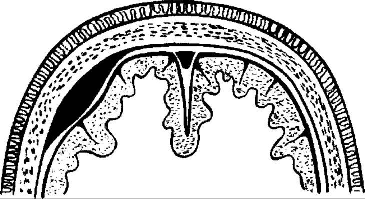
Fig. 31. Compression of the brain by an epidural hematoma.
Epidural hematomas are generally confined to a single lobe.
Clinically, epidural hematomas are characterized by a short lucid interval, a sharp manifestation of signs of brain compression and displacement (progressive impairment of consciousness), and rapidly escalating focal symptoms (ptosis, homolateral mydriasis, heterolateral hemiparesis). Generalized seizures appear early and transition into decerebrate seizures.
Acute subdural hematomas typically form against the background of a brain contusion, resulting from acceleration or deceleration trauma. The source of bleeding is pial veins and cortical arterial vessels associated with the brain contusion (Fig. 32). Subdural hematomas more frequently spread across 2-3 lobes.
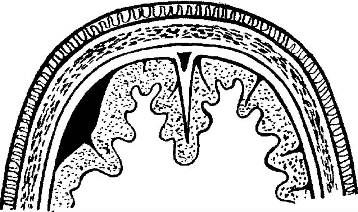
Fig. 32. Compression of the Spinal Cord by a subdural hematoma.
The clinical course may feature a classical lucid interval, though it is more frequently veiled or indistinct. In the clinical picture, generalized cerebral symptoms are more pronounced. Focal symptoms emerge against the background of progressively deteriorating consciousness.
Elevated intracranial pressure leads to a reduction in cerebral perfusion. As a compensatory reaction, tachypnea appears, which, with progressive or prolonged intracranial hypertension, is replaced by a pathological breathing pattern. Concurrently with escalating respiratory disturbances driven by hypoxia and intracranial hypertension, blood pressure rises against a background of bradycardia, which, in the stage of gross clinical decompensation, gives way to arterial hypotension and tachycardia. The latter, occurring alongside profound disturbances of respiration and consciousness, indicate an unfavorable prognosis.
Subacute subdural hematomas typically occur following milder trauma and are characterized by a prolonged "lucid interval," a gradual onset of brain compression symptoms, and a corresponding clinical presentation.
Chronic subdural hematomas develop in cases of mild TBI, often against the backdrop of cerebral atrophy and chronic alcohol abuse, frequently presenting with a pseudotumor-like course. The Emergence of focal and psychopathological symptoms in such cases necessitates brain imaging to rule out a subdural hematoma.
Intracerebral hematomas are a cause of severe TBI, resulting from severe cerebral contusions and lacerations accompanied by vascular damage. These hematomas can be superficial, deep, or deep-seated (Fig. 33).
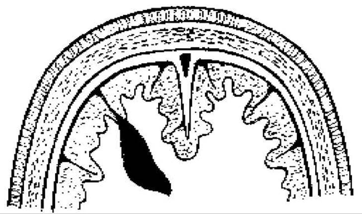
Fig. 33. Compression of the brain by an intracerebral hematoma.
Superficial hematomas are typically directly associated with the contusion-laceration focus of the brain. Deep hematomas are located in the superficial Zones of the white matter, while deep-seated ones are situated in the medial Regions of the brain and subcortical structures. Intracerebral hematomas can form as a result of the coalescence of multiple hemorrhages within contusion zones.
Clinically, intracerebral hematomas are characterized by pronounced both focal and generalized symptoms, with focal symptoms frequently dominating.
Head crushing is a specific type of trauma resulting from the sequential impact of dynamic (short-term) and static (prolonged) mechanical loads, morphologically characterized by injury (prolonged compression) to the soft Tissues of the head, skull, and brain. Clinically, this manifests as the summation and mutual aggravation of systemic, generalized cerebral, cerebral, and extracerebral symptoms. A more precise term is prolonged head compression (PHC). PHC occurs in victims exposed to long-term crushing conditions—such as explosions, structural collapses, or earthquakes. The biomechanics of this injury are characterized by an impact-compression sequence. Prolonged dynamic loading on the head results in a concussion or contusion of the brain. PHC leads to a sharp, sustained increase in intracranial pressure, as well as necrosis of soft tissues and the brain. An additional pathological mechanism is intoxication caused by tissue breakdown products, creating a wide gateway for infection. Pathognomonic symptoms of PHC include head deformation and marked edema of the head's soft tissues, which progresses even after the compromising factor has been removed. PHC is divided into three stages: mild—head compression lasts from 30 minutes to 5 hours, characterized by minor intoxication and complete recovery of trophics; moderate—head compression lasts from 2 to 48 hours, featuring moderate trophic disorders and Intoxication syndrome; severe—compression duration ranges from 1 to 3 days or more, accompanied by total head edema, subsequent necrosis of all soft head coverings, and pronounced intoxication.
Open craniocerebral trauma. The clinical picture of open TBI comprises local signs of skull and brain injury, generalized cerebral manifestations, and focal neurological symptoms. Local signs of open TBI include the presence of brain detritus, cerebrospinal fluid (CSF), and bone fragments within the wound. Alongside local damage to the skull bones and brain, the course of TBI is determined by an existing concussion or contusion of the brain. In open craniocerebral wounds, the clinical course is divided into 5 periods: the initial (acute) period, the period of early reactions and complications, the period of resolution of early complications, the period of late complications, and the period of remote sequelae. The initial period lasts up to 3 days, dominated by generalized cerebral symptoms and brainstem disorders. Primary traumatic soft tissue necrosis, meningeal hematomas, and bone fragments may be observed in the wound. In the first days following injury, intracerebral hematomas frequently develop, accompanied by cerebral edema. The second period—the period of early reactions and complications—begins on days 3–4 and lasts approximately one month. It is characterized by cerebral edema, a decrease in generalized cerebral symptoms alongside an increase in focal symptoms, and the onset of scar formation. Infectious complications (leptomeningitis, encephalitis) arise during this period. The third period—the period of resolution of early complications and containment of the infectious focus—lasts from one to 3–4 months. It is characterized by scar formation and the containment and resolution of infectious processes. The fourth period—the period of late complications—begins 3–4 months after the injury and lasts up to 2–3 years. With a favorable course, this period concludes the scar formation and the resolution of complications from the previous stage. In some patients, infection flare-ups are possible, involving suppuration of the brain scar, brain abscess formation, purulent fistulas, and cranial Osteomyelitis. The fifth period—the period of remote sequelae—begins 2–3 years after the trauma and lasts for many years. It is characterized by processes of ascending and descending nerve fiber degeneration, arachnoiditis, cranial bone defects, hydrocephalus, psychiatric alterations, epileptic seizures, and focal neurological symptoms.
Gunshot wounds are classified into bullet wounds, shrapnel wounds (from landmine, artillery shell, or aerial bomb explosions), as well as injuries caused by pellets, small balls, darts, and other piercing projectiles (see insert, Fig. 34).
The injury zone involves not only soft tissues but also bones, the dura mater, and brain tissue. Non-penetrating gunshot wounds to the soft tissues of the head frequently lead to severe craniocerebral injuries due to brain contusion and intracranial hemorrhage formation. In contrast, penetrating gunshot wounds to the skull invariably cause severe brain damage, including infectious complications.
Blast injuries result in contused and lacerated-contused soft tissue wounds measuring from 3–4 to 18–20 cm. The skin around the wounds is scorched, and the scalp-aponeurosis flap is typically detached from the bone over a significant area and bleeds heavily. Such wounds are contaminated with Hair, technical fluids, metal particles, etc. Gunshot TBI is characterized by comminuted fractures with large bone fragments separated by gaping cracks extending to the Base of the skull.
According to their localization, skull fractures are classified into fractures of the cranial vault and the base of the skull; by type, they are classified as linear, depressed, comminuted, and traumatic suture diastasis. Depressed fractures are characterized by a disruption of the cranial architecture resulting from trauma, with bone fragments displaced below the cranial vault, exerting a focal mass effect on the brain (Fig. 35).
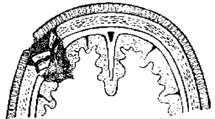
Fig. 35. Compression of the brain by bone fragments from a depressed skull fracture.
They rank second after hematomas in frequency as a factor in brain compression. In turn, depressed fractures are subdivided into depression and impression fractures. Impression fractures are those where bone fragments retain their connection with the cranial vault and are positioned at an angle to it. Depression fractures occur when bone fragments lose their connection with the intact BONES OF THE cranial vault. Depressed fractures are characterized by contusion of the underlying brain tissue. Sharp bone fragments frequently wound blood vessels, damaging major cerebral vessels, the meninges, the venous sinuses of the dura mater, and the brain itself, thereby resulting in epidural, subdural, and intracerebral hematomas.
In open penetrating TBI, particularly gunshot wounds, cerebrospinal fluid leakage is frequently observed. Subarachnoid, ventricular, and cisternal liquorrhea are distinguished, involving damage to the subarachnoid space, ventricles, and brain cisterns, respectively. The presence of liquorrhea, especially ventricular, significantly increases the risk of intracranial infection.
The clinical picture of open TBI is diverse and depends on the nature, severity, and localization of the wound. It is characterized by two main syndromes that dictate treatment tactics and The sequence of therapeutic measures: the "surgical" syndrome (local manifestations of trauma) and the neurological syndrome, which encompasses the entire clinical totality of the traumatic brain disease. The "surgical" syndrome is determined by the type and condition of the craniocerebral wound. Such a wound is characterized by a tissue defect in the injury area, gaping, hemorrhage, pain, leakage of brain detritus from the wound, as well as the length, direction, and trajectory of the wound channel, which can be determined by correlating the entrance and exit openings.
The neurological syndrome consists of generalized cerebral and focal symptoms analogous to those seen in closed TBI. The dynamic clinical course of open craniocerebral trauma features a complex intertwining of generalized cerebral, focal, and brainstem symptoms alongside local wound changes, which together determine the severity of the traumatic brain injury.
General Principles for examining patients with TBI. All patients with TBI, including concussion, require hospitalization in a surgical (neurosurgical) department and necessitate thorough examination and monitoring. When clarifying the presence and nature of brain damage, anamnestic data obtained from accompanying persons or emergency medical personnel—specifically regarding the presence or absence of impaired consciousness or loss of consciousness and the dynamics of these disorders—are of paramount importance. A meticulous physical examination of the victim is crucial to identify tissue damage and subcutaneous hematomas in the head region. A neurological examination, particularly when monitored dynamically over the course of the disease, helps clarify individual generalized cerebral and vegetative symptoms, while in cases of concussion, it helps reveal transient neurological symptoms of Central Nervous System damage. Somatic evaluation is supplemented by routine blood and urine tests.
An important role in TBI Diagnostics is played by skull radiography (craniography), which typically reveals no cranial damage in cases of concussion. The presence of skull fractures indicates a brain contusion, even in the absence of focal neurological symptoms. A fracture of the cranial vault bones in the projection zone of the middle meningeal artery and its branches is a primary indicator of potential epidural hematoma formation.
Echoencephalography is the simplest, most accessible, and safest method, requiring no special preparation of medical personnel or the patient. As an ultrasound pulse passes through a hematoma, it records the appearance of additional echo signals and detects a shift in the midline echo. Two-dimensional and multi-axis echoencephalography provides higher diagnostic value, allowing for the Determination of the hematoma's volume and type.
Cerebral angiography—formerly widely used during the specialized neurosurgical care stage to detect intracranial hematomas, clarify The Nature and localization of brain contusions, and assess cerebral blood flow changes—is now employed only when computed tomography or Magnetic Resonance imaging is unavailable, or when a carotid-cavernous fistula or other vascular pathology (aneurysm, malformation) is suspected.
In the topical diagnosis of brain contusions, radionuclide imaging holds significant value; by utilizing labeled radioactive substances via positron emission tomography, it allows for the detection of cerebral metabolic disturbances following TBI.
A major achievement of modern medical practice is the introduction of advanced in vivo brain imaging methods—computed tomography (CT) and magnetic resonance imaging (MRI). Brain CT makes it possible to detect the presence of a pathological process, determine its nature, localization, and dimensions, identify signs of cerebral edema and the presence and type of dislocations, and track these parameters dynamically. Since the 1980s, diagnostic evaluations have been supplemented by a highly informative visualization method based on the nuclear magnetic resonance phenomenon—magnetic resonance imaging (MRI)—which enables brain slicing in various planes (axial, frontal, sagittal) and the detection of not only morphological brain changes but also, through specialized programs, vascular pathology and metabolic disturbances.
Management of traumatic brain injury. An essential therapeutic measure ensuring a favorable course of severe traumatic brain injury (STBI), rapid healing of sustained wounds, and the patient's recovery is primary surgical debridement (PSD). Wounds of the scalp are subject to PSD. It must be performed after a clinical and radiological examination of the victims. The MAIN OBJECTIVES OF PSD are the definitive cessation of bleeding within the wound, the removal of foreign bodies (bone fragments, hair) and non-viable traumatized tissues that serve as a source of infection and a culture medium for it, and the restoration of the integrity of the integumentary tissues (see insert, Fig. 36).
Bleeding is stopped by ligation, clipping, vessel coagulation, and bandaging. PSD of head wounds is classified as early (performed within the first 24 hours after injury), delayed (during the second day), and late (after 2 days). An important rule for the PSD of soft facial tissues is their conservative (economical) excision. In addition to PSD (when the patient is operated on for the first time), The Need for repeat surgical debridement sometimes arises. It is performed to correct shortcomings of an inadequate primary debridement before the development of purulent infection. Secondary surgical debridement is performed based on secondary indications due to Changes in the wound caused by wound infection. When surgically treating gunshot wounds of The Skull and brain, the following main rules must be guided:
1. Early PSD should be performed. To this end, modern medical transport facilities (helicopter, airplane, resuscitation vehicle) should be used for transportation when necessary.
2. Early implementation of a comprehensive intensive care protocol at the stages of medical evacuation aimed at stabilizing vital function disorders.
3. Prevention of wound infection through the early administration of Antibiotics at the stages of medical evacuation.
4. At the stage of qualified surgical care, wounded patients with TBI are operated on strictly according to life-saving indications (hemorrhage, progressive brain compression).
5. Radical management of skull and brain injuries must be performed by a neurosurgeon at the stage of specialized surgical care.
6. To accelerate the healing of granulating wounds following a purulent process, it is advisable to apply secondary sutures.
7. When removing traumatic intracranial hematomas, three MAIN TYPES OF surgical interventions are used: osteoplastic Craniotomy, decompressive (resection) craniotomy, and the creation of expanded burr holes (trepanation) followed by blood aspiration.
8. In cases of threatening intracranial pressure elevation in TBI, ventricular drainage is performed.
Conservative treatment depends on the severity and type of TBI. The foundation of therapeutic tactics for concussion (mild TBI) consists of bed rest, a protective regimen, and symptomatic therapy. A semi-bed rest regimen for cerebral concussion is prescribed for 5–10 days. Symptomatic therapy is aimed at reducing headache, dizziness, psychomotor agitation, insomnia, and irritability. The prescription of analgesics and sedatives is mandatory.
The normalization of neurodynamic processes is facilitated by the administration of a bromide-caffeine mixture. For dehydration purposes, salutretics (furosemide, lasix, veroshpiron, diacarb) are used once a day for 1–2–3 days post-injury. Additionally, a 25% magnesium sulfate solution, calcium preparations, ascorbic acid, rutin, and desensitizing agents (pipolphen, suprastin) are employed. Autonomic lability is corrected with diazepam or beloid. The improvement of metabolic processes in the brain during the acute period of concussion is facilitated by piracetam (nootropil), aminalon, and B Vitamins.
For mild and moderate closed traumatic brain injury (CTBI), the following measures should be implemented:
- rest (bed rest for 4–10 days, hospital ward regimen for 10–20 days);
- to reduce asthenic syndrome and normalize neurodynamic processes — a bromide-caffeine mixture;
- symptomatic treatment: analgesics for pain, hypnotics in case of insomnia;
- to improve cerebral metabolism — cerebrolysin;
- vitamin therapy is suggested (ascorbic acid, B vitamins);
- for middle-aged and elderly individuals, medications that improve cerebral microcirculation are prescribed (aspirin, cinnarizine, trental, rheosorbilact, sorbilact);
- in case of signs of increased intracranial pressure — magnesium sulfate, saluretics, hypertonic NaCl solution;
- for open/severe TBI — antibiotics;
- hemostatic and general strengthening therapy is carried out.
For severe CTBI, in addition to the aforementioned therapy, neuroleptics and neurovegetative blockade are also used; in the presence of elevated intracranial pressure, not only saluretics but also osmotic Diuretics (mannitol) are administered. Neurovegetative blockade is effective, comprising antihistamines (diphenhydramine, pipolphen, suprastin, diazolin) and neuroleptic agents (chlorpromazine, propazine, thioridazine/tisercin, etaprazine).
In case of hyperthermia, antipyretics are added to the lytic mixture (4% amidopyrine 5.0; acylisine, 50% analgin 2.0–4.0 several times a day). External cooling of the head and major blood vessel projection zones (inguinal region, lateral surface of the neck) is recommended, along with wrapping the patient in wet sheets. General hypothermia can be combined with local brain hypothermia using specialized apparatus such as "Kholod-2f" or "Termokholod-VF".
In patients with moderate and severe contusions, therapy is administered to ensure adequate blood oxygenation, hemodynamic stabilization, and improved cerebral perfusion. The patient is positioned with the head elevated, respiration is normalized, and humidified oxygen is delivered (preferably via a dedicated mask). All patients with an impaired level of consciousness corresponding to a Glasgow Coma Scale (GCS) score of 8 or lower are intubated.
In patients with TBI, blood pressure (BP) must be maintained at an optimal level of 140/80 mmHg or higher. Mean arterial pressure (MAP) should not fall below 90 mmHg. MAP is equal to diastolic pressure plus 1/3 of the pulse pressure. For example, with a BP of 140/80 mmHg, the MAP will be 80 + (140-80)/3 = 80 + 20 = 100 mmHg. In turn, cerebral perfusion pressure (CPP) is the difference between MAP and mean intracranial pressure. In TBI patients, CPP should not drop below 80 mmHg. Autoregulatory mechanisms maintain constant cerebral blood flow when CPP is kept above 70 mmHg. If cerebral perfusion pressure falls below this critical threshold, it leads to a reduction in volumetric cerebral blood flow and the development of brain ischemia.
Adequate sedation of the patient is mandatory, along with the administration of antihypoxants and agents that increase the brain's resistance to hypoxia. Dehydration and anti-edema therapy must be performed with careful monitoring of blood electrolytes and plasma osmolality (290-315 mOsm/L). Agents that improve blood rheology are administered. Vasoactive drugs are not used in the acute period of TBI.
In cases of airway obstruction, the primary objective is to restore airway patency by removing foreign bodies, mucus, and vomitus from the respiratory tract. For central respiratory failure, adequate artificial pulmonary ventilation must be provided, which is achieved through artificial respiration via an endotracheal tube or tracheostomy using ventilators.
Starting from the first day after trauma, if oral intake is impossible, patients receive parenteral Nutrition, and food is administered into The Stomach via a gastric tube in small portions of 100-150 grams several times a day from day one. The tube is inserted through the nose and left in place for 5-7 days.
Organization and content of surgical care at the medical evacuation stage.
First aid is provided at mass casualty sites or at the scene of the incident. It includes applying an aseptic dressing for severe TBI, stopping external bleeding, and managing asphyxia (placing the patient in a stable lateral recovery position, clearing the Oral Cavity of foreign bodies, and inserting an airway tube). For unconscious patients with isolated TBI, the administration of promedol is not advisable. The application of rigid cervical collars is mandatory for such patients.
Pre-hospital care includes management of asphyxia (artificial pulmonary ventilation using a DP-2 apparatus, oxygen inhalation using a KI-4 apparatus), and placing the casualty in a stable lateral recovery position.
First medical care. In cases of isolated TBI, intravenous fluid infusion is not performed, and narcotic analgesics are not administered if consciousness is impaired.
Temporary cessation of external bleeding is achieved using a pressure dressing, suturing, and ligation of bleeding vessels.
Casualties exhibiting signs of brainstem dysfunction are administered intramuscular injections of cordiamine (4.0), caffeine (1.0), and ephedrine (5% - 1.0). For psychomotor agitation, 1.0-3% phenazepam and 2.0-2% chlorpromazine solution are administered. Prevention of wound infection is carried out through intramuscular administration of antibiotics, along with the administration of 0.5 tetanus toxoid.
When there are severe respiratory disorders or continuous leakage of blood and mucus into the Trachea (in fractures of the skull base or facial Skeleton injuries) accompanied by signs of mechanical asphyxia, a tracheostomy is performed.
In the case of a distended Urinary Bladder, urinary catheterization is performed.
Evacuation of casualties with head injuries to a specialized medical facility must be carried out in a supine position.
Qualified surgical care. During medical triage, a group of casualties is identified who require Surgical treatment strictly according to life-saving indications (continuing external bleeding, progressive brain compression).
Primary surgical debridement (PSD) of head wounds consists of 3 main stages: management of the soft tissue scalp wound, bone trepanation in the area of the skull fracture, and Treatment of the dura mater and the brain.
The desire to perform classic PSD of a brain wound at the qualified care stage, lacking the necessary technical equipment and constant neurosurgical skills, only complicates the surgical situation and treatment outlook. Therefore, in some cases, the cessation of external bleeding can be achieved by the topical application of various hemostatic Materials (hemostatic sponge or gauze, a wipe moistened with a 3% hydrogen peroxide solution, a 5% aminocaproic acid solution, or coprofer-applied wipes). However, when surgery is the only means to stop bleeding, it must be considered an intervention aimed at performing the essential elements required to control hemorrhage. Such operations do not replace PSD, but rather help avert the threat of fatal complications and make it possible to defer PSD until the specialized surgical care stage.
Casualties with craniocerebral and brain injuries at the qualified surgical care stage are divided into three groups:
- casualties with external bleeding and progressive brain compression are sent to the operating room;
- casualties in agonal states are sent to the tent for the dying for symptomatic therapy;
- other casualties are evacuated to the forward hospital base.
Casualties who have undergone cranial surgery for penetrating injuries are non-transportable for 14-21 days, but may be evacuated by air transport within a 2-3 day period.
Last update: 08/08/2026
Editorial and Educational Adaptation: This material has been compiled based on the primary/original source text. The project team performed an editorial review, corrected technical inaccuracies, structured sections, and adapted the content for an educational format.
What was processed:
- elimination of formatting defects (OCR errors, structural breaks, corrupted characters);
- editorial organization of content;
- standardization of terminology in accordance with academic sources;
- verification of factual statements against the original source text.
All mentions of the author, publication year, and origin of the primary text have been preserved in accordance with the source.