BIOLOGY Volume 2 - A Guide to General Biology - 2004
14. TRANSPORT IN ANIMALS
14.6. Formation of Tissue Fluid
Tissue fluid, or interstitial fluid, is formed as Blood passes through the capillaries. As previously noted, capillary walls are permeable to small molecules and ions, but do not allow red Blood Cells, platelets, and Plasma Proteins to pass through. Consequently, tissue fluid is an aqueous solution whose composition closely resembles protein-free plasma.
The Water potential (osmotic potential) of plasma is approximately —3.5 kPa, which is much lower than that of tissue fluid. Under these conditions, one might expect water to move from the tissue fluid back into the vessels driven by osmotic pressure. However, the blood (hydrostatic) pressure at the arterial ends of the capillaries is close to 5.2 kPa (Fig. 14.10), acting in the opposite direction. The more negative the osmotic potential of the blood, the greater the tendency for water to enter it from the surrounding Tissues. Conversely, the higher the blood pressure, the stronger the opposite tendency. The same holds true for tissue fluid. Therefore, to determine the net direction of water movement (along with dissolved solutes), one must consider both the osmotic and hydrostatic potentials of these two environments. The figures in Fig. 14.10 show that in the arteriolar half of the capillaries, solution is forced out and fills the microscopic spaces between cells (the interstitial space) as tissue fluid. It is precisely here that the exchange of substances takes place between the blood and the other Tissues of the body.
Class="center">
Fig. 14.10. Formation of tissue fluid and Lymph. Tissue fluid is formed by filtration at the arteriolar end of the capillaries. Some of the fluid lost by the blood here is reabsorbed at the venular end of the capillaries, while the remaining fluid is collected and enters the lymphatic capillaries. The direction of filtration is determined by the balance between the blood (hydrostatic) pressure (HP) generated by The Heart and the osmotic potential of the solution (ψ0). The higher the solute concentration, the more negative this potential. The values in the figure are given in kilopascals. These are average values and should not be applied to all capillaries indiscriminately.
Blood cannot continuously afford to lose such large amounts of fluid. Furthermore, if interstitial fluid were to accumulate unchecked, the tissues would swell—a condition known as oedema. Normally, therefore, water continuously returns to the blood at the same rate at which it leaves. This return occurs via two pathways.
1. During The formation of tissue fluid, protein molecules remain in the blood. Consequently, the blood becomes more concentrated; in other words, its osmotic potential becomes more negative. In addition, the hydrostatic pressure of the blood drops within the capillaries, so at their venular end, the fluid tends to flow back into the blood (Fig. 14.10).
2. The remainder of the tissue fluid diffuses into blind-ending lymphatic capillaries, from which point it is referred to as lymph. Lymphatic capillaries join together to form larger Lymphatic vessels. Lymph is propelled through these vessels by the contractions of surrounding Muscles, while its unidirectional flow is ensured by Valves that function similarly to venous valves (Fig. 14.11).
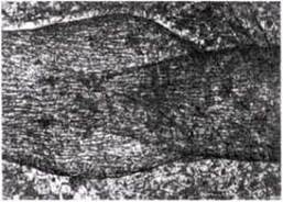
Fig. 14.11. Longitudinal section through a lymphatic vessel showing the internal valve.
From the lymphatic vessels, lymph ultimately enters the Circulatory system; this occurs in the region where the subclavian and jugular Veins merge, just where they drain the arms and HEAD on their way to the heart (Fig. 14.12).
Along the course of the lymphatic vessels, at certain intervals, lie lymph glands, or Lymph Nodes. These store lymphocytes, which circulate in the BLOOD AND LYMPH. Lymphocytes produce Antibodies and form a vital part of the body's immune system. In addition, phagocytes located within the lymph nodes engulf Bacteria and foreign particles from the lymph.

Fig. 14.12. The human Lymphatic system (From E. G. Springthorpe (1973). An Introduction to functional systems in animals, Longman)
Last update: 06/08/2026
Editorial and Educational Adaptation: This material has been compiled based on the primary/original source text. The project team performed an editorial review, corrected technical inaccuracies, structured sections, and adapted the content for an educational format.
What was processed:
- elimination of formatting defects (OCR errors, structural breaks, corrupted characters);
- editorial organization of content;
- standardization of terminology in accordance with academic sources;
- verification of factual statements against the original source text.
All mentions of the author, publication year, and origin of the primary text have been preserved in accordance with the source.