Introduction to Molecular Biology: From Cells to Atoms - Anthony Rees, Michael Sternberg 2002
Cells and molecules at work
Muscles and their contraction
Class="center">Introduction/introduction.files/image098.jpg" width="393"/>
Fig. 37.1.
Vertebrate Muscles are classified into smooth and striated. Smooth muscles, which appear relatively structureless under a microscope, are involuntary, meaning they are not under conscious control; typically, these are slow-contracting muscles associated with Internal Organs. Striated muscles, which appear fibrillar under a microscope, are of two types: skeletal and cardiac. Cardiac Muscle is regulated by nerves and Hormones to produce regular and self-sustaining contractions. Skeletal muscles are voluntary, and although they are also controlled by nerves and hormones, their regulatory mechanism differs from that of cardiac muscle. This section focuses on the Fine Structure of striated skeletal muscles.
STRIATED MUSCLES. The Cells that form these muscles can reach several millimeters in length and are surrounded by an electrically excitable membrane, the sarcolemma. Typically, each such Cell contains multiple nuclei and Mitochondria embedded in a fluid phase known as the sarcoplasm. Each cell contains numerous strands called myofibrils, which are the Organelles responsible for contraction.
A myofibril is a long, thin structure with alternating light and dark regions, which gives the muscle its striated appearance under a phase-contrast microscope. The light regions are the so-called isotropic bands, or I-bands (I-discs), while the dark regions are anisotropic bands, or A-bands (A-discs). In the central part of the A-band, There is a slightly lighter region called the H-zone, and in the middle of this zone runs a narrow dark stripe, the M-line. Each I-band is divided in two by a thin dark Z-line. The segment from one Z-line to the next is considered the structural unit of the myofibril, the sarcomere, which repeats periodically along its entire length.
Myofilaments are the primary Components of the myofibril. They are of two types: thick filaments have a diameter of 15 nm and consist primarily of the fibrous protein Myosin, whereas thin filaments have a diameter of 7 nm and are composed of Actin, Tropomyosin, and troponin. Under an Electron microscope, cross-sections of the I-band reveal only thin filaments, whereas cross-sections of the H-zone within the A-band reveal only thick filaments. In the remaining, very dark Regions of the A-band, both thick and thin filaments are found, forming a regular hexagonal lattice in which each thick filament is surrounded by six thin ones.
Sarcomere contraction can lead to a decrease in its length from approximately 2.5 µm to about 1.7 µm. Meanwhile, the length of the A-band remains constant, while the I-band shortens. This can be explained by the so-called sliding filament model, according to which the length of neither thin nor thick filaments changes during sarcomere contraction, and the approximation of the Z-lines occurs due to the filaments sliding past one another (Fig. 37.2).
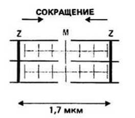
Fig. 37.2.
Myosin, the major component of thick filaments, is constructed from two large and four small polypeptide chains. Each large chain consists of two parts: an extended "tail" with an α-helical conformation, and a globular "HEAD". The tails of both large chains are intertwined around each other, forming a supercoiled structure 140 nm in length. The globular head of each large chain is in a complex with two small chains; the entire complex is also globular. Thus, a myosin molecule has two globular heads and one fibrillar double-stranded tail (Fig. 37.3).
Actin, the primary component of thin filaments, is present in myofibrils as F-actin (filamentous actin). F-actin is a polymer, and the monomeric units from which it is built are called G-actin (globular actin). In its structure, F-actin resembles two strands of beads, where the beads are G-actin molecules; the strands are twisted around each other into a helical structure with a pitch of 36–38 nm.
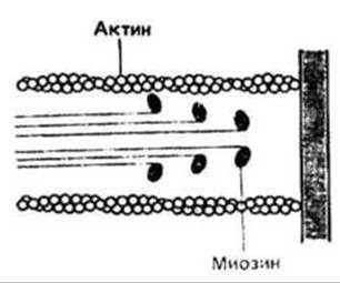
Fig. 37.3.
Tropomyosin is another component of thin filaments. A tropomyosin molecule is a 40 nm long strand formed by two intertwined α-helical polypeptide chains. Tropomyosin is bound to F-actin. Each tropomyosin molecule spans seven G-actin globules, with adjacent molecules slightly overlapping to form a continuous tropomyosin strand running along the F-actin fiber. Since F-actin consists of two strands, it is presumed to associate with two tropomyosin chains as well.
Troponin, yet another component of thin filaments, is a complex of three Proteins: troponin I, troponin T, and troponin C. It has a generally more or less globular shape and is spaced out along F-actin at regular intervals of approximately 38 nm.
ATP provides energy for Muscle contraction. The globular heads of myosin bind ATP and rapidly hydrolyze it, yet they do not readily release the Hydrolysis products, ADP and Pj. F-actin, which binds to myosin to form a complex known as Actomyosin, accelerates the release of ADP and Pj from the myosin heads. The newly vacated ATP-binding sites on the actomyosin complex can bind fresh ATP molecules; as soon as this occurs, however, it induces the dissociation of actomyosin into Actin and myosin. This cycle can repeat indefinitely in the presence of an adequate supply of ATP. The described interaction between actin and myosin forms The basis of the Molecular Mechanism of muscle contraction.
The power stroke cycle of myosin heads During muscle contraction comprises four distinct stages (Fig. 37.4).
1. Myosin in the thick filaments contains bound ADP and Pi, but is not attached to the actin of the thin filaments.
2. Upon receiving a contraction signal, globular myosin heads bound with ADP and Pi attach to actin, forming actomyosin.
3. The formation of actomyosin accelerates the release of ADP and Pi, which is accompanied by the tilting of the myosin heads; this tilt causes the thin actin filament, still attached to the head, to slide along the thick filament, resulting in sarcomere shortening.

Fig. 37.4.
4. ATP binds to the myosin heads within the actomyosin complex, causing actin to detach from myosin; subsequent hydrolysis of ATP by myosin returns the system to The first phase of the cycle.
Muscle activity is regulated by Calcium Ions (Ca2+). At low Ca2+ concentrations, troponin and tropomyosin prevent the interaction between actin and myosin. When a Nerve Impulse arrives (Ch. 36) and muscle cell membrane depolarization occurs, the intracellular level of Ca2+ rises, triggering a Ca2+-dependent conformational change in troponin. This change is transmitted to tropomyosin, causing it to shift its position on the actin filament so that the binding sites become accessible to the myosin heads.
Last update: 13/08/2026
Editorial and Educational Adaptation: This material has been compiled based on the primary/original source text. The project team performed an editorial review, corrected technical inaccuracies, structured sections, and adapted the content for an educational format.
What was processed:
- elimination of formatting defects (OCR errors, structural breaks, corrupted characters);
- editorial organization of content;
- standardization of terminology in accordance with academic sources;
- verification of factual statements against the original source text.
All mentions of the author, publication year, and origin of the primary text have been preserved in accordance with the source.