Molecular Biology of the Cell - Volume 3 - Alberts B., Bray D., Lewis J., Raff M., Roberts K., Watson J. 1994
From Cells to Multicellular Organisms
The Immune System
Fine Structure of Antibodies
Because Antibodies exist in such an enormous variety of forms, in an unimmunized Organism any single type of antibody accounts for less than one-millionth of all Ig molecules present in the Blood. This fact presented immunochemists with an exceptionally difficult Protein Chemistry problem: how is it possible to obtain a sufficient quantity of a specific antibody to determine its Amino Acid Sequence and three-dimensional Structure?
This problem was solved thanks to a specific property of tumor Cells arising in multiple myeloma, a malignant condition characterized by The Development of multiple tumors in the Bone Marrow ("myelogenic" tissue). These cells secrete large quantities of a single type of antibody into the bloodstream. Such antibodies are homogeneous, or monoclonal, because Cancer typically originates from the uncontrolled growth of a single Cell (see Section 21.1.2); in this case, an antibody-producing plasma cell. The antibody that accumulates in the blood is known as myeloma protein.
It has been known since the last century that the urine of patients suffering from this disease frequently contains unusual Proteins, termed Bence-Jones proteins after the English physician who first described them. However, it was not until the 1950s that these proteins were shown to be free immunoglobulin light (L) chains. Initially, the detailed Structure of antibodies was determined by studying myeloma proteins from the urine or blood of patients, or from mice in which analogous forms of cancer had been purposefully induced. Subsequently, it became possible to immortalize antibody-secreting B cells by fusing them with non-secreting myeloma cells. The resulting hybridomas became a convenient source of Monoclonal Antibodies, which can be obtained against any desired antigen in unlimited quantities (Section 4.5.4).
18.3.1. L and H chains consist of constant and variable regions [11, 14]
Comparing the Amino acid sequences of numerous myeloma proteins revealed a striking feature with important and unexpected genetic implications: the N-terminal portion of the sequence in both L and H chains is variable, whereas the C-terminal portion is constant.
For example, if one compares the sequences of A large number of different myeloma $\kappa$ chains (each about 220 Amino Acids long), it turns out that their C-terminal halves are identical or differ only slightly, whereas their N-terminal halves are entirely distinct. Thus, L chains contain a constant region of approximately 110 Amino Acids and a variable region of the same length. The N-terminal variable region of H chains also consists of roughly 110 amino acids, while the constant region comprises 330 or 440 amino acids, depending on the antibody Class (Fig. 18-24).
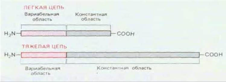
Fig. 18-24. Both light and heavy chains of molecules consist of constant and variable regions. For all light chains of a given type ($\kappa$ or $\lambda$), the entire C-terminal half has an identical amino acid sequence (sometimes with minor variations), whereas the N-terminal halves differ. The N-terminal variable regions of heavy and light chains are similar in length (about 110 amino acid residues), whereas the constant region in heavy chains is three to four times longer than in light chains (depending on the antibody class).
It is precisely the N-terminal portions of the L and H chains that jointly form the antigen-binding site, and the Variability of their amino acid sequences serves as the structural basis for The Diversity of these sites. The existence of Variable and constant regions raises important questions regarding the genetic mechanisms of antibody formation, which we will discuss later. Yet even before these questions could be directly addressed, The Study of myeloma proteins uncovered other crucial features of antibody structure.
18.3.2. Each L and H chain contains three hypervariable regions that together form the antigen-binding site [17]
Only a portion of the variable region participates directly in antigen binding. This Conclusion was initially reached by estimating the maximum dimensions of the antigen-binding site. The first measurements, using oligomers of varying sizes as "molecular rulers," were performed on antibodies directed against dextran, a polymer of D-glucose. When Disaccharides, trisaccharides, and longer Oligosaccharides composed of glucose residues were used to inhibit the binding of dextran to its antibodies, their inhibitory effect increased with chain length up to approximately six monomers; with larger oligosaccharides, the effect increased no further. This suggested that the largest antigen-binding sites can contact at most five or six sugar residues of the antigen. Therefore, it seemed highly unlikely that all 220 amino acids of the L and H chain variable regions directly contribute to building the antigen-binding site.
Indeed, it is now clear that the antibody-binding site is formed by only about 20–30 amino acid residues from the variable region of each chain. The first evidence for this came from amino acid sequence data, which showed that differences among variable regions in both L and H chains are largely restricted to three small hypervariable regions in each chain. The remaining parts, known as framework regions, are relatively constant. These data led to the prediction that the antigen-binding site is formed by just 5–10 amino acids from each hypervariable region (Fig. 18-25), which was subsequently confirmed by X-ray crystallographic analysis of antibodies (see below).

Fig. 18-25. This highly simplified diagram illustrates how the three hypervariable regions of each light and heavy chain jointly form the antigen-binding site of the antibody molecule. Hypervariable regions are sometimes referred to as complementarity-determining regions.
18.3.3. L and H chains are folded into a series of repeating homologous domains [11, 16, 18]
When the complete amino acid sequence of an H chain was first determined in the late 1960s, another important feature of Ig structure became apparent. It turned out that the constant region, which in most H chains is roughly three times longer than in L chains, consists of three homologous segments, each approximately 110 amino acids long and containing a single intrachain disulfide bond. These three segments are homologous not only to one another but also to the constant region of L chains. The single variable domains in L and H chains are also similar to each other, and to a lesser extent, to the constant domains.
These data led to the prediction that both L and H chains are constructed from repeating segments, or domains, each of which folds independently to form a compact functional unit. Indeed (Fig. 18-26), an L chain consists of one variable domain (VL) and one constant domain (CL), whereas most H chains consist of one variable domain (VH) and three constant domains (CH1, CH2, and CH3). (Each $\mu$ and $\varepsilon$ chain contains one variable and four constant domains.) The variable domains are responsible for antigen binding, whereas the constant domains of H chains (with the exception of CH1) form the Fc region, which determines other biological properties of antibodies.
The Homology between domains suggests that Ig chains evolved through a series of successive duplications of an initial ancestral Gene that encoded a single domain of unknown function consisting of 110 amino acids (Section 18.6.20). This hypothesis is supported by evidence that each domain of the H chain constant region is encoded by a separate DNA sequence (exon) (Fig. 18-27).

Fig. 18-26. Each light and heavy chain in an Ig molecule is folded into repeating domains that resemble one another. The variable domains of both chains (VL and VH) form the antigen-binding sites (see Fig. 18-25), whereas the constant domains of the heavy chains (primarily CH2 and CH3) determine other biological Properties of the molecule. The heavy chains of IgM and IgE antibodies possess an additional constant domain, CH4.

Fig. 18-27. Organization of DNA sequences encoding the HEAVY CHAIN CONSTANT region. The sequences encoding each domain and the hinge region (exons) are separated by non-coding sequences (introns). Introns are removed by splicing primary RNA transcripts during mRNA formation. The DNA encoding the heavy chain variable region is not shown. It is possible that the presence of introns in the DNA sequence facilitated random duplications in DNA, leading to The Emergence of antibody genes during evolution (Sections 9.4.12 and 10.5.3).
18.3.4. X-ray crystallographic studies have revealed the three-dimensional structure of immunoglobulin domains and antigen-binding sites [19]
Even when the complete amino acid sequence of a protein is known, its three-dimensional structure cannot be deduced from it alone—this requires X-Ray Diffraction Analysis of protein crystals (Section 4.1.14). Several myeloma protein fragments and one intact IgG have already been obtained in crystalline form. Data from X-ray analysis of these proteins confirmed the immunochemists' predictions. Even more importantly, these studies provided insight into how millions of distinct antigen-binding sites are constructed based on a single structural framework.
As shown in Fig. 18-28, all Ig domains share a very similar spatial architecture based on the so-called immunoglobulin fold. Each domain is roughly a cylinder measuring $4 \times 2.5 \times 2.5$ nm, constructed as a "sandwich" of two protein layers, where one layer is formed by three and the other by four segments of the polypeptide chain. Within each layer, adjacent segments are antiparallel and form a $\beta$-sheet (Section 3.3.2). These two layers lie approximately parallel to each other and are linked by a single intrachain disulfide bond.
Variable domains are unique in that each of them features a distinct set of three hypervariable regions folded into three hypervariable loops (see Fig. 18-28). The hypervariable loops of the L and H variable domains group together to form the antigen-binding site, exactly as predicted. An important principle revealed by these studies is that the variable region of an antibody molecule consists of a highly conserved, rigid framework with hypervariable loops attached to one of its ends. Consequently, an enormous diversity of antigen-binding sites can be generated by altering solely the hypervariable loops—their length and amino acid sequence—without disrupting the overall three-dimensional structure required for antibody function.
X-ray crystallographic ANALYSIS OF ANTIBODY fragments bound to an antigen or an antigenic determinant (hapten) has made it possible to determine precisely how (in A number of specific cases) the hypervariable loops of the L and H variable domains jointly form a single antigen-binding surface. The size and shape of each such surface vary depending on the conformation of the polypeptide chain within the hypervariable loops, which in turn is determined by The sequence of amino acid side chains in these loops. Thus, the General Principles of antibody structure are now well understood.

Fig. 18-28. Three-dimensional structure of an IgG antibody molecule (based on X-ray crystallography data). A. Each amino acid residue of the protein molecule is depicted as a small sphere. One of the heavy chains is shown in white, and the other in dark gray. The light chain domains are shown in color. The antibody molecule is glycosylated: the oligosaccharide chain attached to the CH2 domain is depicted in light gray. B. Three-dimensional folding of the entire light chain. Both the variable and constant domains consist of two ß-sheets (one composed of three strand segments, and the other of four). The polypeptide chains in these two sheets are shown in different colors; the sheets are connected by a disulfide bond. Note that all hypervariable regions form loops at the distal end of the variable domain, where they converge to form part of the antigen-binding site (A - after E. W. Silverton et al., Proc. Natl. Acad. Sci. USA 74, 5140, 1977; B - after M. Schiffer et al., Biochemistry, 12, 4620, 1973. Copyright 1973 Amer. Chem. Soc.)
Conclusion
Each immunoglobulin L and H chain consists of a variable region about 110 amino acid residues long at the N-terminus, followed by a constant region of the same length in the L chain and three to four times that length in the H chain. Each chain is composed of repeating, similarly folded domains: the L chain has one domain in the variable region (VL) and one in the constant region (CL), whereas the H chain has one domain in the variable region (VH) and three or four in the constant region (CH). The amino acid sequence variability in the variable Regions of the L and H chains is largely restricted to a few short hypervariable regions that are spatially proximate to one another and form the antigen-binding site. This site is sufficiently large to contact an antigenic determinant comparable in size to five or six sugar residues.

Fig. 18-29. Three-dimensional structure of an antigen-antibody complex (based on X-ray crystallography data). The antigen—in this case, the enzyme Lysozyme—is highlighted in color. The antigen-binding site of the antibody Fab fragment is formed jointly by two chains, light and heavy (light gray and dark gray in the figure, respectively). B. Here, the antigen and antibody models are separated to reveal their complementary interacting surfaces. The protrusion on the complementary surface of the antigen represents a glutamine residue. In several other antibodies investigated by the same method, the antigen-binding site (for a small hapten) forms a much deeper cleft. (A. Amit et al., Science, 233, 747-753, 1986. Copyright 1986 by the AAAS.)
Last update: 12/08/2026
Editorial and Educational Adaptation: This material has been compiled based on the primary/original source text. The project team performed an editorial review, corrected technical inaccuracies, structured sections, and adapted the content for an educational format.
What was processed:
- elimination of formatting defects (OCR errors, structural breaks, corrupted characters);
- editorial organization of content;
- standardization of terminology in accordance with academic sources;
- verification of factual statements against the original source text.
All mentions of the author, publication year, and origin of the primary text have been preserved in accordance with the source.