BIOCHEMISTRY - V. V. Emelyanov - 2016
SECTION 5. METABOLISM OF PROTEINS AND NUCLEIC ACIDS
5.1. Biological Role of Proteins
Quantitatively, proteins constitute the most important group of macromolecules. Proteins perform a number of unique Functions:
- biocatalytic: all Enzymes are proteinaceous substances;
- structural (plastic): proteins form an integral part of vital cellular and tissue structures;
- regulatory: the majority of Hormones have a protein-peptide nature;
- protective: proteins known as IMMUNOGLOBULINS protect the Organism against foreign Antigens;
- respiratory: the protein Hemoglobin, found in erythrocytes, ensures The transport of oxygen to Tissues and CO2 to the Lungs;
- transport: many substances vital to the organism are carried through the bloodstream in complexes with proteins.
A human body with a mass of 70 kg contains approximately 10 kg of protein, and the recommended daily Dietary Protein Intake is about 100 g. The proportion of other nitrogen-containing compounds in the body is small; therefore, nitrogen balance is determined primarily by Protein METABOLISM. Unlike Introduction/36.html">CARBOHYDRATES and Lipids, proteins and their constituent Amino Acids cannot be stored as reserves in the body.
5.2. Transformations of PROTEINS AND AMINO Acids in the Organism
Proteins ingested with food are broken down into amino acids in the gastrointestinal tract through the action of Proteolytic Enzymes (peptide Hydrolases, peptidases, proteases). Internal peptide bonds are cleaved by Endopeptidases (Pepsin, Trypsin, and Chymotrypsin), whereas terminal bonds are cleaved by exopeptidases (Carboxypeptidases and aminopeptidases). Together, endo- and exopeptidases complete the Hydrolysis of Proteins down to individual amino acids.
The resulting Amino acids are then absorbed by the intestinal wall and, after being transported via the bloodstream, enter Cells to be utilized in Various metabolic pathways, the principal one being the Synthesis of the body's own proteins. A significant portion of amino acids is distributed by the Blood throughout the entire body and used for various physiological needs. Amino acids also participate in the Synthesis of specific nitrogen-containing compounds, such as purine and pyrimidine NUCLEOTIDES, creatine, etc. The Amino Acid Nitrogen, cleaved during Cytology/cytology/16.html">Early stages of Catabolism, can either be reutilized in anabolic processes According to the body's demands or incorporated into the final product of Nitrogen metabolism, urea, and excreted from the organism. The nitrogen-free carbon skeletons of amino acids most commonly form keto acids, which subsequently degrade via the common Catabolic pathways of oxidized Hydrocarbons. By entering these general catabolic pathways, Amino acids can serve as an energy source. The main PATHWAYS OF AMINO acid utilization are illustrated in Fig. 21.
Class="center">Fig. 21. Main Pathways of amino acid utilization

The organism maintains an amino acid pool that is replenished from the following sources:
- dietary amino acids;
- amino acids resulting from the catabolism of the body's own proteins;
- amino acids synthesized de novo by the cells themselves.
However, the capacity for Amino acid synthesis in The Human Body is quite limited. Table 7 lists the Essential Amino Acids, which cannot be synthesized by cells due to the lack of necessary synthetic precursors; consequently, their presence in dietary proteins is mandatory. There are also conditionally essential amino acids, which can be synthesized in small amounts by the organism, and semi-essential amino acids, the synthesis of which requires essential amino acids. The requirement for non-essential amino acids can be met by synthesis from Other Compounds.
Table 7. Non-essential and essential amino acids
Essential |
Conditionally essential |
Semi-essential |
Non-essential |
Valine |
|||
Isoleucine |
Asparagine |
||
Leucine |
Aspartate |
||
Glutamate |
|||
Glutamine |
|||
Phenylalanine |
The Amino Acid Composition determines the Biological value of a protein. The higher the content of essential amino acids, the greater its nutritional value. Dietary proteins are considered complete when they contain essential amino acids in the required quantitative and qualitative proportions.
5.3. General Pathways of Amino acid metabolism
The general pathways of amino acid metabolism include deamination, Transamination, and decarboxylation reactions:

Transamination reactions are among the most critical pathways in general amino acid metabolism. They play a primary role in Gluconeogenesis and the synthesis of new amino acids. The Essence of this reaction is the reversible transfer of an amino group from an amino acid to a keto acid without The intermediate formation of ammonia. Transamination reactions are catalyzed by transaminases (aminotransferases), with Pyridoxal phosphate (the active form of vitamin B6) serving as the coenzyme:

Many amino acids participate in transamination (with the exception of lysine and threonine), with glutamic and aspartic acids being the most active. This is attributed to the high concentration in animal tissues of two specific aminotransferases: aspartate aminotransferase and alanine aminotransferase.
Aminotransferases are involved in the synthesis of new amino acids from α-ketoglutarate. This synthesis can also yield essential amino acids provided that the corresponding keto acids are supplied with the diet.
Deamination is a reaction in which the amino group is released as ammonia. Deamination mechanisms can vary:
a) reductive deamination (resulting in The formation of a saturated fatty acid):

b) hydrolytic deamination (resulting in the formation of a hydroxy carboxylic acid):

c) eliminative deamination (resulting in the Formation of Unsaturated Fatty acids):

d) Oxidative Deamination (resulting in the formation of keto acids). In humans, oxidative deamination serves as the primary pathway of Amino Acid Catabolism:

There are two types of oxidative deamination: Direct and Indirect. If the amino group is directly converted into ammonia during deamination, the process is referred to as direct oxidative deamination. Direct oxidative deamination can occur under both aerobic and anaerobic conditions:
1. Aerobic direct oxidative deamination is catalyzed by D-Amino Acid and L-amino acid oxidases, utilizing FAD and FMN as Coenzymes, respectively. Reactions Catalyzed by these oxidases proceed slowly within cells.

2. Anaerobic direct oxidative deamination exists exclusively for L-glutamic acid and is catalyzed by the enzyme Glutamate dehydrogenase. This enzyme is present in the Cell/35.html">Mitochondria of all body cells (except for Muscle cells). The glutamate dehydrogenase reaction requires the coenzymes NAD+ or NADP+:

During the deamination of glutamate, the amino group is immediately converted into ammonia.
The glutamate deamination reaction catalyzed by glutamate dehydrogenase is reversible. The reverse reaction—the reductive amination of α-ketoglutarate—serves as a source of glutamate in cells and detoxifies ammonia.
All amino acids except glutamic acid undergo deamination via indirect pathways. Indirect oxidative deamination is highly active in all body cells and consists of two stages:
1. Transamination with α-ketoglutarate to form glutamate;
2. Direct oxidative deamination of glutamate.
As a result of transamination, α-ketoglutarate is converted into glutamate, while amino acids lose their amino groups and are transformed into α-ketoacids. Subsequently, their carbon Skeleton undergoes catabolism via specific pathways and enters the Krebs cycle, where it is oxidized to carbon dioxide and Water.
Glutamic acid acts as a common collector for all amino groups, and it alone undergoes direct oxidative deamination, yielding ammonia and α-ketoglutaric acid.

Thus, the direct oxidative deamination reaction is closely linked to AMINO ACID TRANSAMINATION. The coupling of transamination and deamination reactions directs the excess amine nitrogen to the Liver for urea synthesis and to the Kidneys for the synthesis of ammonium salts.
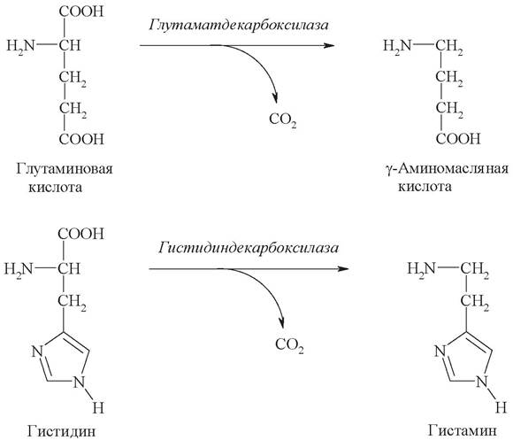
The Decarboxylation of amino Acids is catalyzed by Decarboxylases, which require Pyridoxal phosphate as a coenzyme. The products of decarboxylation exhibit high biological activity, hence their designation as "biogenic amines" (Table 8):

Table 8. Biogenic amines
Amino acid |
Amine |
Function |
Serine |
Ethanolamine |
A constituent of Phospholipids |
Histidine |
Histamine |
Produced in mast cells; acts as a potent vasodilator |
Threonine |
Aminopropanol |
A constituent of vitamin B12 |
Tryptophan |
Serotonin |
Produced in hypothalamic Neurons; functions as an excitatory neurotransmitter |
Cysteine |
Cysteamine |
A constituent of CoA |
Glutamic acid |
y-Aminobutyric acid (GABA) |
Produced in Brain tissue; functions as an inhibitory neurotransmitter |
Tyrosine |
Dopamine |
Produced in the kidneys, Adrenal Glands, synaptic ganglia, and nerves. Functions as a neurotransmitter in the motor structures of the brain |
Examples include the decarboxylation reactions resulting in the formation of GABA and histamine:
5.4. Mechanism of Ammonia Toxicity
One of the final products of amino acid metabolism is ammonia, which is highly toxic to the body. Even a slight increase in its concentration exerts adverse effects on the organism, particularly on the Central Nervous system:
1. Ammonia easily penetrates cell membranes and shifts the glutamate dehydrogenase-catalyzed reaction in mitochondria toward glutamate synthesis:

A decrease in α-ketoglutarate concentration leads to the inhibition of amino acid transamination, as well as a slowdown of the Krebs cycle and The Development of energy deficiency.
2. Ammonia is a basic compound; an increase in its blood concentration shifts the pH toward the alkaline range, causing alkalosis. This increases the oxygen affinity of hemoglobin, thereby hindering oxygen release. As a result, tissue Hypoxia and energy deficit develop, which primarily affect the brain.
3. Being a lipophilic substance, ammonia readily crosses the blood-brain barrier into Cells of the central nervous system. Elevated concentrations of ammonia stimulate the Synthesis of Glutamine from glutamate in neural tissue, mediated by Glutamine Synthetase:

The accumulation of glutamine in neuroglial cells leads to an increase in osmotic pressure, Swelling of astrocytes, and, at high concentrations, brain edema. A decrease in glutamate concentration disrupts amino acid and neurotransmitter metabolism—specifically, the synthesis of y-aminobutyric acid (GABA), the major inhibitory neurotransmitter. A deficiency in GABA and other Neurotransmitters impairs Nerve Impulse Conduction and triggers seizures.
4. Upon dissolving in water, ammonia forms the ammonium ion (NH+4), which is virtually impermeable to cytoplasmic and mitochondrial membranes. Excess blood NH+4 disrupts the Transmembrane Transport of monovalent cations Na+ and K+ by competing with them for Ion Channels, which also affects nerve impulse transmission.
5. Low concentrations of ammonia stimulate the respiratory center, whereas high concentrations inhibit it.
5.5. Detoxification of Ammonia
5.5.1. Urea Synthesis
Virtually all ammonia is eliminated from the body through the kidneys as urea—synthesized in the liver—and as ammonium ion salts formed in the epithelial cells of the renal tubules.
Urea is the primary end product of nitrogen metabolism, accounting for up to 90% of total nitrogen excretion from the body. Normal urea excretion is approximately ~ 25 g/day. An increase in dietary protein intake leads to elevated urea excretion. Urea is synthesized exclusively in the liver.
The Urea Cycle (urea cycle, Ornithine cycle, Krebs–Henseleit cycle) is a cyclic metabolic process. It proceeds in 3 stages comprising 5 reactions, each catalyzed by a specific enzyme:
- synthesis of The amino acid citrulline (two reactions);
- synthesis of the amino acid arginine (two reactions);
- formation of urea (one reaction).
Urea synthesis begins in the mitochondria (the First and Second reactions), while the remaining three reactions take place in the Cytosol (Fig. 22).
Fig. 22. The urea cycle

The First stage—the synthesis of the amino acid citrulline—takes place in the liver mitochondria, where ammonia is detoxified by combining with carbon dioxide to form carbamoyl phosphate, a reaction mediated by the enzyme carbamoyl phosphate synthetase (1).
This is followed by a Condensation reaction between the resulting carbamoyl phosphate and the amino acid ornithine, catalyzed by ornithine transcarbamylase; this reaction yields citrulline and regenerates a molecule of inorganic phosphate (2).
The Second Stage—the synthesis of arginine from citrulline and aspartate (the amino group donor)—takes place in the liver cytosol and involves two reactions. The first is the condensation of citrulline and aspartic acid to form argininosuccinate, catalyzed by argininosuccinate synthetase (3). In the second reaction, argininosuccinate is cleaved into arginine and fumaric acid by the action of argininosuccinate lyase (4).
In the Third Stage, arginine is cleaved into urea and ornithine by the enzyme arginase (5).
The overall net reaction of urea synthesis can be represented as follows:

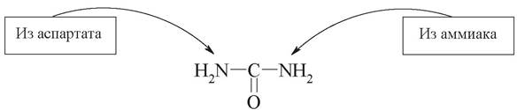
Analysis of the urea cycle shows that nitrogen atoms are incorporated in two separate reactions. One nitrogen atom enters in the form of NH3 in reaction 1 as a product of AMINO ACID DEAMINATION, while the other is incorporated as part of aspartate (reaction 3). This second nitrogen atom can be supplied to aspartate from any amino acid via transamination with oxaloacetate. Consequently, the nitrogen atoms in urea have different origins:
Interconnection Between the Urea Cycle and the Krebs Cycle
Complex relationships exist between the Krebs cycle and the urea cycle, which to a certain extent determine reaction rates depending on the energy demands of The Cell and the concentrations of end metabolites. The initiating reactions of both the urea cycle and the Krebs cycle take place within the mitochondrial matrix.
Urea synthesis is linked to the Krebs cycle through three main pathways:
1. Fumarate serves as a shared metabolite of both the urea cycle and the Krebs cycle. In the Krebs cycle, fumarate is converted into malate and then into oxaloacetate. Oxaloacetate undergoes transamination to form aspartate, which then enters the urea synthesis pathway. Oxaloacetate is a vital metabolite involved in both the TCA cycle and gluconeogenesis.
2. The oxidation of malate to oxaloacetate in the Krebs cycle generates 3 molecules of ATP, which are subsequently utilized to drive urea synthesis.
3. The supply of CO2 required for urea formation is provided by The activity of the Krebs cycle:
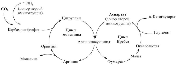
Thus, through this unusual coupled mechanism, the reactions of both cycles are intricately intertwined. This mechanism is commonly referred to as the "Krebs bicycle" or the "Krebs twin-cycle mechanism."
5.5.2. Synthesis of Ammonium Salts
The direct synthesis of ammonium salts takes place in the lumen of the renal tubules from ammonia and hydrogen ions secreted therein, combined with organic anions (acetic, oxalic, and lactic acids) and inorganic anions (phosphates, chlorides, and sulfates) filtered in the primary urine. Approximately 10% of total ammonia is excreted by The Kidneys in the form of ammonium salts.
5.6. Fate of the Nitrogen-Free Amino Acid Residue
The catabolism of carbon skeletons derived from amino acid deamination results in the formation of either acetyl-CoA (which is further converted into fats or Ketone Bodies, known as ketogenic amino acids) or metabolites capable of entering gluconeogenesis (glucogenic amino acids).
There are several specific pathways of amino acid catabolism that converge into 6 products entering the Krebs cycle, where they are completely oxidized to CO2 and H2O.
Glucogenic amino acids include alanine, glycine, threonine, serine, cysteine, methionine, aspartate, asparagine, glutamate, glutamine, arginine, proline, and histidine. Their breakdown yields gluconeogenesis substrates such as Pyruvate or Krebs cycle metabolites (oxaloacetate, α-ketoglutarate, succinyl-CoA).
Strictly ketogenic amino acids are lysine and leucine; their oxidation yields exclusively acetyl-CoA, which subsequently participates in the synthesis of ketone bodies, fatty acids, and Cholesterol.
There is a small group of mixed amino acids that give rise to both pyruvate or Krebs cycle metabolites and acetyl-CoA. Mixed amino acids include phenylalanine, tyrosine, isoleucine, and tryptophan.

For A number of amino acids, specific metabolic pathways exist due to the peculiarities of their chemical Structure and physiological role in the body.
5.7. Amino acid Biosynthesis
Plants and many species of Bacteria possess the enzymes required to synthesize all necessary α-keto acids. Animals have lost The ability to synthesize certain α-keto acids from which essential amino acids are derived. Other α-keto acids can be formed through the metabolism of other compounds (primarily glucose) and utilized to synthesize non-essential amino acids.
Humans and animals are capable of synthesizing only 8 out of the 20 amino acids required for Protein Synthesis from non-amino acid precursors (non-essential amino acids); 2 amino acids (semi-essential amino acids) are synthesized in limited quantities, and another 2 are formed from essential amino acids (conditionally essential amino acids). The carbon skeleton of amino acids is derived from glycolytic intermediates, the Pentose Phosphate Pathway, and the Krebs cycle. The primary pathways of amino acid biosynthesis include: direct amination of α-keto acids or unsaturated organic acids, transamination reactions, and enzymatic interconversions of individual amino acids—both essential and non-essential.
Below is a diagram illustrating the synthesis of eleven non-essential amino acids whose carbon skeletons can be formed from glycolytic and Krebs cycle metabolites:

It should be noted that the source of the sulfur atom in the conditionally essential cysteine molecule is essential methionine. The twelfth amino acid, tyrosine, is synthesized via the hydroxylation of the essential amino acid phenylalanine and is not shown in the diagram.
5.8. Digestion AND ABSORPTION of Nucleic Acids
The human body ingests approximately 1 g of nucleic acids daily through food. The Digestion of nucleic acids takes place in the Small Intestine (Fig. 23). Dietary nucleic acids undergo hydrolysis through the action of pancreatic juice enzymes. The enzymes catalyzing nucleic acid breakdown are called Nucleases or phosphodiesterases. Based on their action Specificity, they are divided into deoxyribonucleases (DNases, which cleave DNA) and ribonucleases (RNases, which cleave RNA).
Fig. 23. Schematic representation of nucleic acid Cleavage (hydrolysis)

Endonucleases and exonucleases are distinguished. Endonucleases act on internal phosphodiester bonds within DNA and RNA molecules, resulting in The breakdown of nucleic acids primarily into oligonucleotides. Exonucleases cleave nucleotides from the 3'- or 5'-end of the polynucleotide chain, leading to the formation of free mononucleotides.
As a result of intracellular endo- and exonuclease activity, nucleic acids are broken down into mononucleotides, which are subsequently hydrolyzed by small intestinal enzymes known as nucleotidases (Phosphatases) to yield the corresponding nucleoside and orthophosphoric acid.
Nucleosides are further broken down by nucleosidases into nitrogenous bases and pentoses (ribose or deoxyribose).
The products of nucleic acid digestion enter the bloodstream and are subsequently transported to The Liver and other Organs. In living cells, RNA turnover is considerably more active than DNA turnover.
Ultimately, nucleic acids are broken down into nitrogenous bases, pentoses, and phosphoric acid.
Nitrogenous bases are converted into End products of Metabolism and excreted in the urine. During catabolism, purine nitrogenous bases lose their amino group as ammonia, become oxidized, and are converted into uric acid. Pyrimidine bases undergo more extensive degradation into carbon dioxide, water, and ammonia.
Pentoses enter The pentose phosphate pathway and can be oxidized to carbon dioxide and water.
Phosphoric acid does not undergo degradation and is utilized for the phosphorylation of Organic compounds or excreted from the body via urine.
The products of nucleic acid hydrolysis enter the body's cells, where they are used to synthesize nucleotides and nucleic acids, as well as to meet the organism's Energy Requirements.
5.9. Catabolism of Purine Bases
The cleavage reactions of purine nucleotides—adenosine monophosphate (AMP) and guanosine monophosphate (GMP)—proceed via different pathways, yet they converge on a single common product: xanthine (Fig. 24). Initially, nucleotides undergo hydrolytic Cleavage of the phosphate group to yield their respective nucleosides, adenosine and guanosine. Guanosine is subsequently hydrolyzed into the free base guanine, which is directly converted into xanthine. The formation of xanthine from adenosine occurs through a series of intermediate steps: the deamination of adenosine to form inosine, the hydrolysis of inosine into the free base hypoxanthine, and, finally, the oxidation of hypoxanthine to xanthine. The conversion of hypoxanthine to xanthine is catalyzed by xanthine oxidase. This enzyme is found in significant quantities in the liver and intestines, where it oxidizes Purines using molecular oxygen, generating a highly toxic superoxide radical (O2-) as a byproduct.
Fig. 24. Degradation pathways of purine nucleotides

The ultimate metabolic fate of xanthine varies and depends on the specific organism. In humans, as in most primates, birds, certain reptiles, and the majority of insects, xanthine is converted into uric acid by xanthine oxidase. Uric acid is synthesized predominantly in the liver. The human body produces about 0.5–1 g of uric acid daily, which is excreted primarily through urine and, to a lesser extent, via feces. Uric acid has low solubility in water; consequently, when NUCLEIC ACID METABOLISM is impaired, it crystallizes and deposits in the finger joints, Cartilage, Skin, and Muscles, forming nodules. A painful inflammatory lesion develops around these deposits, a condition clinically known as Gout.
In all other terrestrial animals, the end product of purine catabolism is allantoin, a more soluble compound formed through the further oxidation of uric acid.

In certain animals, allantoin can be degraded further into urea and ammonia. In amphibians and fish, allantoin is hydrolyzed to allantoic acid. In many organisms, allantoic acid is further cleaved into urea and glyoxylate.
5.10. Catabolism of pyrimidine Bases
The main catabolic pathway for pyrimidine bases, generated via the hydrolysis of pyrimidine nucleotides, proceeds through the reduction of uracil and thymine to fully hydrogenated structures—dihydrouracil and dihydrothymine, respectively. Cytosine is also degraded via this pathway following its deamination to uracil (Fig. 25).
Fig. 25. Catabolism of pyrimidine bases

Ring opening of dihydrouracil followed by hydrolysis yields β-Alanine, carbon dioxide, and ammonia. Analogous cleavage reactions of thymine produce β-aminoisobutyric acid, carbon dioxide, and ammonia.
Pyrimidine catabolites are either excreted from the body or recycled into other metabolic pathways. For instance, ammonia enters the urea cycle (ornithine cycle). β-Alanine is utilized by the intestinal microflora in The biosynthesis of pantothenic acid (vitamin B3), which is essential for the synthesis of coenzyme A and the acyl carrier protein involved in fatty acid synthesis. β-Aminoisobutyric acid participates in transamination reactions with α-ketoglutarate to form methylmalonate semialdehyde, which is subsequently used in the synthesis of propionate and succinate.
5.11. Anabolism of Nucleotides
Almost all organisms possess the ability to synthesize pyrimidine and purine nucleotides from simple precursors, such as CO2, NH3, aspartate, glycine, glutamine, and ribose.
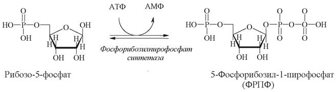
During the synthesis of both nucleotide types, the phosphoribosyl moiety is transferred as 5-phosphoribosyl-1-pyrophosphate (PRPP), which is produced through the phosphorylation of ribose-5-phosphate, an intermediate of the pentose phosphate pathway:
Distinct pathways exist for the Synthesis of purine and pyrimidine nucleotides. The metabolic routes leading to purine and pyrimidine nucleotides differ primarily in the stage at which the β-glycosidic bond is formed (Table 9). In purine synthesis, this bond is established at the very first step, and the ring system is subsequently assembled onto it. In contrast, the assembly of the pyrimidine ring is completed prior to the Formation of the bond between the ring and ribose-5-phosphate.
Table 9. Comparative characteristics of purine and pyrimidine nucleotide synthesis pathways
Synthetic features |
Biosynthetic pathway |
|
Purines |
||
Sequence of synthesis |
1. Formation of the N-glycosidic bond. 2. Assembly of the ring structure. |
1. Assembly of the ring structure. 2. Formation of the N-glycosidic bond. |
Key reaction |
Formation of phosphoribosylamine (phosphoribosylamido-transferase) |
Formation of carbamoyl phosphate (carbamoyl phosphate synthetase) |
Cellular localization |
Cytosol |
Mitochondria and cytosol |
Regulation |
Multi-level feedback inhibition by IMP, AMP, and GMP |
Inhibition of carbamoyl phosphate synthetase by UTP |
The initial nucleotide product of the purine pathway is inosine 5'-monophosphate (IMP), whereas that of the pyrimidine pathway is uridine monophosphate (UMP). All other purine and Pyrimidine nucleotides are subsequently derived from IMP and UMP, respectively.
5.11.1. Biosynthesis of Purine Mononucleotides
The biosynthesis of the first purine nucleotide, IMP, involves 10 enzymatic reactions and proceeds with the expenditure of ATP energy (Fig. 26). The formation of the purine ring begins directly on ribose-5-phosphate, to which nitrogen and carbon atoms are attached. The sources of these atoms are the amino acids glycine, glutamine, and aspartic acid. Some of the carbon atoms are supplied by coenzymes containing Folic acid and biotin. As a result of subsequent reactions, the purine ring is sequentially assembled on this foundation.
Fig. 26. Biosynthesis of purine mononucleotides
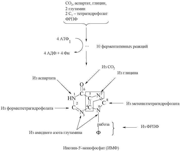
THE ORIGIN OF each atom of the purine heterocycle has been established experimentally using isotopes. As can be seen, two nitrogen atoms (N3 and N9) of the purine ring originate from the amide group of glutamine, the third nitrogen atom (N1) from aspartate, and finally, the fourth (N7) from glycine. Glycine also provides the fourth and fifth carbon atoms. The carbon atoms (C2 and C8) originate from formate, and the sixth carbon atom comes from CO2.
Other nucleoside monophosphates, such as AMP and GMP, are formed from IMP.
An alternative pathway for the synthesis of purine nucleotides exists. This is due to the fact that the synthesis of the cyclic purine structure requires a significant amount of energy in the form of ATP. Therefore, cells contain enzymes that utilize ("salvage") purine bases formed during nucleotide catabolism before they are converted into xanthine and uric acid:
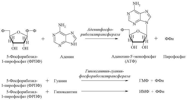
The enzymes adenine phosphoribosyltransferase and hypoxanthine-guanine phosphoribosyltransferase salvage free purines, converting them back into nucleotides through interaction with PRPP.
5.11.2. BIOSYNTHESIS OF PYRIMIDINE mononucleotides
The biosynthesis of pyrimidine mononucleotides occurs in stages and includes 6 enzymatic reactions (Fig. 27). In the synthesis of uridine monophosphate, the cyclic pyrimidine structure is formed prior to attachment to ribose-5-phosphate.
Fig. 27. Biosynthesis of pyrimidine mononucleotides

In the first step, carbamoyl phosphate is formed from CO2, glutamine, and ATP. Then, an unusual nitrogenous base—orotic acid, which contains a pyrimidine ring—is synthesized from carbamoyl phosphate and aspartic acid. Orotic acid attaches to ribose-5-phosphate, yielding the pyrimidine nucleotide orotidine monophosphate. Subsequently, orotic acid within the nucleotide is converted into uridine pyrimidine nucleotides.
5.11.3. Biosynthesis of nucleoside triphosphates
Nucleoside triphosphates are formed with the participation of ATP from nucleotide monophosphates (NMPs) as a result of two consecutive phosphorylation reactions:

Uridine monophosphate (UMP) serves as a precursor for the triphosphate nucleotides UTP and CTP. Furthermore, the formation of UTP occurs via the two-stage phosphorylation described above, whereas CTP is formed from UTP by amination at position 4 of the pyrimidine ring.

The precursor of GTP and ATP is inosine-5'-monophosphate (IMP). In both cases, the hypoxanthine ring of IMP is first modified to yield the corresponding monophosphonucleotides GMP and AMP, which are then phosphorylated.
5.11.4. Biosynthesis of deoxynucleotides
Deoxynucleotides are formed from the corresponding ribonucleoside phosphates by reducing their constituent ribose to deoxyribose with the participation of the enzyme Ribonucleotide reductase.
The source of reducing equivalents is a heat-stable protein, thioredoxin, which contains two free SH groups. Thioredoxin reductase catalyzes the NADPH-dependent reduction of thioredoxin (Fig. 28).
Fig. 28. Biosynthesis of deoxynucleotides

If nucleoside diphosphates enter the reaction, deoxynucleotides are synthesized: dUDP, dCDP, dGDP, and dADP. The deoxyribonucleoside diphosphates dCDP, dGDP, and dADP can be further phosphorylated to the corresponding deoxyribonucleoside triphosphates dCTP, dGTP, and dATP. dUDP serves as the precursor for dTTP.

The biosynthesis of all deoxyribonucleoside 5'-triphosphates and ribonucleoside 5'-triphosphates is regulated within the cell so that they are produced dependently upon one another in strictly defined proportions.
5.12. Nucleic Acid Synthesis
Nucleic acid synthesis requires mononucleotides strictly in their triphosphate form. These nucleotides contain three phosphoric acid residues in their molecules and possess a high energy reserve. RNA Synthesis utilizes ATP, GTP, UTP, and CTP, whereas DNA Synthesis uses dATP, dGTP, dTTP, and dCTP, respectively.
DNA Replication, or reduplication, can also be described as DNA doubling. It occurs prior to Cell Division. Successful replication requires a template—an unwound DNA strand; substrates involved in DNA polymerization (deoxyribonucleoside triphosphates); enzymes that catalyze the process; Mg2+ ions; and protein factors that facilitate the uncoiling of the double-stranded DNA. DNA synthesis is carried out by the enzyme DNA polymerase, which builds a complementary strand for each of the parental strands. Consequently, a single molecule yields two identical daughter molecules, with both original DNA strands serving as templates for the daughter strands. This process is semi-conservative, meaning that upon completion of replication, each daughter DNA molecule contains one parental strand and one newly synthesized strand. As nucleotides attach to the template, they link together into polynucleotide chains that immediately twist into a double helix. The Biological Significance of replication is that it produces two complete copies from a single DNA molecule. This process operates with extremely high fidelity, making errors exceedingly rare.
The process of synthesizing RNA on a DNA template is known as Transcription, which is driven by the enzyme RNA polymerase. This enzyme links together the ribonucleotides that form the backbone of the RNA molecule. It accomplishes this by reading The sequence of the DNA molecule and assembling a complementary sequence. Research has shown that only one of the two DNA strands acts as a template during this process.
Questions for Knowledge Assessment
1. Amino acids: definition and biological significance. Classification of amino acids by structure, side-chain polarity, and physiological properties. Amino acid isomerism and its biological significance. Solubility of Amino acids, acid-base properties, and THE CONCEPT OF the isoelectric point.
2. BIOLOGICAL FUNCTIONS OF proteins: enzymatic, structural, regulatory, protective, receptor, transport, motor, and energetic. Structural Features of structural proteins (Collagen, keratin, Fibroin) and transport proteins (hemoglobin).
3. Nucleic acids. Nucleotides and nucleosides: structure and biological functions. Key representatives of mononucleotides and oligonucleotides. DNA and RNA: structure and biological functions.
4. Pathways of amino acid metabolism in the cell: decarboxylation, transamination, and deamination reactions, and their biological significance. The Fate of the carbon skeleton of amino acids; glucogenic and ketogenic amino acids. Utilization of amino acids for the synthesis of BIOLOGICALLY ACTIVE SUBSTANCES.
5. Generation, toxicity, and detoxification of ammonia. The urea cycle: reactions, regulation, and biological significance. The interconnection of amino acid metabolism with the Krebs cycle, Carbohydrate Metabolism, and Lipid Metabolism.
6. Stages of Protein Biosynthesis (recognition, transcription, Translation): core mechanisms, substrates, enzymes, and intracellular localization. Protein Catabolism in Lysosomes and proteasomes: mechanisms and biological significance. The Role of hormones and The Nervous System in regulating protein metabolism.
Written Homework
Mandatory
1. Protein Digestion in The Stomach is facilitated by pepsin, a gastric juice enzyme. Trypsin, the primary proteolytic enzyme of pancreatic juice, continues this digestive process in the duodenum. Pepsin is known to preferentially hydrolyze peptide bonds formed by the amino groups of aromatic amino acids, whereas trypsin cleaves bonds formed by the carboxyl groups of basic amino acids. What fragments will be produced when the polypeptide of the following structure
H2N-Ala-Gly-Phe-Leu-Ser-Lys-Ala-Val-Ile-Met-Arg-Ser-Glu-Tyr-Gly-COOH is treated with pepsin, with trypsin, and with both enzymes sequentially?
2. Write the Chemical Reactions for amino acid metabolism in cells and name the enzymes that catalyze them:
a) decarboxylation of histidine;
b) oxidative deamination of glutamate;
c) transamination of glutamate and oxaloacetate;
d) hydroxylation of phenylalanine to form tyrosine;
e) decarboxylation of tryptophan;
f) hydrolysis of arginine to form urea.
Which Vitamins serve as precursors for the coenzymes participating in these reactions? Which of the listed amino acids are essential?
3. Amino acid metabolism was studied in an animal experiment. To do this, the animals were fed aspartic acid labeled with radioactive nitrogen at the amino group. After a while, the radioactive label was detected in the urine of the animals within two low-molecular-weight substances. The first substance turned out to be highly water-soluble, while the second readily formed a precipitate. The experiment was repeated under conditions of protein starvation, which resulted in a sharp increase in The excretion of radioactive metabolic products in the urine. What substances are discussed in this problem? How can the observed patterns be explained? In what other substances could the labeled nitrogen atom have been detected?
4. Write the structural formula for a region of a DNA molecule containing the nitrogenous bases A - G - C, and for the complementary strand. What types of bonds are formed between nucleotides within a single strand and between two complementary strands?
Supplementary
1. Write the chemical reactions for amino acid metabolism in cells and name the enzymes that catalyze them:
a) decarboxylation of cysteine;
b) intramolecular deamination of histidine;
c) transamination of leucine and α-ketoglutarate;
d) hydroxylation of tryptamine to form serotonin;
e) methylation of norepinephrine to epinephrine;
f) oxidation of arginine to citrulline with the formation of nitric oxide (NO).
2. Medical practice employs various drugs that affect Nucleotide Metabolism: potassium orotate, inosine (riboxin), and 5-fluorouracil. Write their structural formulas. Indicate which metabolic pathways they activate or inhibit, and describe their therapeutic effects.
Standard Multiple-Choice Test on the Topic “Metabolism of Amino Acids, Proteins, and Nucleic Acids”
Instructions: Unless otherwise specified in the test question, select a single correct answer.
1. Choose the name of the enzyme that catalyzes the following reaction:
a) carbamoyl phosphate synthetase;
b) argininosuccinate synthetase;

c) argininosuccinate lyase;
d) arginase;
e) urease.
2. Indicate the products of AMINO ACID DECARBOXYLATION:
a) a new amino acid and a keto acid;
b) carbon dioxide and water;
c) a new amino acid and ammonia;
d) carbon dioxide and an amine;
d) ammonia and water.
3. Choose the enzyme that catalyzes the oxidative Deamination of Amino acids:
a) aspartate aminotransferase;
b) alanine aminotransferase;
c) glutaminase;
d) glutamate dehydrogenase;
e) glutamate decarboxylase.
4. The carbon skeletons of the amino acids Asp and Asn are converted into the Krebs cycle substrate oxaloacetate; therefore, these amino acids are classified as:
a) conditionally essential;
b) essential;
c) glucogenic;
d) ketogenic;
e) proteinogenic.
5. Indicate The properties of urea that render it non-toxic:
a) strong base, soluble in water, easily excreted in urine;
b) weak base, soluble in water, easily excreted in urine;
c) strong base, soluble in lipids, easily excreted in urine;
d) weak base, soluble in lipids, not excreted in urine;
e) weak base, soluble in water, not excreted in urine.
6. The region of a DNA molecule complementary to the sequence AAGCCT is:
a) AAGCCT;
b) TTAGGU;
c) TTCGAA;
d) CCAGUU;
e) TAGCTU.
7. What type of bond is formed between two phosphate residues in a nucleotide:
a) peptide bond;
б) phosphoanhydride bond;
в) phosphodiester bond;
г) glycosidic bond;
д) Hydrogen bond.
8. Which processes in protein biosynthesis occur during transcription:
а) ribosome assembly and Polypeptide chain synthesis;
б) mRNA synthesis and maturation;
в) interaction of tRNA with an amino acid;
г) attachment of a non-protein component to the polypeptide chain;
д) none of the above.
9. Fill in the missing words (3 Answers) in the sentence: "Uric acid is the end product of the breakdown of ... nucleotides, ... soluble in water, and can form ... in the kidneys."
10. Write the names and class numbers of the enzymes (6 answers) that catalyze reactions 1, 2, and 3:

Last update: 06/08/2026
Editorial and Educational Adaptation: This material has been compiled based on the primary/original source text. The project team performed an editorial review, corrected technical inaccuracies, structured sections, and adapted the content for an educational format.
What was processed:
- elimination of formatting defects (OCR errors, structural breaks, corrupted characters);
- editorial organization of content;
- standardization of terminology in accordance with academic sources;
- verification of factual statements against the original source text.
All mentions of the author, publication year, and origin of the primary text have been preserved in accordance with the source.