Fundamentals of Biochemistry - Filippovich, Y. B. 1999
Nucleic Acids and Their Metabolism
Nucleic Acid Metabolism
The Study of NUCLEIC ACID METABOLISM is fundamentally important for teacher training programs in the chemical-biological and biological-chemical departments of pedagogical institutes. The range of topics covered extends far beyond the Biochemistry of Nucleic Acids, offering a modern interpretation of problems related to heredity, Variability, natural and artificial mutagenesis, systematics, and evolution.
The study of nucleic acid metabolism is also critically important from another perspective, as it is exceptionally vital for a profound understanding of life processes in organisms. Research into the MOLECULAR MECHANISMS OF purine and pyrimidine Biosynthesis has uncovered key regularities in The regulation of de novo synthesis of these compounds and helped formulate General Principles governing Metabolic Regulation. Elucidating the mechanism behind the specific biosynthesis of gigantic polynucleotide molecules—where The sequence of constituent mononucleotide units is maintained with astonishing precision—has highlighted the leading role of complementary purine and pyrimidine base interactions in this process.
This, in turn, provided the first insights into the intimate mechanism ensuring the precise Replication of the Introduction/19.html">Primary Structure of macromolecules during their biosynthesis. Data concerning the regulation of nucleic acid synthesis have driven fundamental discoveries, taking initial steps toward explaining the principles not only of specific macromolecule reproduction, but also of morphogenesis.
Pathways of nucleic acid degradation. Nucleic acids undergo active degradation within the living Organism. For instance, the half-life of DNA molecules in mouse Tissues ranges from 1 to 5 days; the half-life of most mRNAs in eukaryotes spans several days, whereas in prokaryotes it is merely a matter of seconds.
Nucleic acids (RNA and DNA) are degraded in the organism through the action of specialized Enzymes known as Nucleases. They accelerate the Cleavage of internucleotide phosphodiester bonds within nucleic acid molecules and thus belong to the broader category of phosphodiesterases.
Nucleases that target internal internucleotide bonds in DNA and RNA molecules are termed endonucleases. They mediate the depolymerization of nucleic acids primarily into oligonucleotides. Nucleases that catalyze the successive cleavage of NUCLEOTIDES from RNA, DNA, or their fragments starting from the end of the polynucleotide chain are called exonucleases. They ensure The breakdown of nucleic acids down to free nucleotides.
Depending on their reaction Specificity, nucleases are divided into ribonucleases and deoxyribonucleases. The former accelerate the cleavage of both internal and terminal internucleotide bonds in RNA molecules, while the latter perform an identical function regarding DNA. Additionally, There is a large group of non-specific endo- and exonucleases that act simultaneously on both RNA and DNA.
Based on their mode of action on phosphodiester bonds within nucleic acid molecules, nucleases are categorized into two groups. Some accelerate the Hydrolysis of the ester bond linking the nucleoside phosphate to the 3'-carbon atom of the ribose or deoxyribose residue, whereas others target the 5'-carbon atom. Consequently, the nomenclature of these enzymes always highlights which specific bond's hydrolysis is catalyzed. However, the primary Classification criterion is the localization of the phosphate within the resulting oligo- or mononucleotides produced by nucleic acid hydrolysis. Therefore, nucleases cleaving the P—5'C bond are designated as 3'-nucleases, while those cleaving the P—3'C bond are termed 5'-nucleases:
Class="center">
The most important groups of nucleases are detailed below.
Deoxyribonucleases I (deoxyribonucleate 5'-oligonucleotidohydrolases) catalyze the hydrolysis of phosphodiester bonds in DNA between the phosphate residue and the 3'-carbon atom of the deoxyribose residue: DNA+(n — 1) H2O→n oligodeoxyribonucleotides.
Representative Examples of DNase I include pancreatic DNases (M~31,000). The primary structure of bovine pancreatic DNase has been fully sequenced: it consists of a single polypeptide chain comprising 257 amino acid residues. Its pH optimum lies between 6.8 and 8.0. The enzyme is activated by Mn2+ and Mg2+ ions and inhibited by anions that bind these cations, as well as by oligonucleotides. The MECHANISM OF ACTION of pancreatic DNase on DNA initially involves preferential single-strand breaks in the DNA duplex. Double-strand breaks are very rare, meaning depolymerization does not occur instantaneously. The final product of DNA Digestion by pancreatic DNase contains traces of deoxyribonucleoside 5'-phosphates, small amounts of dinucleotides, and a high proportion of oligodeoxyribonucleotide 5'-phosphates averaging 4 monomer units in length. Bovine pancreatic DNase exists in four multiple forms.
Deoxyribonucleases II (deoxyribonucleate 3'-oligonucleotidohydrolases) yield oligodeoxyribonucleotide 3'-phosphates with an average chain length of 6 residues and minor amounts of deoxyribonucleoside 3'-phosphates as end products. Spleen DNase II has a Molecular Weight of 38,000 and a pH optimum of 4.5–5.5. Its molecule consists of 343 amino acid residues and contains a carbohydrate component whose structural unit is glucosamine. The enzyme is activated by Mg2+ ions and inhibited by SO2-4, РО3-4, AsO3-4 ions, AU copolymer, and tRNA. DNA depolymerization proceeds via double-strand breaks in the DNA molecule, leading to the rapid accumulation of large fragments; bonds composed of paired GG and AC combinations are attacked with particular efficiency. DNases II are hypothesized to be dimers.
Alongside DNases I and II, a large group of endonucleases attacks both double- and single-stranded DNA. For instance, Escherichia coli possesses 7 such enzymes, comprising 4 low-molecular-weight (12–33 kDa) and 3 high-molecular-weight (68–114 kDa) variants.
Exodeoxyribonucleases catalyze the hydrolysis of DNA molecules to yield deoxyribonucleoside 5'-phosphates. Eight exodeoxyribonucleases (I–VIII) with varying degrees of purity and distinct characteristics have been isolated from Escherichia coli. Notably, exodeoxyribonuclease III (M = 30,000) possesses The ability to cleave a 3'-phosphate group from the terminal nucleotide of DNA fragments, if present.
Restriction Endonucleases (restriction enzymes) are bacterial DNases that cleave foreign (phage) DNA at strictly defined sites recognized by the enzyme. These recognition sites possess a palindromic structure. Two examples are shown below, with arrows indicating the sites of phosphodiester bond cleavage:

Depending on the relative position of the recognition site and the cleavage site, restriction Enzymes are classified into Type I (recognition site located far away, hundreds of Base Pairs from the cleavage point), Type II (cleavage occurs within the recognition site), or Type III (cleavage occurs near the recognition site). By 1993, 2,393 Type I, 188 Type II, and only 4 Type III restriction enzymes had been isolated and characterized, bringing the total number of studied restriction enzymes to 2,585.
Because restriction enzymes cleave DNA into a limited number of fragments, they have found application in determining primary DNA Structure. However, an even more significant area of practical use is Genetic Engineering; it is precisely through the action of restriction enzymes that specific DNA fragments are excised, subsequently integrated into bacterial DNA to become an integral part of the bacterial genome, and confer novel biochemical traits upon the bacterium that it did not previously possess—such as the ability to synthesize interferon, Insulin, and other Proteins widely used in medicine (Fig. 81). In principle, such processes are also feasible within the genomes of higher organisms. The seamless incorporation of restriction fragments into recipient DNA (i.e., DNA incorporating these fragments) is attributed to the generation of "sticky," complementary ends at the cleavage sites produced by restriction enzymes. Given the immense theoretical and practical importance of this work, comprehensive research under the "Restrictase" project has been underway in our country since 1975, with plans to expand it into a broader "Nuclease" project involving various research institutes in subsequent stages. In 1980, a large team of Soviet scientists was awarded the State Prize for developing protocols to produce 30 restriction enzymes and introducing them into genetic engineering practice.
Ribonucleases I (ribonucleate 3'-pyrimidine-oligonucleotidohydrolases) are endonucleases that catalyze the hydrolysis of RNA at pyrimidine nucleotide residues. Previously, they were classified as cyclizing ribonucleate nucleotide-2'-transferases, but since 2',3'-cyclic phosphates are formed as intermediates during their action—the subsequent hydrolysis of which to 3'-phosphates is catalyzed by the same enzyme—ribonucleases (RNases) I are now included in the class of Hydrolases.
Representative examples of RNases I include pancreatic ribonucleases isolated from numerous species. The primary structure has been established for pancreatic RNase from cattle, pigs, sheep, giraffes, red deer, roe deer, reindeer, fallow deer, chinchillas, mice, rats, guinea pigs, dromedary camels, and nutrias. In all cases except the last two, the enzyme consists of 124 amino acid residues. The Tertiary Structure of several pancreatic RNases has been elucidated. Pancreatic RNases can exist as single-component (Ribonuclease A) or two-component systems (ribonuclease B, containing a carbohydrate moiety with M = 1,350). Their mechanism of action on RNA has been studied in detail and serves as a striking Complement to the material in Chapter III concerning enzyme catalysis.
The polypeptide chain of RNase, cross-linked by four disulfide bridges (Fig. 82, A), adopts a spatial conformation (Fig. 82, B) that largely dictates the tertiary structure (Fig. 82, C) of this enzyme. Understanding the Catalytic Mechanism of pancreatic RNase has been facilitated by data regarding The structure of its Active Site (Fig. 82, D). This site binds Regions of the RNA molecule containing pyrimidine (cytidylic and uridylic) nucleotide residues, with paired combinations of CA, CG, CC, and CU being attacked in a ratio of 3,000:500:240:27. Within the enzyme's active site, the pyrimidine nucleotide residue is positioned such that the pyrimidine base is anchored to the substrate binding pocket via Hydrogen Bonds with Threonine and Serine radicals located at positions 45 and 123 of the polypeptide chain, as well as through hydrophobic interactions with a phenylalanine radical (amino acid residue 120). Consequently, the phosphate group located between the 3'-carbon of the pyrimidine nucleotide ribose and the 5'-carbon of the adjacent nucleotide in the RNA chain is fixed between Histidine residues 12 and 119 of the catalytic center. This stabilization of the internucleoside phosphate is further assisted by a Lysine residue at position 41 of the RNase molecule, which is situated in the active site at a distance of 0.5 nm from the aforementioned histidine residues. This is followed immediately by the catalytic act itself, carried out via concerted proton transfer resulting in the Cleavage of the internucleotide phosphate bond, The formation of a 2',3'-cyclic phosphate on the pyrimidine nucleotide ribose, and the subsequent hydrolysis of the 2',3'-cyclic phosphate bond with the restoration of the active center's initial structure (see reaction scheme).

Fig. 81. Schematic Overview of genetic engineering workflows
Genes responsible for The biosynthesis of biologically significant proteins are utilized as foreign DNA fragments; their expression can be harnessed for Protein Synthesis following the integration of plasmid DNA into the bacterial genome, or for Gene cloning and subsequent expression in non-bacterial protein-synthesizing systems.
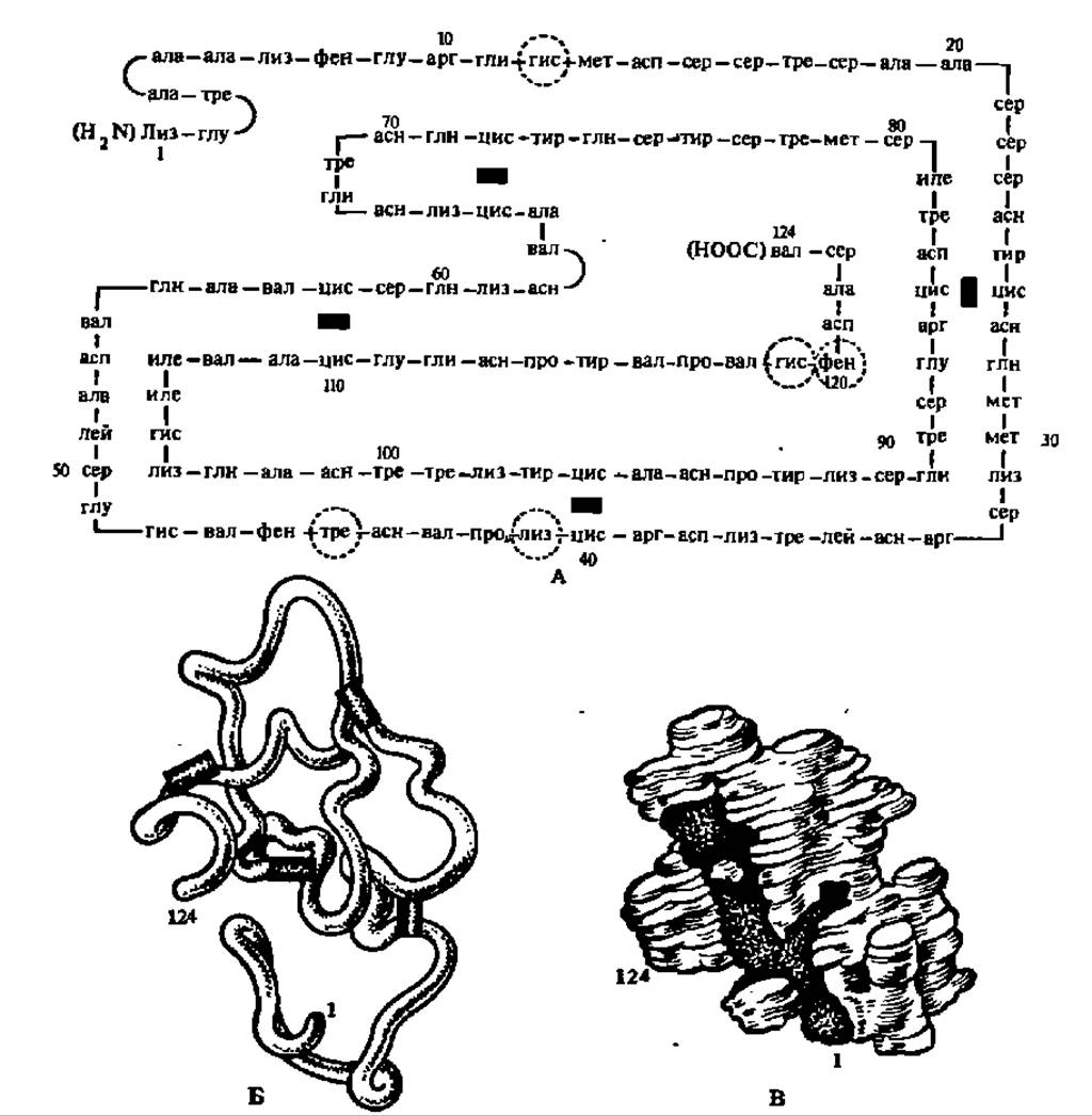
Fig. 82. Structure of bovine pancreatic ribonuclease and its active center:
A — primary structure; black rectangles indicate —S—S-bonds, Arabic numerals indicate amino acid residue numbers, dashed lines outline amino acid residues that make up the active center; B — spatial arrangement of the polypeptide chain; C — three-dimensional model of the molecule at a resolution of 0.5 nm; the darkened depression houses the active center at its bottom; D — STRUCTURE OF THE active center, in which the residues of His, Lys, Phe, Ser, and Thr (their ordinal numbers
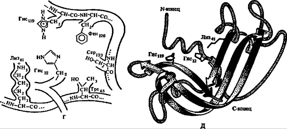
Fig. 82. Continued (in the polypeptide chain indicated by numerical indices) are brought together to a distance of several tenths of a nanometer; E — Spatial Structure of the active center
Crucially, it is specifically the His residues located at positions 12 and 119 in the polypeptide chain, brought into proximity within the catalytic center of the enzyme, that facilitate proton transfer.

This function of histidine imidazole radicals is replicated when examining The Mechanism of action of many Other Enzymes, especially hydrolases (see the section on peptide bond hydrolysis — p. 263, and glycosidic bond hydrolysis — p. 331).
Guanylyl ribonucleases (ribonucleate 3'-guanylyl-oligonucleotide hydrolases) accelerate the hydrolysis of bonds at the 5'-carbon atom of ribose of the guanylic acid residue and the internucleotide phosphate in the RNA molecule, yielding guanosine 3'-phosphate and oligonucleotides with a guanosine 3'-phosphate residue as the terminal nucleotide. The primary structure of guanylyl ribonuclease isolated from the mold fungus *Aspergillus* (T1-RNase) has been deciphered (M = 11,000; 104 amino acid residues).
The enzyme has found widespread application for RNA degradation in determining their primary structure, and it was precisely through the intermediacy of T1-RNase that R. Holley et al. (1965) first obtained large fragments of tRNAala (see p. 213).
Another 10 endoribonucleases isolated from Bacteria, microscopic Fungi, plants, and animals have been characterized.
As in the case of DNases, there is a large group (over ten) of exoribonucleases that accelerate the cleavage of ribonucleotides from the terminal residues of RNA and of oligoribonucleotides arising from the selective hydrolysis of RNA by endoribonucleases. Thus, As a result of The activity of various nucleases, nucleic acids upon degradation yield a complex mixture of individual ribo- and deoxyribonucleoside 3'- and 5'-phosphates.
In addition to the enzymes listed, some other enzymes that are not hydrolases of phosphodiester internucleotide bonds also participate in the Degradation of Nucleic acids, for example, polynucleotide phosphorylase and uracil-DNA glycosylase.
Polynucleotide phosphorylase (polynucleotide: orthophosphate nucleotidyltransferase), unlike all previously discussed enzymes involved in nucleic acid degradation, is a nucleotidyltransferase, i.e., it transfers nucleotide residues from the 3'-end of RNA to inorganic phosphate with the formation of nucleoside diphases (NDPs):

The enzyme was discovered by M. Grunberg-Manago and S. Ochoa (1955) and isolated from many sources. It is concentrated mainly in the microsomal and ribosomal fractions of The Cell contents. By rough estimation, its molecular mass is close to 230,000. The phosphorolysis reaction rate depends on the conformation and Nucleotide Composition of RNA: double-stranded and methylated regions are resistant to the action of the enzyme. It is hypothesized that in vivo polynucleotide phosphorylase ensures the degradation of cellular RNAs, especially mRNA, to nucleoside diphases, regulates the concentration of inorganic phosphate in the cell, and supplies the necessary amount of NDPs for their conversion into deoxyNDPs.
The enzyme possesses a remarkable feature: from nucleoside diphases and their mixtures in vitro, it ensures the synthesis of polyribonucleotides with a ratio of monomeric units in their composition identical to that in the initial solution. Therefore, polynucleotide phosphorylase has been widely used for the synthesis of polyribonucleotides of various compositions, which played an outstanding role in deciphering The Genetic Code of protein synthesis.
Uracil-DNA glycosylase accelerates the cleavage of the U residue from damaged DNA where deamination of the C residue has occurred. At the resulting apyrimidinic site of one of the DNA strands, the phosphodiester bond is hydrolyzed with the elimination of deoxyribose, the 3'-phosphate is cleaved with the participation of exodeoxyribonuclease III, and instead of the missing nucleotide residue, a new one—in this case, a cytidylic acid residue—is incorporated via DNA polymerase and DNA ligase reactions (see p. 251 and Fig. 84).
DNA glycosylases represent a new group of enzymes involved in DNA metabolism. Through their intermediacy, other modified purine and pyrimidine bases are also removed, after which the original DNA structure is restored in a series of subsequent reactions; i.e., this group of enzymes is of essential importance in DNA Repair (restoration of structure). This occurs, in particular, during the replacement of methylated purine and pyrimidine bases, since along with uracil-DNA glycosylase, 3-methyladenine-DNA glycosylase has been studied. A total of 8 DNA glycosylases have already been discovered.
Metabolism of nucleoside phosphates. Deoxyribonucleoside phosphates and ribonucleoside phosphates, which represent the End products of the enzymatic degradation of nucleic acids, break down further into even simpler compounds. The First stage of this breakdown consists in the cleavage of the phosphoric acid residue:

In the second stage of degradation, the ribose residue is transferred from the nucleoside to phosphoric acid. This reaction is accelerated by ribosyltransferases specific to each type of nucleoside. An example is the phosphorolysis of uridine:

Uridine phosphorylase has an M = 165 kDa, contains 6 subunits of 27.5 kDa each, composed of 253 amino acid residues, including seven histidine residues; two of them (the 8th and 122nd) are part of the active center of the enzyme.
Consequently, as a result of nucleoside phosphate degradation, ribose 1-phosphate and all types of purine and pyrimidine bases involved in the construction of nucleic acids are released in a free state. The presented scheme of nucleoside degradation is not the only one. Other pathways of nucleoside degradation are also possible. One of them consists in the hydrolysis of nucleosides, for example:

In turn, both CARBOHYDRATES and nitrogenous bases undergo further modifications. Ribose and ribose-1-phosphate enter into metabolic pathways typical of carbohydrates, which will be discussed below. Purine and pyrimidine bases undergo further degradation, converting into various simple nitrogen-containing products that are either excreted from the organism or stored within it.
Degradation of purine and pyrimidine bases. The first phase in the degradation of purine and pyrimidine bases involves the deamination of those bearing amino groups. This process is mediated by specific aminohydrolases. As a result, adenine is converted into hypoxanthine:

Guanine is converted into xanthine:

Cytosine is transformed into uracil:

Deamination occurs not only at the level of free purine and pyrimidine bases, but also at the level of Nucleosides and Nucleotides. The latter occurs with greater intensity because the corresponding nucleoside and nucleotide aminohydrolases are more active than purine or pyrimidine aminohydrolases. Thus, adenosine and adenosine phosphate are converted into inosine and inosine phosphate much more vigorously than adenine is into hypoxanthine:

During the further degradation of deaminated nucleosides and nucleotides, hypoxanthine, xanthine, or uracil are released from their structure.
The subsequent fate of the deaminated purine and pyrimidine bases varies. Hypoxanthine and xanthine are oxidized to uric acid:

The oxidation reaction of hypoxanthine to xanthine, and the latter to uric acid, is accelerated by xanthine oxidase—a broad-spectrum oxidoreductase that is a molybdenum-containing flavoprotein.
The enzyme from various sources has a molecular weight ranging from 280,000 to 360,000 Da and, upon Treatment with dissociating agents, breaks down into two identical subunits, each containing one FAD molecule, one Mo atom, and 4 non-heme iron atoms linked to labile sulfur atoms in Fe2S2-type clusters. Activated hypoxanthine and xanthine reduce molybdenum, which rapidly reduces the Fe2S2 cluster, and finally, flavin reduces molecular oxygen.
In A number of animals (great apes, birds, reptiles, silkworms) and in humans, the final product of purine base degradation is uric acid, which is excreted from the body. However, most animals and plants possess enzymes and enzyme systems capable of accelerating the further breakdown of uric acid. Derived from the term for uric acid (*acidum uricum*) and reflecting their cleavage (lysis) action, these enzymes are designated as uricolytic enzymes. In some cases (mammals, insects), uricolysis amounts to The oxidation of uric acid to allantoin; in others (teleost fish), the process is more complex, with allantoin converting into allantoic acid, which subsequently breaks down (in amphibians and most plants) into urea and glyoxylic acid:

Unlike hypoxanthine and xanthine, deaminated pyrimidine bases undergo reduction. For instance, uracil is converted into dihydrouracil, with NADH serving as the H atom donor in this reaction. In turn, dihydrouracil undergoes hydrolysis to form N-carbamoyl-β-Alanine, which is further hydrolyzed into β-alanine and carbamic acid. The latter is either utilized for urea synthesis or degraded into СО2 and NH3. All of these reactions are accelerated by corresponding enzymes:

Carbamic acid and β-alanine are the end products of the degradation of the two pyrimidine bases, U and C. In the case of T, which degrades via a similar pathway, β-aminoisobutyric acid is formed instead of β-alanine.
Thus, during degradation in animal and plant organisms, the huge, complex molecules of polydeoxyribonucleotides and polyribonucleotides are converted into very simple compounds—primarily phosphoric acid, СО2, and NH3. Organisms positioned lower on the evolutionary ladder retain a full complement of enzymes ensuring the degradation of nucleic acids down to these simplest products. With the transition to more highly organized forms, a number of enzymes involved in The conversion of purine and pyrimidine bases are lost, so that the end products of nucleic acid metabolism in certain groups of organisms are more complex compounds than NH3 and СО2, namely urea, allantoic acid, allantoin, and uric acid.
Mechanism of nucleoside monophosphate biosynthesis. To support nucleic acid biosynthesis, the organism must maintain a full supply of deoxyribo- and ribonucleoside triphosphates. Consequently, in any cell of any organism, regardless of its position on the evolutionary ladder, the de novo synthesis of all types of nucleoside triphosphates, nucleoside diphosphates, and nucleoside monophosphates proceeds unimpeded.
Of the three Main Components of a nucleotide—the nitrogenous base, pentose, and phosphoric acid—the latter is normally always present in Cells, while the second inevitably arises during Carbohydrate Metabolism. Thus, only the first constituent part of the nucleotide, the purine or pyrimidine base, must be synthesized via a specific pathway.
The pathways for the formation of purine and pyrimidine bases differ, yet their synthesis mechanisms share several common features. These include: 1) the extensive use of gly, asp, and gln as sources of nitrogen for the heterocyclic rings; 2) the incorporation of carbon atoms from СО2 and formate into the purine and pyrimidine rings; 3) the assembly of the purine base and the completion of pyrimidine base synthesis directly on ribose-5-phosphate, resulting in nucleoside-5'-phosphates rather than free A, G, U, C, and T as the direct end products of biosynthesis; 4) the enzymatic nature of all reactions involved in nucleotide formation; and 5) the emergence at a specific stage of biosynthesis of precursors from which individual nucleoside-5'-phosphates are subsequently formed.
Let us first examine the mechanism of pyrimidine base biosynthesis. The preparatory reaction that initiates this synthesis is the formation of carbamoyl phosphate from NH3 and СО2 with the participation of ATP:

Next, with the participation of a specific enzyme, the carbamic acid residue (carbamyl) is transferred to the amino group of aspartic acid, yielding carbamylaspartic acid. This reaction is considered the first specific step in the synthesis of pyrimidine nucleotides:

When the NH2 and COOH groups in the carbamylaspartic acid molecule approach each other, they interact with the release of Water molecules. This reaction is catalyzed by an enzyme belonging to the hydrolase class—dihydroorotase, named after dihydroorotic acid, the hydrolysis of which it accelerates due to the reversibility of this reaction:

Dihydroorotic acid undergoes enzymatic oxidation. The removal of two H atoms is carried out by a primary dehydrogenase using either NAD+ or NADP+, or FAD as a coenzyme:

As can be seen from its formula, the structure of one of the pyrimidine bases—specifically uracil—is already pre-formed in the orotic acid molecule. A simple decarboxylation reaction is sufficient to convert orotic acid into uracil. However, this process takes place only after orotic acid binds with ribose to form a nucleoside in which the aglycone is the orotic acid residue. A nucleoside of this structure is called orotidine (by analogy with cytidine and uridine). Since the reaction occurs directly between orotic acid and 5-phosphoribosyl pyrophosphate, it results in the formation of orotidine-5'-phosphate. The process is accelerated by a corresponding transglycosidase. The reaction equations leading to the synthesis of orotidine-5'-phosphate are as follows:

The final transformation consists in the decarboxylation of orotidine-5'-phosphate:
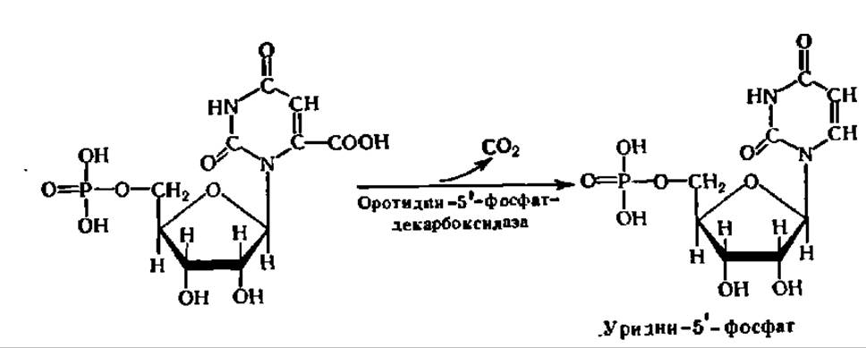
As a result, one of the pyrimidine nucleotides—uridine-5'-phosphate—is formed. Uridine-5'-phosphate occupies a central place in the BIOSYNTHESIS OF PYRIMIDINE nucleotides, as it can subsequently be converted into other pyrimidine nucleotides According to the following scheme:

Scheme 2. Pathways of pyrimidine nucleotide transformations
These transformations of Pyrimidine nucleotides are carried out via reduction, amination, and methylation reactions of nucleoside mono-, di-, and triphosphate esters. The latter are formed through the interaction of nucleoside monophosphates with ATP, the reserves of which in cells are continuously replenished via Oxidative Phosphorylation. For example:

The reduction proceeds at the hydroxyl group at the 2nd carbon atom of ribose, thereby converting the ribose residue into a deoxyribose residue. This reaction is characteristic of nucleoside diphosphates:

A special protein, thioredoxin, serves as the donor of H atoms for ribose reduction during the conversion of ribonucleoside diphosphate to deoxyribonucleoside diphosphate. Composed of 108 amino acid residues, thioredoxin contains Cys residues at positions 32 and 35, thus possessing two HS groups. These very groups supply the H atoms by forming a disulfide bridge. Oxidized thioredoxin is immediately converted back to its reduced form by receiving H atoms from NADH via the enzyme thioredoxin reductase.
In addition to providing H atoms for the reduction of the ribose residue, reduced thioredoxin is capable of binding with two other catalytically active proteins, B1 and B2. The latter are thereby activated and directly accelerate The process of ribose residue reduction. They are also strongly influenced by other allosteric activity regulators, notably ATP, GTP, TTP, dATP, dGTP, etc.
Thus, the conversion of ribonucleoside diphosphates into deoxyribonucleoside diphosphates proceeds according to the following scheme:

As for the amination reactions (transition from UTP to CTP) and methylation reactions (transition from dUMP to dTMP), in the first case the amino group donor is NH3 in bacteria and Gln in mammals, with the introduction of the amino group being coupled with ATP breakdown; in the second case, the methyl group donor is N5-methyltetrahydrofolic acid, and its transfer reaction is accelerated by thymidylate synthase (a dimer; each polypeptide chain contains 316 amino acid residues; its primary and tertiary structures have been elucidated).
All these reactions collectively ensure the maintenance of a pool of free pyrimidine nucleoside triphosphates (UTP, CTP, dCTP, dTTP) within the organism, which are necessary for DNA and RNA Synthesis.
An important feature of Pyrimidine Nucleotide Biosynthesis is its self-regulation. It has been established that end products of pyrimidine nucleotide biosynthesis, such as CTP and dCTP, inhibit the activity of aspartate carbamoyltransferase—the enzyme that accelerates the first reaction in the chain of interactions leading to the Formation of the pyrimidine ring. It has been revealed that the decrease in enzyme activity is caused by the binding of CTP to the allosteric site of the enzyme. Thus, the intracellular accumulation of an excess of CTP and dCTP immediately affects the activity of aspartate carbamoyltransferase, slowing down the biosynthesis of pyrimidine nucleotides. The antagonist of CTP in inhibiting this enzyme is ATP, which acts as an activator. Consequently, the inhibition or stimulation of pyrimidine nucleotide biosynthesis depends on the intracellular ratio of ATP to CTP—that is, on the cellular Energy balance, the level of metabolism, and specifically The rate of oxidative phosphorylation reactions, through which The energy released during the oxidation of organic substances is stored in the high-energy bonds of ATP.
The dual control mechanism governing the first enzyme in a biosynthetic reaction chain provides a highly precise way to regulate metabolism and is utilized in numerous biosynthetic pathways within the organism. This is most thoroughly illustrated by the biosynthesis of pyrimidine nucleotides.
In Conclusion, it should be noted that one of the pyrimidine nucleotide derivatives, specifically 3'-azido-2',3'-dideoxythymidine, inhibits the replication of Retroviruses, including those responsible for Acquired Immunodeficiency Syndrome (AIDS):

Other nucleotide derivatives have also been proposed for combating this formidable disease.
Let us now turn to the mechanism of purine base biosynthesis. The formation of the purine ring begins directly on ribose-5-phosphate. Therefore, the initial reaction involves the interaction of glutamine with 5-phosphoribosyl-1-pyrophosphate, catalyzed by a glycosyltransferase that accelerates The transfer of the 5-phosphoribose moiety to the amide group of glutamine. Concurrently, the resulting 5-phosphoribosylglutamine apparently undergoes hydrolysis, yielding 5-phosphoribosylamine:

In the presence of ATP and with the participation of a specific ligase (aminosynthetase), Glycine is attached to 5-phosphoribosylamine, forming a peptide bond:

The 5-phosphoribosylglycinamide molecule is elongated by one carbon atom through the action of phosphoribosylglycinamide formyltransferase. Its coenzyme is tetrahydrofolic acid, which attaches a formyl group to the nitrogen atom occupying the 5th position of the coenzyme molecule.
The formyl residue is transferred from N-formyltetrahydrofolic acid to the H2N group of 5-phosphoribosylglycinamide. The resulting 5-phosphoribosylformylglycinamide then interacts with glutamine in the presence of concomitantly cleaved ATP and the corresponding ligase (see the first two equations in Scheme 3, p. 242).
The product of this reaction is a derivative in which the carbonyl oxygen of the peptide bond is replaced by an imino group. Subsequently, a series of transformations of the resulting compound leads to the closure of the imidazole ring. This process also proceeds concurrently with ATP cleavage and is accelerated by a specific enzyme which, once isolated and purified, exhibits relatively low resistance to Denaturation.
It is highly significant that the reaction forming the imidazole portion of the future purine moiety is practically irreversible, unlike the vast majority of other steps in the process under consideration.
Following this, the pyrimidine ring is constructed on the imidazole ring via a series of ENZYMATIC REACTIONS OF the aforementioned type, utilizing aspartic acid, СО2, and formate, thereby ultimately completing the formation of the purine nucleotide. The scheme outlining the MAIN STAGES OF this synthesis is as follows:
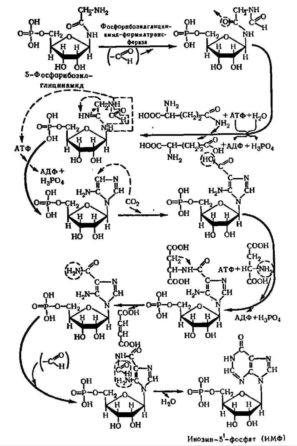
Scheme 3. Mechanism of purine nucleotide biosynthesis
To summarize the compounds from which the purine ring is constructed, it turns out to be assembled from very simple precursors:

As evident from the provided scheme, the 1st nitrogen atom of the purine ring originates from aspartic acid, the 3rd and 9th from glutamine, and the 7th from glycine. Regarding the Origin of the carbon atoms in the purine ring, their sources are formate (2nd and 8th carbon atoms), glycine (4th and 5th), and СО2 (6th atom). The chemical equations presented in Scheme 3 illustrate the details of the incorporation of specific N and C atoms from the listed compounds into the purine moiety of the nucleotide during its biosynthesis.
The pyrimidine ring of pyrimidine nucleotides is synthesized in the organism from similar building blocks: NH3, СО2, and aspartic acid. Thus, the starting Materials for the Biosynthesis of Purine and pyrimidine bases in the organism are exceptionally accessible and constantly present, since ammonia, the formyl group, and carbon dioxide are generated during the degradation of various Organic compounds or enter the organism from outside, while glutamic and aspartic acids and their amides represent primary Amino Acids synthesized in large quantities—a factor of great importance for ensuring the unobstructed synthesis of these vital cellular components.
As follows from Scheme 3, Purine nucleotides are formed as inosine-5'-phosphate through successive reactions of purine ring elongation on ribose-5-phosphate. The latter can be oxidized to xanthzoin-5'-phosphate. Amination of the former yields adenosine-5'-phosphate, while amination of the latter yields guanosine-5'-phosphate. Both processes are accelerated by specific enzymes (see the reaction equation on p. 244).
In turn, purine nucleoside monophosphates are further converted into nucleoside triphosphates. Specifically, guanosine-5'-phosphate is converted into guanosine-5'-triphosphate via a double displacement reaction with ATP:

The interaction of adenosine-5'-phosphate with ATP yields adenosine diphosphate; this reaction was discovered by A. V. Kotelnikova in the early 1960s:


It serves as a substrate in oxidative phosphorylation, which continuously replenishes the body's ATP reserves, ensuring a sufficient supply to convert all other nucleoside monophosphates into nucleoside triphosphates.
The synthesis of deoxyadenosine 5'-triphosphate (dATP) and deoxyguanosine 5'-triphosphate (dGTP) proceeds via the reduction of the ribose moiety at the C2' hydroxyl group. In the case of dGTP, the reduction occurs at the GDP level, followed by the subsequent conversion of dGDP to dGTP:

The mechanism by which the ribose residue is reduced to a deoxyribose residue in purine ribonucleoside diphosphates is identical to that previously discussed for pyrimidine nucleotides. This reduction process is stimulated by dGTP and dTTP, but inhibited by dATP.
Similar to pyrimidine nucleotides, the end products of purine nucleotide biosynthesis (IMP, AMP, ADP, ATP, GMP, GDP, and GTP) inhibit amidophosphoribosyltransferase, the enzyme that catalyzes the first step in the de novo purine ring synthesis pathway. This ensures the autoregulation of purine nucleotide production. Furthermore, through the reciprocal Cross-Regulation where ATP and GTP participate in the reactions leading to the conversion of IMP into GMP and AMP, respectively, these nucleotides are consistently synthesized in the body in a strictly defined ratio:
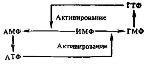
This pathway diagram is highly illustrative, demonstrating the Principles of Metabolic self-regulation that ensure a set of compounds is synthesized in precise proportions relative to one another. Maintaining a strict ratio of nucleoside triphosphates in the organism is critically important, as they serve as the building blocks for nucleic acids. Similar regulatory mechanisms are also observed in the synthesis of pyrimidine nucleotides.
There is an alternative pathway for purine and pyrimidine nucleotide synthesis in living organisms, utilizing free purine and pyrimidine bases along with 5-phosphoribosyl-1-pyrophosphate. While not a de novo synthesis pathway—since it reuses preformed purine and pyrimidine rings derived from nucleic acid degradation—it effectively rescues these bases from being catabolized into their respective end products. This reaction is catalyzed by specific enzymes known as phosphoribosyltransferases:
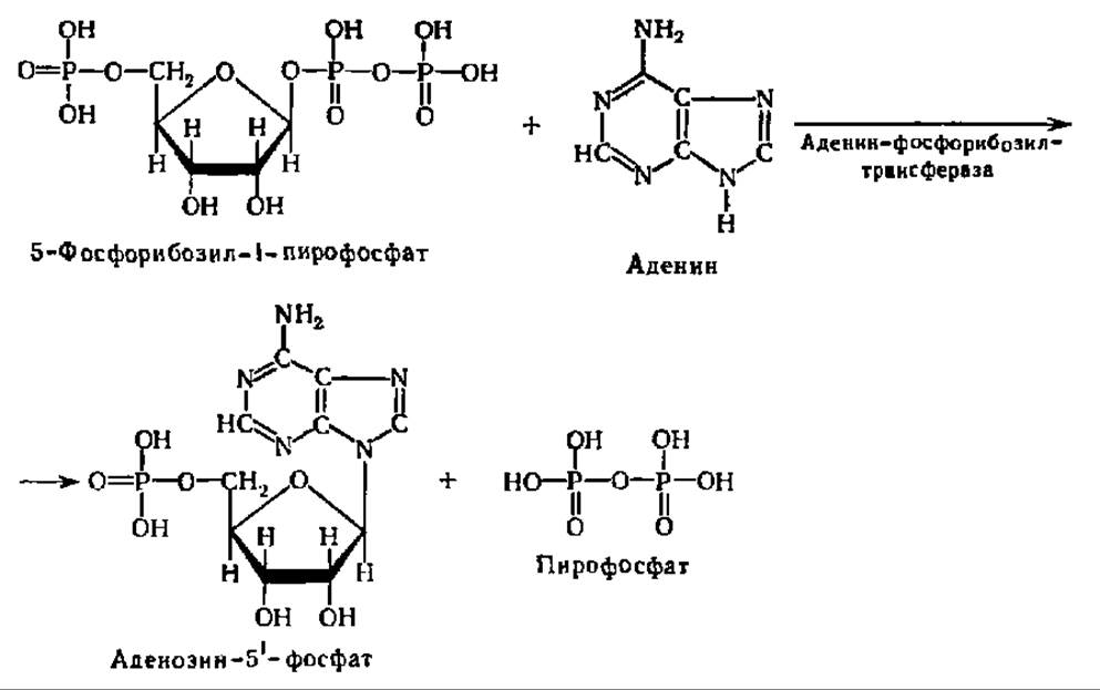
This salvage pathway is particularly prominent in malignant tumors.
Last update: 06/08/2026
Editorial and Educational Adaptation: This material has been compiled based on the primary/original source text. The project team performed an editorial review, corrected technical inaccuracies, structured sections, and adapted the content for an educational format.
What was processed:
- elimination of formatting defects (OCR errors, structural breaks, corrupted characters);
- editorial organization of content;
- standardization of terminology in accordance with academic sources;
- verification of factual statements against the original source text.
All mentions of the author, publication year, and origin of the primary text have been preserved in accordance with the source.