Human Biochemistry, Volume 2 - Murray R. 1993
Biochemistry of Intra- and Intercellular Communication
Membranes: Structure, Assembly, and Function
Transmembrane Transport of Small Molecules
Molecules can passively cross the bilayer along an electrochemical gradient via simple or Facilitated Diffusion. This spontaneous transport, which leads to equilibrium, is opposed by Active Transport, which requires an energy input because it occurs against the electrochemical gradient. These mechanisms are schematically illustrated in Fig. 42.13.
Passive Diffusion
As we have already discussed, certain substances, such as gases, can penetrate The Cell via transmembrane diffusion along an electrochemical gradient without any Energy Expenditure. The rate of simple diffusion of dissolved substances across a membrane is determined by the thermal motion of the moving molecules, the transmembrane concentration gradient of the substance, and its solubility (permeability coefficient; Fig. 42.6) in the hydrophobic layer of the membrane. Solubility is inversely proportional to the number of Hydrogen Bonds that must be broken for a substance dissolved in an aqueous medium to become incorporated into the hydrophobic layer. Electrolytes, which are poorly soluble in Lipids, do not form hydrogen bonds with Water, but they possess an aqueous shell formed As a result of Electrostatic Interactions. The size of this shell is directly proportional to the charge density of the electrolyte. Electrolytes with a higher charge density have a larger Hydration shell and, consequently, a lower rate of diffusion. For instance, Na+ ions are characterized by a higher charge density than K+ ions. Therefore, hydrated Na+ is larger than K+, and its rate of passive diffusion is lower.
Unlike synthetic bilayer membranes, natural membranes contain transmembrane channels—pore-like structures composed of Proteins. Cation-conducting channels have an average diameter of ~5–8 nm and are lined with negatively charged groups. Channel conductance depends on the size, degree of hydration, and charge density of the ion. Specialized channels for Na+, K+, and Ca2+ have been discovered.
The membranes of Nerve Cells contain well-studied Ion Channels responsible for the GENERATION AND PROPAGATION of action potentials along the membrane. The activity of some of these channels is controlled by Neurotransmitters, meaning their operation can be regulated. In addition, one ion can regulate the activity of a channel permeable to another ion. For example, a decrease in the concentration of Ca2+ in the extracellular fluid increases membrane permeability and the diffusion of Na+. As a result, the membrane depolarizes and a Nerve Impulse is generated. This explains the numbness, tingling, and Muscle cramps observed when plasma Ca2+ levels drop.
Class="center">Table 42.4. Transport of substances and Information Across Membranes
|
Transmembrane Movement of small molecules Diffusion (passive and facilitated) Active transport |
|
Transmembrane movement of large molecules Endocytosis Exocytosis |
|
Signal Transduction across membranes Cell surface receptors 1. Signal transduction (e.g., Glucagon→cAMP) 2. Signal internalization (coupled with endocytosis, e.g., LDL receptor) Movement of extracellular receptors (Steroid Hormones; a type of diffusion) |
|
Intercellular junctions and communications |

Fig. 42.13. Many small uncharged molecules freely pass through The Lipid Bilayer. Charged molecules, large uncharged molecules, and some small uncharged molecules cross membranes via channels or pores, or with the help of specific carrier proteins. Passive transport is always directed down the electrochemical gradient toward the establishment of equilibrium. Active transport, on the other hand, occurs against the electrochemical gradient and requires an energy expenditure. (From Alberts B. et al.: Molecular Biology of the Cell. Garland, 1983.)
Channels open only for a specific duration, meaning they possess a gating mechanism. In the case of Ligand-gated channels, a specific molecule binds to a receptor and opens the "gate." Voltage-gated channels open (or close) in response to Changes in the Membrane Potential.
Some microorganisms synthesize small organic molecules called ionophores, which shuttle ions across membranes. These ionophores contain hydrophilic cores that bind specific ions. The periphery of these cores is surrounded by hydrophobic regions, allowing the molecule to easily dissolve in the membrane and diffuse through it. Other ionophores, such as the well-studied polypeptide gramicidin, form channels. Certain microbial toxins (e.g., diphtheria toxin) and components of activated serum Complement are capable of forming large pores in cell membranes through which macromolecules can pass.
To summarize, the diffusion of substances is determined by the following factors:
1) the transmembrane concentration gradient of the substances (dissolved substances move toward lower concentration); 2) the transmembrane electrical potential difference (dissolved substances move toward the solution with the opposite charge); 3) the permeability coefficient of the membrane for the given substance; 4) the hydrostatic pressure gradient across the membrane (an increase in pressure increases the rate of collisions between molecules and the membrane); 5) Temperature (the higher the temperature, the greater the particle velocity and, consequently, the frequency of collisions between particles and the membrane).
Facilitated Diffusion and Active Transport
Transport systems can be described based on the number of transported molecules and the direction of movement (Fig. 42.14), or according to whether the transport occurs toward or against equilibrium. In a uniport system, a single type of molecule is transported in both directions. In cotransport systems, The transport of one solute is accompanied by the transport (simultaneous or sequential) of a stoichiometric amount of another. In symport, both substances move in the same direction. Examples of such systems include H+/sugar and Na+/sugar (glucose, galactose, xylose, and arabinose) transport in Bacteria, and Na+/Amino Acid Transport in mammalian cells. In antiport, substances are transported in opposite directions (e.g., Na+ into the cell and Ca2+ out of the cell).
Molecules that cannot cross the lipid bilayer on their own utilize carrier proteins to which they bind. This movement can occur in two ways: via facilitated diffusion or active transport using highly specific transport systems.
Facilitated diffusion and active transport have much in common. Both processes appear to be carried out with the participation of specialized carrier proteins, and both are characterized by Specificity for ions, sugars, and Amino Acids. This is evidenced by the Analysis of the consequences resulting from Mutations in bacterial and animal cells (including certain mutations causing human diseases). Facilitated diffusion and active transport resemble an enzyme-substrate reaction, yet they occur without The formation of covalent bonds. This similarity is indicated by the following points:
1) There is a specific binding site for the solute; 2) the transport process is characterized by saturation, i.e., a maximum transport velocity Vmax exists (Fig. 42.15); 3) the process is characterized by a definite binding constant, so that the system as a whole has its own Km (Fig. 42.15); 4) substances structurally similar to the transported compound act as Competitive Inhibitors and block transport.
The main differences between facilitated diffusion and active transport are as follows:
1) facilitated diffusion can occur in both directions, whereas active transport usually proceeds in only one; 2) active transport always goes against an electrical or chemical gradient and requires an energy expenditure.
A. Facilitated diffusion. Certain substances diffuse across membranes along an electrochemical gradient faster than would be expected based on their size, charge, or partition coefficient. This facilitated diffusion differs in its properties from simple diffusion. The rate of facilitated diffusion via a uniport mechanism reaches a plateau, meaning the number of binding sites for a given substance is limited. Many facilitated diffusion systems are stereospecific, but, as in simple diffusion, transport occurs without energy expenditure.

Fig. 42.14. Schematic representation of transport system types. Carriers can be classified According to the direction of substance movement and The Diversity of transported molecules. (From Alberts B. et al.: Molecular Biology of the Cell. Garland, 1983.)
As we have already mentioned, the asymmetric distribution of Membrane Proteins between the inner and outer sides of the membrane is quite stable, and spontaneous movement of proteins across the membrane occurs extremely rarely. Consequently, transmembrane movement of carrier proteins is unlikely to underlie facilitated diffusion, with the exception of ionophores present in bacterial cell membranes.

Fig. 42.15. Comparison of The kinetics of carrier-mediated (facilitated) diffusion and simple diffusion. In the latter case, the rate of substance transport is directly proportional to its concentration in solution, whereas carrier-mediated transport exhibits saturation. Vmax is the maximum velocity. The constant Km is defined as the concentration of a substance at which the velocity is half of the maximum.
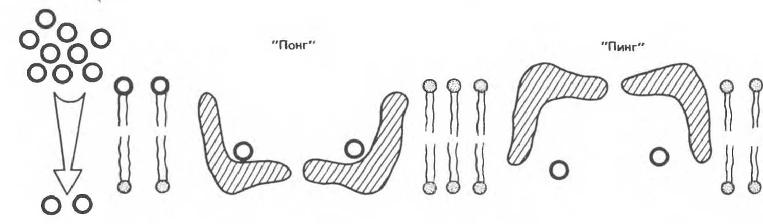
Fig. 42.16. Facilitated diffusion, the ping-pong mechanism. The carrier protein (shaded) binds a substance present at a high concentration in the solution on one side of the membrane. The carrier then undergoes conformational changes ("pong"→"ping"), resulting in the release of this substance on the opposite side of the membrane. The free carrier returns to its initial state ("ping"→"pong"), and the cycle is completed.
The process of facilitated diffusion can be explained using the ping-pong mechanism (Fig. 42.16). According to this model, the carrier protein can exist in two main Conformations. In the "pong" state, it is exposed to a solution with a high concentration of the substance, and the molecules of the latter can bind to specific sites. As a result of Conformational Changes in the protein, the binding sites along with the transported substance are exposed to a solution with a low concentration of the substance (the "ping" state). This process is fully reversible, and the net flux of the substance across the membrane is determined by its concentration gradient. The rate at which a solute enters a cell depends on the following factors: (1) the transmembrane concentration gradient; (2) The amount of carrier (the key to regulation); (3) the rate of substance binding to the carrier; (4) the rate of conformational changes of both the loaded and unloaded carrier.
Hormones regulate facilitated diffusion by altering the number of available carriers. Insulin increases the rate of glucose transport in adipose and Muscle Tissues by inducing the recruitment of new carriers from an intracellular pool (see Fig. 51.13). It also enhances amino acid transport into The Liver and other tissues. One of the many coordinated effects of glucocorticoid hormones is an increase in amino acid transport into the liver, where they serve as substrates for Gluconeogenesis. Growth Hormone enhances amino acid transport into all cells, whereas estrogens stimulate this process in the Uterus. Animal cells contain at least five distinct amino acid carrier systems. Each of these systems is specific for a particular group of closely related Amino Acids and can function as a Na+ symport system (Fig. 42.13).
B. Active transport. Active transport differs from diffusion in that it is accompanied by a Displacement of the system away from thermodynamic equilibrium and, consequently, requires energy expenditure. The source of energy can be ATP Hydrolysis, an Electron Transport Chain, or light. The maintenance of electrochemical gradients plays such a vital role in biological systems that it consumes about 30–40% of the total energy utilized by the cell.
Cells generally maintain a low intracellular Na+ concentration and a high K+ concentration (Table 42.1), alongside a net negative electrical potential. The pump that maintains these gradients is the Na+, K+-activated ATPase (Fig. 42.17). This ATPase is an integral membrane protein that requires Phospholipids for its catalytic activity. The catalytic sites of the ATPase for ATP and Na+ are located on the cytoplasmic face of the membrane, whereas the K+-binding site is located on the outer face. Ouabain inhibits ATPase activity by binding to its extracellular domain. This inhibition can be partially reversed by extracellular K+.

Fig. 42.17. Stoichiometry of the Na+, K+ pump. The pump extrudes three Na+ ions from the cell and imports two K+ ions into the cell for every molecule of ATP hydrolyzed to ADP by the membrane-bound ATPase. Ouabain and other cardiac Glycosides block the pump when introduced into the extracellular medium. (Courtesy of R. Post.)
Propagation of the Nerve Impulse
The membranes bounding nerve cells maintain an electrical potential difference (transmembrane electrical potential difference); these membranes are electrically excitable. Upon chemical stimulation mediated by a specific synaptic membrane receptor (see the section "Biochemical Signal Transduction"), gating mechanisms are triggered, and Na+ and Ca2+ rapidly rush into the cell (while K+ may not exit), causing the membrane voltage to drop sharply and the corresponding membrane patch to depolarize. However, the electrochemical gradient is rapidly restored through the action of ion pumps.
When large areas of the membrane are depolarized in this manner, the electrochemical disturbance propagates along the membrane like a wave, generating a nerve impulse. Myelin sheaths, formed by Schwann cells, wrap around nerve fibers and act as electrical insulators. This insulating layer covers most nerve fibers and greatly accelerates the propagation of the electrical wave (signal), with ions entering and exiting the cell only at the nodes where the insulator is absent. The myelin membrane consists of phospholipids, notably sphingomyelin and Cholesterol, as well as proteins and glycosphingolipids. Associated with it are only a few integral and peripheral proteins that presumably hold together the numerous membrane bilayers, forming a hydrophobic insulating Structure impermeable to ions and water. Certain diseases, such as multiple sclerosis and Guillain-Barré syndrome, are characterized by demyelination and impaired Nerve Impulse Conduction.
Glucose Transport
Using glucose transport as an example, we can summarize several Key Concepts introduced in this chapter. Glucose Transport into the cell is the first step in energy utilization. An exception to this general rule is the liver, in which no such specific process has been found. Glucose enters liver cells via simple diffusion down a concentration gradient, which is always extremely steep due to the rapid intracellular conversion of glucose to glucose-6-phosphate. Into other cells (adipose and, to an even greater extent, muscle cells), glucose enters via a specific transport system regulated by insulin (see Fig. 51.13). Changes in transport are primarily driven by variations in Vmax (largely owing to an increase or decrease in the number of carriers), though they may also be associated with variations in Km. The structure of the erythrocyte glucose carrier was deduced from its corresponding cDNA sequence (Fig. 42.7). Cells transformed with this cDNA synthesize the protein and incorporate it into the membrane in a functionally active state; further studies utilizing Site-Directed Mutagenesis may help elucidate how this Protein Functions.
In examining glucose transport, we encounter various aspects of transport mechanisms discussed previously. Glucose and Na+ bind to different sites on the glucose carrier. In this process, Na+ enters the cell driven by the electrochemical gradient and "drags" glucose along with it (Fig. 42.18). Thus, the steeper the Na+ gradient, the more glucose is transported, and if the extracellular Na+ concentration decreases, glucose transport is inhibited. To maintain the Na+ gradient required for the operation of the Na+/glucose carrier, the Na+, K+ pump is utilized to maintain a low intracellular Na+ concentration. Similar mechanisms are employed by cells to transport other sugars and amino acids.

Fig. 42.18. Transcellular movement of glucose across an intestinal epithelial cell. Glucose crosses the luminal epithelial membrane coupled to the influx of Na+. The Na+ gradient driving this symport is established by Na+, K+ exchange across the basolateral membrane facing the extracellular fluid. The glucose, now concentrated within the cell, then moves down its gradient into the extracellular fluid via facilitated diffusion (via a uniport mechanism).
The transcellular movement of sugars involves an additional component—a uniporter, through which glucose that has entered the cell via one surface can exit through the opposite surface; this process is observed in Kidney and intestinal cells.
Last update: 06/08/2026
Editorial and Educational Adaptation: This material has been compiled based on the primary/original source text. The project team performed an editorial review, corrected technical inaccuracies, structured sections, and adapted the content for an educational format.
What was processed:
- elimination of formatting defects (OCR errors, structural breaks, corrupted characters);
- editorial organization of content;
- standardization of terminology in accordance with academic sources;
- verification of factual statements against the original source text.
All mentions of the author, publication year, and origin of the primary text have been preserved in accordance with the source.