BIOCHEMISTRY AND MOLECULAR BIOLOGY - W. ELLIOTT - 2002
CHAPTER 4. DIGESTION AND ABSORPTION OF FOOD
In this chapter, we examine the processes of food Digestion and Absorption—the very starting point of METABOLISM—discuss the Chemical Nature of food, how it is transformed before entering the bloodstream, and the mechanisms by which it is absorbed.
Food contains three major components: Proteins, CARBOHYDRATES, and Lipids.
Proteins are high-molecular-weight polymers made up of twenty Different types of Amino Acids linked together by peptide bonds.
Carbohydrates are sugars and their derivatives. The term originates from the general formula for common sugars, Cn(H2O)n, which implies they are composed of carbon and Water. Although food does contain Monosaccharides such as glucose, the vast majority of dietary carbohydrates occur as Disaccharides (including sucrose) and Polysaccharides such as starch.
Lipids are represented primarily by triglycerides, or neutral fats. Typical Examples include butter and olive oil, as well as the fat layers in beef or mutton. Polar lipids are also present in food, but generally in much smaller amounts.
Digestion and Absorption
With the exception of monosaccharides like glucose, all dietary components undergo hydrolytic Cleavage in the intestine, breaking down into their elementary building blocks: proteins into amino acids, carbohydrates into monosaccharides, and neutral fats into Fatty acids and monoglycerides. Food components cannot cross the epithelial Cells lining the digestive tract in any other form.
Anatomy of the Digestive Tract
Let us examine the individual sections of the digestive tract.
In the Oral Cavity, food is chewed and moistened, which facilitates swallowing. Starch digestion begins here to a minor extent.
The Stomach contains Hydrochloric acid, which "sterilizes" the food and denatures proteins; protein breakdown also begins here.
The Small Intestine makes the primary contribution to the digestion and assimilation of all food components. Its inner surface is formed by tiny, finger-like projections called villi, which are covered with epithelial cells. The surface of the epithelial cells facing the intestinal lumen features numerous microvilli that, together with the cells, form the so-called brush border. These microvilli substantially increase the contact area between the intestine and its contents (Fig. 4.1).
Class="center">Fig. 4.1. Structure OF THE inner surface of the small intestine (schematic diagram)

In the Large Intestine, excess water is removed from the food residue.
Energetics of Digestion and Absorption
The hydrolytic Breakdown of Proteins into amino acids, carbohydrates into monosaccharides, and triglycerides into glycerol and fatty acids is an exergonic process, in which large negative values of ∆G completely shift the equilibrium toward The formation of Hydrolysis products.
Absorption is frequently coupled with the Transport of substances against their concentration gradients and therefore requires an input of energy.
Why Doesn't the Body Digest Itself?
In terms of composition, food differs very little from the Tissues of the animals that consume it. The Enzymes responsible for digesting food are synthesized within cells without subjecting the cells themselves to destructive degradation. Two lines of defense prevent this.
Formation of Zymogens (Proenzymes)
Many digestive enzymes are synthesized as inactive precursor proteins called zymogens, or proenzymes. They are secreted by cells in this form, thereby protecting their Cytoplasm from contact with the active enzyme form. Amylase, which breaks down starch, is secreted
in the active state, perhaps because starch is absent inside the producer cells. The synthesis of digestive enzymes and their secretion are not directly related to digestion and will be discussed in Chapter 22.
Protection of epithelial cells by mucus
The mucus layer covering the inner surface of the intestine is the primary defense of epithelial cells against active digestive enzymes. The Main Components of mucus are mucins. These are high-molecular-weight Glycoproteins, a significant portion of which consists of Oligosaccharides composed of fucose, glucosamine, sialic acid residues, etc. Through intermolecular interactions, mucins form three-dimensional molecular networks in water, resulting in a gel that protects the epithelial cells. The protein moiety of mucins is relatively resistant to digestion due to the protective effect of carbohydrates. Mucins are synthesized and secreted by specialized goblet Cells of the intestinal epithelium according to digestive demands.
In native proteins, the polypeptide chain is tightly folded into a compact globule, so that a significant portion of the peptide bonds is inaccessible to hydrolytic enzymes. Hence, preliminary Protein Denaturation is necessary. This occurs in The Stomach, where the contents have a pH of ~2 due to HCl secretion. In such an environment, many weak bonds stabilizing the protein globule are disrupted, causing it to unfold and expose the internal Regions of the polypeptide chain to proteolysis—the enzymatic Hydrolysis of Proteins. In addition, hydrochloric acid destroys microorganisms entering with food.
HCl production in the stomach
Hydrochloric acid is secreted by parietal cells of the gastric epithelium. This process is driven by the outward pumping of hydrogen ions from the cells against their concentration gradient. It occurs via transmembrane H+/K+ exchange, powered by ATP hydrolysis (compare the Na+/K+ exchange described on p. 65). Acid-secreting cells contain H+/K+-ATPase. Utilizing the energy of ATP hydrolysis, they pump out H+ in exchange for inward-moving K+, which is subsequently released from The Cell (Fig. 4.2). Where do the protons come from? Carbon dioxide (CO2) entering the cell from the Blood is converted into carbonic acid by the enzyme Carbonic anhydrase, which then dissociates to form bicarbonate ions.

Bicarbonate ions are exchanged for Cl- ions in the blood via an anion transport protein (see Fig. 4.2) and then pass into the gastric lumen, forming HCl.
Fig. 4.2. Mechanism of HCl formation in the stomach

Pepsin: a gastric proteolytic enzyme
Enzyme names typically include the suffix -ase, but digestive enzymes traditionally retain names such as pepsin, Chymotrypsin, and Trypsin. Their inactive precursors are designated by adding the suffix -ogen: pepsinogen, chymotrypsinogen, and trypsinogen.
Gastric epithelial cells secrete pepsinogen. This secretion is stimulated by the hormone gastrin, which gastric cells release into the bloodstream in response to food intake. Pepsinogen is converted into pepsin upon the cleavage of a 44-amino-acid fragment from its polypeptide chain, which
shields and thereby blocks the Active Site. Upon entering a strongly acidic environment, pepsinogen undergoes conformational changes sufficient to unblock the active site and "cleave off" the additional chain fragment. This enzyme activation is autocatalytic, because as soon as a small amount of pepsin appears, it also participates in the activation process, converting more pepsinogen into the active enzyme.
A strongly acidic environment is optimal for pepsin activity. In this respect, it differs from the vast majority of enzymes that are active under neutral conditions (see Fig. 1.3, a). Pepsin hydrolyzes peptide bonds located within the polypeptide chain of the target protein, resulting in a mixture of Peptides. Such enzymes are termed Endopeptidases, as opposed to exopeptidases, which cleave only the terminal amino acid peptide bonds.
Proteolytic Enzymes (proteinases or proteases) typically exhibit Specificity, cleaving peptide bonds formed only by specific amino acids. This is also true for pepsin, which is why only partial protein digestion occurs in the stomach. Pepsin most readily cleaves peptide bonds whose NH groups belong to aromatic amino acids: Tyrosine, phenylalanine, or Tryptophan.
Mammalian infants obtain most of their protein from maternal milk. In their stomachs, it is curdled by the action of the enzyme rennin. The formation of a clot delays the passage of insoluble casein through the gastrointestinal tract, thereby prolonging its exposure to proteases.
Protein Digestion in the small intestine
Partially digested food from the stomach (chyme) then enters the duodenum. Acidic chyme stimulates the release of Hormones (secretin and cholecystokinin) into the blood by intestinal cells, which prompts the Pancreas to secrete pancreatic juice. This juice is alkaline (as is Bile) and neutralizes the chyme, thereby halting pepsin activity and facilitating the action of pancreatic enzymes, which are most active in a slightly alkaline environment.
Pancreatic cells secrete a variety of proteinases. Via pancreatic juice, they enter small ducts that merge into a single main duct connected to the duodenum. Among the pancreatic proteinases are three endopeptidases: trypsin, chymotrypsin, and Elastase. They enter the intestine as inactive precursors: trypsinogen, chymotrypsinogen, and proelastase. Carboxypeptidase—an exopeptidase that sequentially cleaves amino acids from the C-terminus of the polypeptide chain—is also secreted as an inactive precursor.

Activation of pancreatic zymogens
Like pepsinogen, pancreatic zymogens are activated through Limited proteolysis. This is also an autocatalytic process triggered by a specialized proteolytic enzyme, enteropeptidase, which is produced in its active form by cells of the small intestine. Enteropeptidase cleaves a single peptide bond in trypsinogen, and the resulting trypsin is then able to activate not only trypsinogen but also other zymogens, triggering a cascade reaction (Fig. 4.3). This intricate yet highly sophisticated system serves a single purpose: to prevent protease activation before they reach the intestinal lumen. If premature activation occurs, a pathological condition called pancreatitis develops. Its cause may be duct obstruction or inflammatory damage to the pancreas itself. Following synthesis in the exocrine pancreatic cells, zymogens are packaged into membrane-bound vesicles. Under hormonal or neural stimulation, the membranes of these vesicles fuse with The Plasma Membrane, releasing their contents from the cell. If zymogens escape from the vesicles into the cytoplasm, a specialized cellular protein—trypsin inhibitor—comes into play. It forms such a stable non-covalent complex with trypsin that inhibition becomes virtually irreversible.
Fig. 4.3. Activation of pancreatic proteinases. Active proteinases are highlighted in color

In addition to pancreatic proteinases, the small intestine contains aminopeptidase, which belongs to the exopeptidases. This enzyme sequentially cleaves N-terminal amino acids from peptides produced by the partial hydrolysis of proteins by other proteinases. Thus, protein DIGESTION IN THE intestine involves three endopeptidases, which cleave peptide bonds within the protein molecule, and two exopeptidases, which remove terminal amino acids from the peptides. The combined action of all these enzymes leads to the complete breakdown of dietary proteins into free amino acids.
Transport of Amino Acids from the Intestine into the Bloodstream
To enter the blood, the amino acids formed in the intestine As a result of Protein Hydrolysis must first cross the brush border membrane into the epithelial cells (see Fig. 4.1) and then penetrate the blood capillaries of the villi.
The First stage of this process involves the accumulation of amino acids within the cells, which is achieved via the cotransport of Amino Acids and sodium ions (see Chapter 3). Figure 3.11 illustrates The Mechanism of sodium ion and sugar cotransport; this mechanism also applies to amino acids, although entirely different transport proteins are involved in their translocation.
Carbohydrate Digestion
The main dietary carbohydrates are Starch and other polysaccharides, as well as the disaccharides sucrose and lactose. Among monosaccharides, only glucose and fructose are commonly found. It has been established that only monosaccharides are absorbed in the intestine; therefore, during digestion, all carbohydrate components must be broken down into free monosaccharides.
Polysaccharides consist of monosaccharide residues linked by glycosidic bonds. The structure of glucose can be described by two formulas.

In α-D-glucose, the hydroxyl group at the C-1 atom lies below the plane of the ring, whereas in β-D-glucose, it lies above. In solutions, both forms typically exist in a state of equilibrium, which is achieved through the sequential opening and closing of the pyranose ring (a phenomenon known as mutarotation). Suppose we have two glucose molecules that become joined by a glycosidic bond during a chemical reaction.
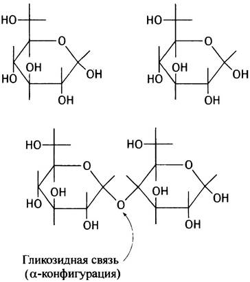
The glycosidic bond fixes the C-1 atom of the first residue in a specific position, thereby preventing mutarotation. As follows from the structure shown, the glycosidic bond between the C-1 and C-4 atoms of the two glucose residues has the α-configuration. The compound formed in this way can be written as glucose-α-(1 —> 4)-glucose. This is a disaccharide whose trivial name is maltose. Disaccharides with a β-glycosidic bond also exist.
Starch Digestion
It follows from the structure of maltose that long polysaccharide chains can be formed via glycosidic bonds. Such a chain, containing from 100 to several thousand glucose residues (glucosyl residues), is amylose, the primary component of starch. If we denote a single α-glucosyl residue as , amylose can be represented as a chain:

where n corresponds to the number of glucosyl residues.
Another component of starch, amylopectin, is also a glucose polymer. It forms highly branched structures and consists of numerous short chains (each containing about 30 residues) linked by α-(1 —> 4)-glycosidic bonds and interconnected by α-(1 —> 6)-glycosidic bonds.

Using the same designations as for amylose, the structure of amylopectin can be represented schematically:

Starch digestion is carried out by the enzyme α-amylase, which is present in saliva and pancreatic juice. It attacks internal glycosidic bonds within the chain and can therefore be classified as an endoenzyme. α-Amylase does not cleave (1 —> 6)-glycosidic bonds; consequently, when acting on amylopectin, a significant portion of the molecule remains intact. This partially digested amylopectin (known as dextrin) is broken down in the small
intestine by the enzyme amylo-α-(1 —> 6)-glucosidase. As a result, the (1 —> 6)-bonds are hydrolyzed, yielding di- and trisaccharides. In turn, these are acted upon by another enzyme, α-glucosidase (or maltase), producing free glucose. Salivary amylase acts on starch for only a brief period because, after swallowing, it is inactivated by the acidic environment of the stomach. Therefore, the primary digestion of starch takes place in the small intestine. Disaccharides, such as lactose and sucrose, are also hydrolyzed there.
Lactose is galactose-β-(1 —> 4)-glucose. It is the principal carbohydrate of milk. Recall that galactose differs from glucose only in the optical configuration of the C-4 atom.
In the structural formula of lactose presented below, the bends in the bonds have no specific chemical meaning and serve only to simplify the visualization of the structure.

In the intestine, lactose is broken down into monosaccharides by the enzyme lactase, which is located on the outer membrane of epithelial cells. Since lactose is a β-galactoside, lactase is also referred to as β-galactosidase. Many adults, particularly those of Asian descent, lose The ability to synthesize lactase with age. As a result, the dietary lactose they consume is not digested in the small intestine; instead, upon reaching the large intestine, it is degraded by bacterial action. This is accompanied by such unpleasant symptoms as excessive thirst and diarrhea. Another important dietary disaccharide, sucrose, is cleaved in the intestine into glucose and fructose through the action of the enzyme sucrase.

Glucose is absorbed by epithelial cells along with sodium ions (Fig. 4.4). The driving force for this glucose transport is the Na+ ion concentration gradient generated by the Na+/K+-ATPase. The subsequent fate of glucose is as follows: it exits the epithelial cell through the membrane facing the blood capillary and is transported by the blood via the portal vein to the Liver. The exit of glucose from the cell occurs down its concentration gradient via Facilitated Diffusion, which is mediated by a specific transport protein (see Fig. 4.4). Unlike glucose, fructose is absorbed by cells passively, without the involvement of sodium ions.
Fig. 4.4. Co-transport of glucose and sodium ions during Glucose Absorption from the lumen of the small intestine

Glucose and other monosaccharides, as well as amino acids and other molecules (except lipids) absorbed in the small intestine, are delivered directly to the liver via the portal Circulation system. This protects the Organism not only from an excess of primary digestion products, which are subsequently processed in the liver, but also from toxic compounds absorbed in the intestine, which are detoxified in the liver.
Digestion and Absorption of Fats
Dietary lipids are primarily neutral fats, or triglycerides (see Chapter 3). They are insoluble in water and form relatively large droplets in it. In this state, they cannot be assimilated because they are inaccessible to enzymes.
Fat digestion occurs mainly in the small intestine through the action of the pancreatic enzyme lipase, which hydrolyzes the ester bonds in triglycerides, predominantly those formed by the primary hydroxyl groups of glycerol.

The rate of triglyceride breakdown is limited by their accessibility to lipase. It increases as the size of the fat droplets decreases and, consequently, as the surface area of contact with the aqueous environment increases. Fat emulsification is promoted both by monoglycerides and free fatty acids—produced by the lipase-catalyzed cleavage of triglycerides—and by bile acid salts. Furthermore, monoglycerides are sufficiently water-soluble to be freely absorbed by epithelial cells. Their solubility is also facilitated by bile acids and their salts.
Bile acids are synthesized in The Liver and stored in the Gallbladder, from which they are subsequently released into the duodenum. They are derived from Cholesterol, with the Introduction of hydroxyl and carboxyl groups into its molecule.
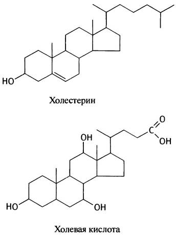
Cholic acid is the predominant bile acid in humans; other acids differ from it in the number and position of their hydroxyl groups. A significant fraction of cholic acid in bile occurs as amides formed with Glycine (glycocholic acid) or its sulfo analogue, taurine (taurocholic acid). These are termed conjugates, or conjugated bile acids, as they consist of two components: cholic acid and glycine or taurine.
The ionic formula of glycine is NН+3СН2СОО-, and that of taurine is NН+ 3СН2СН2SO3-.
If the cholic acid residue is denoted as RСОO-, the structures of glycocholic and taurocholic acids will be RСONНСН2СОO- and RСONНСН2СН2SO3-, respectively. Both of these conjugates have lower pKa values (3.7 and 1.5) compared to cholic acid (5.5). It is highly likely that conjugation is facilitated by the complete ionization of bile acids in the intestine.
In the presence of bile acids, monoglycerides and fatty acids form mixed micelles—disc-shaped particles whose edges are lined with bile acid molecules, while the more Hydrophobic core is composed of fat breakdown products, cholesterol, and Phospholipids. Micelles are much smaller than the smallest fat droplet. The concentration of fat breakdown products in the micellar solution can be quite high; nevertheless, the solution remains homogeneous and even transparent. There is no consensus regarding the mechanism by which fat micelles are absorbed. Fat digestion products may enter epithelial cells either as intact micelles or following the disintegration of these micelles at the cell surface, releasing fatty acids and monoglycerides as individual molecules that readily diffuse into the cells. Bile acids are also partially reabsorbed and transported back to the liver.
What happens to fatty acids and monoglycerides inside the epithelial cells of the brush border?
Resynthesis of Neutral Lipids
Triglycerides are synthesized anew from monoglycerides and fatty acids within the epithelial cells.

The resynthesis of triglycerides poses a problem: how to export them (along with cholesterol) out of the epithelial cells and deliver them to other tissues of the body? Within the epithelial cells, Neutral Fats and cholesterol are assembled into small particles called chylomicrons. They are surrounded by a membrane envelope and exported from the cells via exocytosis.
What are chylomicrons?
Chylomicrons are spherical particles whose core is filled with hydrophobic molecules of neutral fats and cholesterol esters (Fig. 4.5).
Fig. 4.5. Structure of a chylomicron

The latter differ from cholesterol in that their hydroxyl group is esterified with fatty acids.
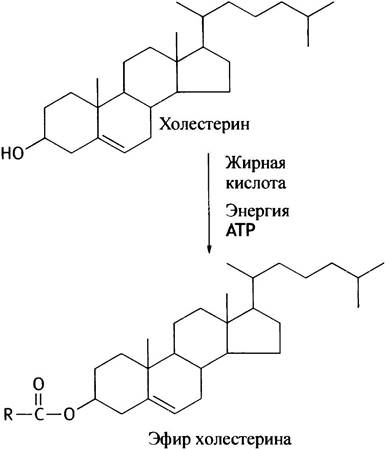
This modification ensures the packaging of cholesterol into the hydrophobic core of the chylomicron. The surface of the chylomicron is formed by phospholipid molecules, cholesterol, and specific proteins.
Several such proteins are known. The primary and essential protein required for chylomicron formation is the glycoprotein apolipoprotein B. The prefix apo- indicates that this protein in its Native State is a component of a lipoprotein. In this case, that lipoprotein is the chylomicron. The hydrophilic shell stabilizes chylomicrons to such an extent that they are distributed throughout the body as intact particles via the BLOOD AND Lymph. Consisting of more than 90% fats, they are characterized by a very low density.
Unlike monosaccharides and amino acids, chylomicrons pass from epithelial cells not into the blood, but into the lymphatic capillaries that also permeate the cellular villi. It is helpful at this point to say a few words about lymph. As blood flows through the capillaries, a clear lymphatic fluid containing proteins, electrolytes, and other substances is filtered into the interstitial fluid. All tissues are permeated by a network of lymphatic capillaries, which collect lymph from the intercellular spaces through their blind ends. The system of lymphatic capillaries and vessels culminates in the Thoracic duct, through which the lymph enters the neck Veins. Due to the high chylomicron content, upon mixing with lymph, the blood becomes turbid and begins to opalesce.
Digestion of Other Dietary Components
Digestion is a process of enzymatic hydrolytic cleavage, and nearly all dietary components undergo this process. Thus, dietary phospholipids are broken down by phospholipases, and Nucleic Acids by Nucleases. Plant-based foods contain Cellulose, a polysaccharide that resists animal digestive enzymes. However, herbivores are still able to utilize cellulose thanks to microorganisms inhabiting their digestive tract that break it down. For humans, dietary cellulose serves merely as a fibrous bulking agent beneficial for normal bowel function.
Chapter 4 Questions
1. Which digestive enzymes are synthesized as inactive precursors?
2. What is the purpose of this? Why is amylase synthesized directly in its active form? Why do active enzymes not digest the walls of the small intestine?
3. How are proenzymes activated in the small intestine?
4. What are the consequences for the organism of premature activation of pancreatic proenzymes?
5. Why is it necessary to digest food?
6. Sugars and many Amino acids are transported into epithelial cells against their concentration gradient; however, this occurs without The Use of specialized transport systems, ATP, or other high-energy phosphates. How can this seemingly paradoxical phenomenon be explained?
7. Why do many people, particularly those of Asian descent, poorly tolerate milk and dairy products?
8. What are neutral fats?
9. Dietary fats are insoluble in water. How do digestive enzymes manage to process them?
10. Amino acids and sugars absorbed in the small intestine are subsequently transported by the bloodstream to the liver. What is The Fate of the products of fat digestion?
Last update: 06/08/2026
Editorial and Educational Adaptation: This material has been compiled based on the primary/original source text. The project team performed an editorial review, corrected technical inaccuracies, structured sections, and adapted the content for an educational format.
What was processed:
- elimination of formatting defects (OCR errors, structural breaks, corrupted characters);
- editorial organization of content;
- standardization of terminology in accordance with academic sources;
- verification of factual statements against the original source text.
All mentions of the author, publication year, and origin of the primary text have been preserved in accordance with the source.