Orthopedics - Oleksa A.P. 2006
Metabolic Bone Diseases
Skeletal Changes in Thyroid Disorders
Myxedema
Pathological bone changes typically occur when thyroid function is significantly reduced, that is, in the presence of myxedema.
Myxedema is a congenital condition caused by hypoplasia or aplasia of The Thyroid Gland, and therefore manifests itself from early childhood. However, acquired myxedema in a young child (following an infection or trauma) is indistinguishable from the congenital form and presents with the exact same signs.
Clinically, affected children present as lethargic and drowsy, with intellectual impairment resulting from neuropsychic deceleration. Metabolic Regulation is disrupted, yet the hallmarks of childhood myxedema (Fig. 93) are marked delays in growth, as well as in physical, sexual, and mental development. All ossification centers appear with a significant delay and are somewhat altered, though their sequential appearance remains undisturbed. Instead of the single ossification centers normally found in bones, small foci emerge within the Cartilage, eventually forming fragmented ossification centers with uneven radiographic density and intervening shadow clefts.
The deceleration of bone growth leads to dwarfism. Radiographically, the hands of a ten-year-old child have a characteristic appearance resembling those of a normal three-year-old. The medullary cavities in the long bones are narrow, while the cortical layer is thickened due to normal periosteal activity. Bone changes are symmetrical, and their articular ends are thickened and even deformed.
Characteristic changes are also evident in the heads of the humerus and Femur. The humeral HEAD is slightly rotated laterally, flattened, flexed at the neck at a somewhat narrower angle, and its upper margin lies level with the tip of the greater tubercle. Initially, the femoral head exhibits fragmented ossification centers—resembling changes seen in osteochondropathy—and subsequently becomes deformed during development, most frequently resulting in coxa plana and coxa vara. Radiolucencies are observed in the femoral neck and along the ossification border, indicating incomplete cartilage ossification. The acetabulum is also deformed, as it develops to match the shape of the femoral head; it tends to be wider and deeper, which later in life predisposes to The Development of coxarthrosis.
Furthermore, the epiphyses of the radius and ulna may appear widened and coarse.
The BONES OF THE feet also develop abnormally As a result of static weight-bearing on unossified epiphyses. The calcaneus is shortened, the FOOT is flat, and its dorsal surface is deformed.
Significant radiographic changes are detected in the Skull bones. Due to the delayed growth of the skull base bones and delayed Ossification of the synchondrosis intersphenoidalis et sphenooccipitalis—while the cranial vault develops normally—the Base of the skull becomes flexed. On lateral radiographs, the skull resembles brachycephaly.
Class="center">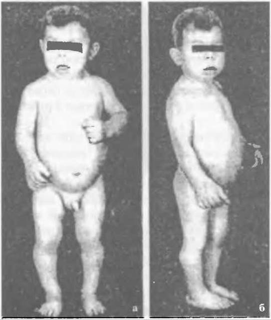
Fig. 93. Hypothyroidism in a 9-year-old boy: a - short stature, b - bone age corresponds to 3 years.
The anterior and middle cranial fossae exhibit a height discrepancy in their floor levels, and the clivus is shortened. Forward protrusion of the facial bones is also characteristic. The Nose develops a saddle shape due to the shortening of the Sphenoid bone. The vertebral bodies may be mildly flattened, with widened intervertebral spaces.
All of these radiographic abnormalities become most pronounced between the ages of 30 and 40, owing to the extremely slow progression of growth and ossification processes. There is no other condition that delays ossification to the extent seen in childhood myxedema.
It is very often necessary to differentiate myxedema from Pituitary dwarfism. In such cases, radiographic evaluation of the sella turcica and assessment of thyroid function are helpful. Radiographically, the sella in pituitary dwarfism is enlarged and deepened, whereas thyroid function and mental development are normal.
Occasionally, myxedema must be differentiated from osteodystrophy. Unlike osteodystrophy, myxedema does not present with Changes in the vertebral bodies or a disproportionate lag in lower limb growth. In osteodystrophy, endocrine disorders are absent, whereas in myxedema, they are predominant.
Similarly, radiographic differentiation of femoral head changes between myxedema and osteochondropathy (Legg-Calvé-Perthes disease) presents no diagnostic difficulty.
Treatment. Due to thyroid hypofunction, replacement therapy is required, which was first introduced by Murray. Today, thyroidin, L-thyroxine, and other agents are used to treat myxedema. Patients are managed by endocrinologists.
Attempts at thyroid transplantation have been made, but they have proven ineffective to date because the graft undergoes resorption.
Corrective osteotomies are employed to address extremity bone deformities. Surgical Procedures are occasionally performed to improve the shape of the nose and face.
Last update: 10/08/2026
Editorial and Educational Adaptation: This material has been compiled based on the primary/original source text. The project team performed an editorial review, corrected technical inaccuracies, structured sections, and adapted the content for an educational format.
What was processed:
- elimination of formatting defects (OCR errors, structural breaks, corrupted characters);
- editorial organization of content;
- standardization of terminology in accordance with academic sources;
- verification of factual statements against the original source text.
All mentions of the author, publication year, and origin of the primary text have been preserved in accordance with the source.