Review of Medical Physiology - William F. Ganong 2002
Endocrine System, Metabolism, and Reproduction
Hormonal Regulation of Calcium Metabolism and Bone Physiology
Vitamin D and Hydroxycholecalciferols
Chemistry
Active Transport of Ca2+ and PO43 across the intestinal mucosa is enhanced by vitamin D metabolites. The term vitamin D refers to a group of closely related sterols produced by the action of ultraviolet light on certain provitamins (Fig. 21-7). Vitamin D3, also known as cholecalciferol, is synthesized in mammalian Skin from 7-dehydrocholesterol under METABOLISM/18.html">The Influence of sunlight. The reaction involves the rapid formation of provitamin D3, which subsequently converts more slowly into vitamin D3 (cholecalciferol). Vitamin D3 and its hydroxylated derivatives circulate in plasma bound to the vitamin D-binding protein (DBP), which is also known as Gc-protein and binds G-Actin (see Chapter 1). Additionally, DBP enhances the Complement-induced chemotactic activity of neutrophils. Its affinity for provitamin D3 is low, whereas its affinity for vitamin D3 is high, allowing DBP to transport vitamin D3 from the skin into the bloodstream. Vitamin D3 is also obtained from dietary sources.
Vitamin D3 is metabolized by Enzymes belonging to the cytochrome P450 (CYP) superfamily (see Chapters 17 and 20). In the Liver, vitamin D3 is converted into 25-hydroxycholecalciferol (calcidiol, 25-OHD3), which is then converted in the Cells of the renal proximal tubules into the more active metabolite 1,25-dihydroxycholecalciferol, also referred to as calcitriol or 1,25-(OH)2D3. 1,25-Dihydroxycholecalciferol is also synthesized in the Placenta, skin keratinocytes, and macrophages. In patients with sarcoidosis, 1,25-dihydroxycholecalciferol is produced by pulmonary alveolar macrophages, presumably upon stimulation by y-interferon. The normal plasma concentration of 25-hydroxycholecalciferol is approximately 30 ng/mL, whereas that of 1,25-dihydroxycholecalciferol is 0.03 ng/mL (100 pmol/L). A less active metabolite, 24,25-dihydroxycholecalciferol, is also produced in the Kidneys (see Fig. 21-7).
Vitamin D3 and its derivatives are secosteroids—that is, Steroids in which one ring, in this case the B-ring, is opened (see Fig. 21-7). Furthermore, 1,25-dihydroxycholecalciferol Functions as a hormone because it is synthesized endogenously, acts on target cells, and is transported via the Blood.
Given that 1,25-dihydroxycholecalciferol is a steroid, it is not surprising that it acts through a receptor belonging to the nuclear receptor superfamily through which steroids, THYROID Hormones, and many Other Compounds mediate changes in Gene Expression (see Chapter 1). Binding of the steroid to the receptor affects the DNA-binding domain, resulting in the upregulation of Transcription for certain mRNAs and the downregulation of others.
Actions
The action of 1,25-dihydroxycholecalciferol induces the synthesis of mRNAs that control The production of the calbindin-D family of Proteins. These proteins belong to the troponin C superfamily of Ca2+-binding proteins, which also includes calmodulin (see Chapter 1). Calbindin-Ds are found in the human intestine, Brain, and Kidney, as well as in various Tissues of rats. In the intestinal epithelium and other tissues, two calbindins are induced: calbindin D9K, which has a molecular mass of 9,000 and binds two Ca2+ ions, and calbindin D28K, with a molecular mass of 28,000, which typically binds four Ca2+ ions despite possessing six Ca2+-binding sites. In the intestine, the elevation of calbindin D9K and calbindin D28K levels is dependent on Ca2+ transport; however, the precise mechanism by which they facilitate Ca2+ passage across the intestinal epithelium remains unclear. There is also evidence that 1,25-dihydroxycholecalciferol increases the number of Ca2+-H+-ATPase molecules in intestinal cells, which are required for the extrusion of Ca2+ into the interstitial space.
Class="center">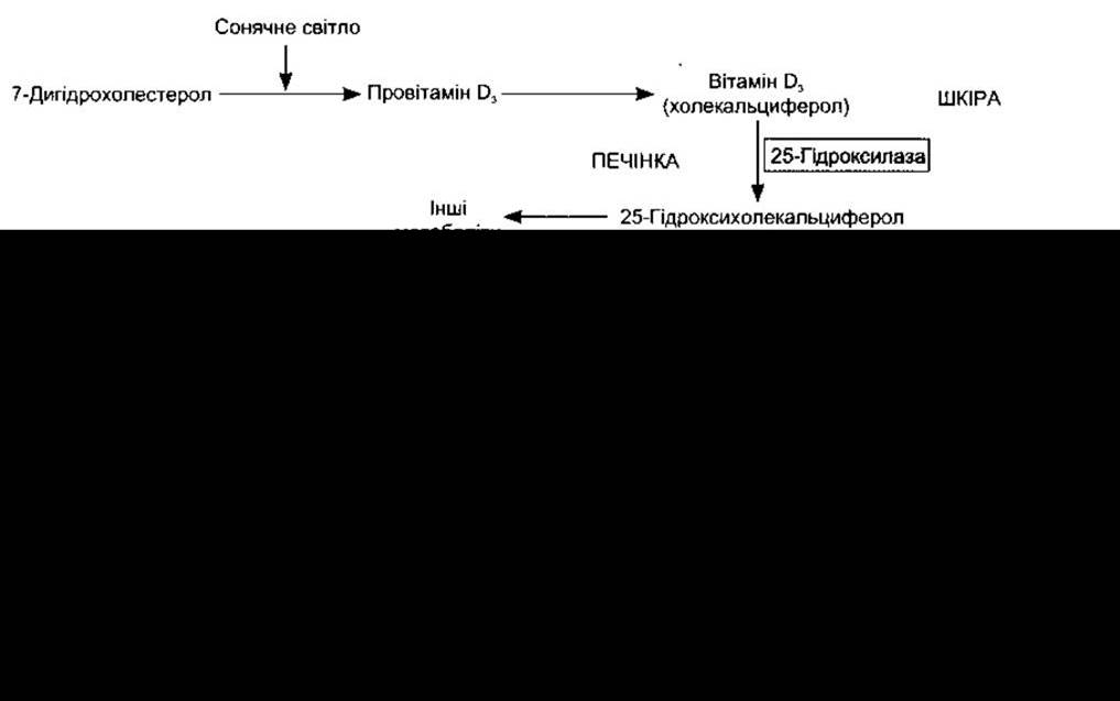
Fig. 21-7. Biosynthesis and hydroxylation of vitamin D3; 25-hydroxylation occurs in the liver, whereas other hydroxylations take place primarily in the kidneys. The chemical structures of 7-dehydrocholesterol, vitamin D3, and 1,25-dihydroxycholecalciferol are also shown.
In addition to increasing intestinal Ca2+ absorption, 1,25-dihydroxycholecalciferol promotes renal Ca2+ reabsorption. It acts on bone, where it mobilizes Ca2+ and PO43- by increasing the number of mature osteoclasts, and it also stimulates osteoblasts, though the net physiological outcome is bone resorption and Ca2+ mobilization.
Receptors for 1,25-dihydroxycholecalciferol have been identified in numerous tissues beyond the intestine, kidney, and bone, including the skin, lymphocytes, monocytes, skeletal and cardiac Muscle, mammary gland, and anterior pituitary. Accumulating evidence indicates that 1,25-dihydroxycholecalciferol stimulates the differentiation of immune system cells and cutaneous keratinocytes. Consistent with this, Vitamin D deficiency is associated with an increased incidence of infections. Furthermore, 1,25-dihydroxycholecalciferol has been implicated in the Regulation of Cell growth and the production of growth factors; however, its precise physiological role in these processes requires further investigation.
The synthesis of 25-hydroxycholecalciferol does not appear to be tightly regulated. In contrast, The formation of 1,25-dihydroxycholecalciferol in the kidneys, catalyzed by 1a-hydroxylase, is feedback-regulated by plasma levels of Ca2+ and PO43- (Fig. 21-8). Its production is stimulated by parathyroid hormone (PTH); when plasma Ca2+ levels are low, PTH secretion increases. Conversely, when plasma Ca2+ levels are high, little 1,25-dihydroxycholecalciferol is produced, and the kidneys instead generate the relatively inactive metabolite 24,25-dihydroxycholecalciferol. This effect of Ca2+ on 1,25-dihydroxycholecalciferol synthesis provides a mechanism for adapting intestinal Ca2+ absorption (see above). The formation of 1,25-dihydroxycholecalciferol is also stimulated by low plasma PO43- levels and inhibited by high levels through a direct action of PO43- on 1a-hydroxylase. Additional control over 1,25-dihydroxycholecalciferol synthesis is exerted via direct negative feedback of the metabolite on 1a-hydroxylase, positive feedback on the formation of 24,25-dihydroxycholecalciferol, and a direct inhibitory effect on parathyroid gland synthesis of PTH mRNA. Prolactin enhances 1a-hydroxylase activity, leading to elevated circulating levels of 1,25-dihydroxycholecalciferol during Lactation. Estrogens increase total circulating 1,25-dihydroxycholecalciferol, although this effect is likely mediated by an increase in the synthesis of its binding protein without significant changes in free 1,25-dihydroxycholecalciferol. Hyperthyroidism is associated with decreased circulating 1,25-dihydroxycholecalciferol and an increased prevalence of Osteoporosis. The synthesis of 1,25-dihydroxycholecalciferol is inhibited by metabolic acidosis. Growth Hormone, prolactin, and Calcitonin also stimulate 1,25-dihydroxycholecalciferol production.
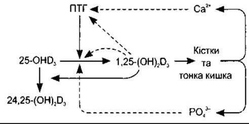
Fig. 21-8. Feedback regulation of 1,25-dihydroxycholecalciferol (1,25-[OH]2D3) synthesis from 25-hydroxycholecalciferol (25-OHD3) in the kidneys. Solid arrows indicate stimulation, and dashed lines indicate inhibition.
Rickets and Osteomalacia
Vitamin D deficiency impairs the mineralization of the bone matrix, resulting in rickets in children and osteomalacia in adults. In severe cases, childhood rickets causes weakness and bowing of weight-bearing bones, dental defects, and hypocalcemia. In adults, the condition is subtler and has historically been linked to inadequate sunlight exposure in smoggy urban environments, though today it predominantly stems from insufficient Dietary intake of provitamins subject to cutaneous photoactivation. In such cases, vitamin D supplementation is required. These disorders can also be caused by inactivating Mutations in the gene encoding renal 1a-hydroxylase, a condition characterized by a lack of response to vitamin D coupled with a normal response to 1,25-dihydroxycholecalciferol (type I vitamin D-resistant rickets). Rarely, the disease may arise from inactivating mutations in the 1,25-dihydroxycholecalciferol receptor gene (type II vitamin D-resistant rickets), which presents with unresponsiveness to both vitamin D and 1,25-dihydroxycholecalciferol.
Last update: 10/08/2026
Editorial and Educational Adaptation: This material has been compiled based on the primary/original source text. The project team performed an editorial review, corrected technical inaccuracies, structured sections, and adapted the content for an educational format.
What was processed:
- elimination of formatting defects (OCR errors, structural breaks, corrupted characters);
- editorial organization of content;
- standardization of terminology in accordance with academic sources;
- verification of factual statements against the original source text.
All mentions of the author, publication year, and origin of the primary text have been preserved in accordance with the source.