Biochemistry - The Chemical Reactions of Living Cells, Volume 3 - D. Metzler 1980
Biochemical Genetics and the Synthesis of Nucleic Acids and Proteins
Transcription of RNA Molecules
RNA Polymerases and Their Regulation
RNA polymerases are Enzymes that catalyze the METABOLISM/31.html">Transcription of Genetic information from a DNA template strand. Since cellular DNA is double-stranded, the question naturally arises: how can a single-stranded RNA be produced from a double-stranded template? A partial answer comes from the fact that purified RNA polymerases can also synthesize RNA from four ribonucleoside triphosphates using single-stranded DNA AS A template. This suggests that the transcription mechanism, much like DNA Replication, involves base pairing. This Conclusion is further supported by the ability of RNA polymerase to convert single-stranded DNA from bacteriophage φX174 (Supplement 4-B) into a double-stranded RNA–DNA hybrid molecule. However, when double-stranded DNA serves as the template, free single-stranded RNA molecules are released. It follows that at the site of RNA polymerase action, the double-stranded DNA temporarily separates into individual strands, one of which is then copied by the enzyme.
Surprisingly, within The Cell, typically only one of the two strands of the DNA double helix is transcribed for any given genetic locus. This has been clearly demonstrated using the double-stranded replicative form of DNA from phage φX174. This DNA is a circular molecule, and only one of its strands is actively transcribed in E. coli Cells infected with this bacteriophage. Interestingly, mRNA transcribed in this manner and isolated from Bacteria does not hybridize with single-stranded phage φX174 DNA isolated from mature Viral Particles. This DNA strand is commonly referred to as the "plus" strand. This implies that the transcribed RNA is also a "plus" strand and, consequently, is transcribed from the "minus" strand of the replicative DNA form. This conclusion is confirmed by the observation that the isolated RNA readily hybridizes with denatured double-stranded replicative DNA, which contains both "plus" and "minus" strands.
RNA polymerases are large protein molecules; some of these Proteins consist of multiple subunits [40, 51–53]. Notably, the mitochondrial RNA polymerase of Eukaryotic cells can be a monomeric protein with a Molecular Weight of 64,000, possessing a simpler Structure than bacterial enzymes. Bacterial Viruses sometimes induce their own RNA polymerases, which are also monomeric. For instance, phage T7 induces an RNA polymerase with a molecular weight of 100,000, consisting of a single long polypeptide chain. The best-characterized RNA polymerase is that of E. coli, an oligomer with a molecular weight of ~500,000 composed of four types of subunits (α, β, β', σ) and containing bound Zn2+ ions. The subunit COMPOSITION OF THE oligomer is represented by the formula α2ββ'σ. The molecular weights of the α, σ, β, and β' subunits are approximately 39,000, 86,000, 155,000, and 165,000, respectively. The σ subunit plays a crucial role in the initiation of transcription. It is essential for the precise recognition of promoter sites and the onset of synthesis, but is no longer required for RNA chain elongation once transcription has begun. In eukaryotic cells, at least three distinct classes of RNA polymerases have been identified. Polymerase I (or A) is localized in the nucleolus and likely transcribes pre-rRNA genes (Section B, 3). Polymerases II (or B) and III (or C) are found in the nucleoplasm. Evidence indicates that type II enzymes transcribe the majority of genes, whereas type III enzymes transcribe Transfer RNA genes. Similar to bacterial polymerases, eukaryotic RNA polymerases possess a complex oligomeric structure [53, 53a].
a. Initiation of Transcription
According current models of initiation, RNA polymerase repeatedly binds to and dissociates from random sites on DNA until it encounters a promoter region. The enzyme is believed to recognize the promoter through specific interactions with Base Pairs in the major groove of the DNA double helix (Fig. 2-23). Calculations suggest that The formation of a unique sequence recognized by RNA polymerase requires a specific combination of approximately 12 base pairs, the random occurrence of which in the E. coli chromosome is highly improbable [40].
The initial specific polymerase–promoter complex is termed the closed complex, because the DNA base pairs are thought to remain intact. The closed complex is postulated to be in equilibrium with an open complex, which is ready to initiate mRNA synthesis; this transition is accompanied by significant conformational changes [39, 40]. In the open complex, the Hydrogen Bonds between complementary bases of the DNA template are disrupted, making the template bases accessible for pairing with incoming ribonucleoside triphosphates.
Returning to Fig. 15-4, A, let us examine the sequence GAAATGTGAAAT (in the bottom strand). While this sequence lacks regions of local Symmetry, it contains a repeated pentamer, GAAAT. Positioned centrally within the putative polymerase-binding site, it may serve as a promoter recognition element. Structural Asymmetry might be required for the binding of a protein whose function involves directional movement. Furthermore, this region is enriched in A-T base pairs, and consequently, the helix unwinds more easily here than in G-C-rich regions (Chap. 2, Sec. D, 6). Thus, this specific segment may serve as the attachment site for RNA polymerase during the Formation of the open complex [39].
Dickson et al. suggested that the enhanced binding of RNA polymerase to the adjacent promoter region in the presence of the CAP–cAMP complex is likely due to the ability of this complex to facilitate the destabilization of G-C-rich sequences located between the CAP and polymerase binding sites [39]. Once the open complex is formed, the polymerase must (as indicated in Fig. 15-4) shift a short distance along the DNA template to reach the start site of mRNA synthesis.
The initiation of RNA chain synthesis begins with the reaction of ATP or GTP with a second ribonucleoside triphosphates molecule [Equation (15-5)], yielding a dinucleotide whose 5' end retains a triphosphate group.
Class="center">ATP (GTP) + XTP → PPi + pppPupX. (15-5)
Subsequently, nucleotide units are added to the 3' ends via reactions of this type. The rate of transcription is approximately 50 NUCLEOTIDES per second at 25 °C, which is roughly 30 times slower than the rate of DNA replication.
b. Action of Antibiotics
The antibiotic rifamycin appears to inhibit initiation by competing with the initiating purine nucleoside 5'-triphosphate for its binding site (Supplement 15-A). In bacterial cells, the same RNA polymerase that synthesizes mRNA also catalyzes the synthesis of rRNA and tRNA. Consequently, rifamycin inhibits the synthesis of all RNA species in bacteria. When a bacterial population is treated with this antibiotic, some individuals survive. These rifamycin-resistant mutants have lost their sensitivity to the drug. Among the resistant mutants are strains that produce an RNA polymerase with an altered β subunit. Because such mutant polymerases fail to bind rifamycin, it was concluded that rifamycin binds to the β subunit, and that the Gene conferring resistance to rifamycin—the rpoB or rif gene (map position 89 min; Fig. 15-1)—encodes the β subunit of RNA polymerase.
Several Other Antibiotics also interfere with transcription. Streptolydigin inhibits both the initiation and elongation of RNA chains. Actinomycin D inhibits both DNA and RNA polymerases, the latter even at concentrations as low as 10-6 M (Supplement 15-B). Eukaryotic RNA polymerases are insensitive to rifamycin; however, two of them, namely RNA polymerases II and III, are completely inhibited by α-amanitin, a potent toxin found in certain mushrooms (Supplement 15-C).
Supplement 15-A
Rifamycin and Rifampicin Antibiotics

Rifamycin, an antibiotic produced by Streptomyces mediterranei, is widely used in medicine because it is active against both acid-fast and Gram-positive bacteria. The semisynthetic derivative rifampicin has proven particularly effective in the Treatment of tuberculosis. Note the right-hand ring in the structural formula: the ether linkage is cleaved, and the resulting hydroquinone is oxidized to a quinone within the bacterial cell.
Rifampicin is an extremely potent inhibitor of bacterial RNA polymerase, achieving 50% inhibition at an antibiotic concentration of 2 × 10⁻⁸ M. Rifampicin does not prevent polymerase binding to DNA, but it blocks Transcription initiation. E. coli mutants resistant to rifampicin (carrying the rif mutation) produce an RNA polymerase with an altered β subunit (sometimes manifested as A change in electrophoretic mobility). A related antibiotic, streptolydigin, also binds to the β subunit of RNA polymerase and blocks elongation. On the chromosome map, Mutations conferring resistance to this antibiotic map very close to the rif mutations.
a Goldberg, J. H., & Friedman, P. A. (1971). Annu. Rev. Biochem., 40, 775–810.
Supplement 15-B
Actinomycin D, a Toxic Antibiotic

Actinomycins, antibiotics produced by Streptomyces species, not only kill bacteria but also exhibit potent antitumor activitya. However, due to their exceptionally high toxicity, they cannot be used in Cancer Chemotherapy. Actinomycin finds widespread application in biochemical research owing to its ability to inhibit RNA polymerases with a high degree of Specificity.
The actinomycin D (actinomycin C1) molecule contains a planar phenoxazone chromophore bearing two carboxyl groups. Both groups are linked to identical cyclic Peptides composed of L-Threonine, D-valine, L-Proline, Sarcosine (N-methylglycine), and L-N-methylvaline residues.
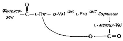
The methylvaline residue of the peptide is linked by an ester bond to the hydroxyl group of the threonine side chain.
In addition, There are two more cis-peptide bonds. Disregarding the obvious structural differences on either side of the phenoxazine ring, actinomycin can be considered to possess approximate twofold symmetry. Biochemical studies have established that this antibiotic binds tightly to double-stranded DNA at guanine-rich sites.
The (2:1) deoxyguanosine–actinomycin complex was obtained in crystalline form, and its structure was investigated by X-Ray Diffraction Analysisb–g. The phenoxazine ring was found to lie in the center of the complex, with one peptide loop positioned above it and the other below it (as shown in the right-hand drawing). The structure of the peptide chain is most easily traced for the upper loop. The dideoxyguanosine complex retains the same twofold symmetry as actinomycin itself. The phenoxazine ring is held between the flat guanosine rings through Van der Waals forces. Note that the two amino groups of the guanine rings form strong hydrogen bonds with the carbonyl groups of the threonine residues. Alongside these, There are also weaker, nonlinear hydrogen bonds between the N-3 atoms of the guanines and the NH groups of the same threonine residues. A symmetric pair of hydrogen bonds connects the two carbonyl and NH groups of the D-valine residues within the peptide loops.
Molecular modeling studies have shown that a similar complex can be formed by actinomycin with double-stranded DNA as well. Note that while the two amino groups of the guanine rings shown in the figure form hydrogen bonds with actinomycin, the other hydrogen atoms of those same amino groups, as well as the hydrogen atoms at N-1 and the carbonyl groups of the guanine ring, remain available for hydrogen bonding that forms G-C pairs. Thus, the structure shown in the figure can be readily converted into part of a double-stranded DNA molecule in which the phenoxazine ring of actinomycin is intercalated between two GC pairs (Fig. 2-21). For this to occur, the DNA must unwind by 180° at the site of insertion of the extra ring.
The Study of the crystal STRUCTURE OF THE actinomycin–deoxyguanosine complex is significant primarily because it provides graphical proof of the physical feasibility of intercalating planar rings into nucleic acid molecules. This method also demonstrates how a specialized reagent possessing approximate twofold symmetry can bind selectively to regions of a DNA molecule that also exhibit a twofold axis of symmetry.
These structural studies are likewise of great importance to medicine. By understanding The Mechanism of actinomycin's toxic action, we may eventually be able to chemically modify the antibiotic molecule in such a way that its toxicity toward normal cells is reduced while remaining unchanged for tumor cellsb. Another approach, proposed by Sobell, is to produce an actinomycin-like antibiotic capable of interacting specifically with The Double Helix of viral RNA.
a Perlman D., in: Medicinal Chemistry (A. Burger, ed.), 3rd ed., Part I, pp. 309—316, Wiley (Interscience), New York, 1970.
b The structure, as sketched here, is slightly distorted from the computer-generated drawings of Sobell to show the bonding arrangement more clearly. H. M. Sobell, Prog. Nucleic Acid Res. Mol. Biol., 13, 153—190 (1973).
c Sobell H. M., Jain S. C., Sakore T. D., Ponticello G., Cold Spring Harbor Symp. Quant. Biol., 36, 263—270 (1971).
d Sobell H. M., Sci. Am., 231, 82—91 (Aug. 1974).
Supplement 15-B
A Potent Mushroom Poison: α-Amanitin

Certain lethal toadstools of the genus *Amanita* produce colorless, toxic octapeptides known as amanitinsa. The molecule of the highly toxic α-amanitin contains two Glycine residues, one L-isoleucine residue, one residue of the unusual 1-dihydroxyisoleucine, one L-asparagine residue, and one L-hydroxyproline residue. A modified Tryptophan residue located in the center is linked via oxidation to the S–H group of a Cysteine residue. The LD50 value for mice is 0.3 mg∙kg-1 (ingesting 50 g of fresh *Amanita phalloides* mushrooms can be fatal to humans). Amanitins act slowly: a mouse cannot be killed in less than 15 hours, regardless of the dose. Replacing the dihydroxyisoleucine residue in α-amanitin with unhydroxylated leucine yields amanullin, a completely nontoxic compound.
α-Amanitin completely blocks transcription mediated by eukaryotic RNA polymerases II and III. Polymerase II appears to be the principal nuclear polymerase, and its inhibition virtually shuts down cellular Protein Synthesis. Note that, much like actinomycin (Supplement 15-A), the amanitin molecule
overall exhibits a semisymmetrical structure with an aromatic group projecting from the center. It is quite possible that amanitins, similarly to actinomycin, interact with a symmetrical region of the polymerase or the polymerase–DNA complex.
The same mushrooms contain A number of fast-acting toxic heptapeptides, the phalloidins, whose structure closely resembles that of the amanitins. However, unlike amanitins, they contain a reduced sulfur atom (—S—) acting as a bridge. The mechanism of phalloidin action remains unknown. In addition, these mushrooms contain antamanide, an antidote to phalloidin. Like the toxins, this cyclic decapeptide is composed entirely of L-Amino Acids. Antamanide competes with phalloidins for binding sites, though unfortunately, it proves ineffective in cases of Mushroom poisoning.

Antamanide is an ionophore that specifically binds sodium (Fig. 5-5).
a Wieland T., Wieland O., Microb. Toxins, 8, 249—280 (1972).
B. Nucleotide Selection in Base Pairing
How do cells achieve such a high degree of fidelity in selecting the correct base during replication and transcription, as well as during codon–anticodon pairing in protein synthesis? Early researchers often suggested that base-pairing specificity is governed exclusively by the formation of two (or three, respectively) Hydrogen bonds and stabilization via neighboring base-stacking interactions. However, it turned out that the Free energy of base-pair formation is small (Chap. 2, Sec. D, 6), and the additional free energy resulting from base binding to the end of a pre-existing chain is insufficient to ensure pairing specificity. Based on current enzymological data, one can surmise that the enzyme itself plays a critical role in ensuring pairing fidelity. RNA and DNA polymerases are quite large molecules; consequently, the binding site of the enzyme can completely encircle the double helix. If so, it is easy to conceptualize that the base selection process might proceed as illustrated in Fig. 15-5. The figure depicts a guanine base on the template DNA strand located at the site of 3'-terminal elongation of the complementary chain (DNA or RNA). To form a correct base pair, the appropriate nucleoside triphosphate must be accommodated prior to the displacement reaction that attaches the nucleotide to the growing chain. Suppose the enzyme possesses specific binding pockets for the deoxyribose moiety of the template nucleotide and for the sugar component of the incoming nucleoside triphosphate, and that these sites are held at a fixed distance from one another. As shown in Fig. 15-5, each binding
site may also feature an H–Y group that forms a Hydrogen bond with a nitrogen or hydrogen atom (indicated by the bold arrows). It should be noted that such hydrogen bonds in the exact same relative position to the deoxyribose or ribose rings can be formed by all four bases. Furthermore, we can imagine that a hydrophobic region (represented by the hatched areas in the figure) provides additional stabilization. This ordered structure vividly illustrates how the correct nucleoside triphosphate can be selected regardless of which of the four bases occupies the binding site on the left side of the diagram. (The outlines of the thymine and adenine rings are drawn with dashed lines: the dashed line denotes thymine, and the dashed-dotted line denotes adenine.)

FIG. 15-5. Selection of the correct nucleotide for the next structural unit of a growing RNA or DNA chain. A template strand deoxyguanosine is shown bound to a hypothetical DNA polymerase site.
An essential detail of the scheme shown in Fig. 15-5 is that if a purine is located on the left side (as illustrated), only enough room remains on the right side for a pyrimidine ring. Thus, the presence of A and G on the right is ruled out, leaving a choice only between C and U (or T). However, U is unsuitable because the dipole required for hydrogen bonding is oriented in the wrong direction within this base. In solution, these dipolar groups are hydrated. It is unlikely that these groups would shed their bound Water molecules prior to hydrogen bond formation within a base pair. Furthermore, binding would be hindered not only by the inability of U (or T) molecules to form strong hydrogen bonds within the vacant site shown in Fig. 15-5, but also by electrostatic repulsion between similarly charged dipole ends. As a result, the affinity of RNA polymerase for incorrectly paired bases is diminished. This reduction in affinity (an increase in the apparent Km value) has been observed experimentally, at least for bacteriophage T4 DNA polymerase, for which mutant forms are known. One of these, a "mutator" polymerase, makes far more errors during base pairing precisely because it lacks the wild-type polymerase's high-fidelity base discrimination during the pairing process. Conversely, an "antimutator" polymerase makes fewer errors than the wild-type enzyme, binding complementary base pairs even more tightly relative to non-complementary ones than the wild-type enzyme does [53b]. This issue is discussed in greater detail by Drake and Baltz [53c].
Base pairs exhibit interesting tautomeric properties, suggesting the hypothesis that the multiplicity of tautomeric forms facilitates correct base pairing and transmits a signal to the Active Site of the polymerase where the formation of a new phosphodiester bond takes place. A purely hypothetical scheme of how this might occur is shown in Fig. 15-6. In this figure, the H—Y group depicted in Fig. 15-5 corresponds to an imidazole group hydrogen-bonded to another protein residue capable of tautomeric transitions (Ch. 6, Sec. D, 5, e). As indicated by the curved arrows, electron pairs can shift away from the ring nitrogen atoms. Such a shift may be driven by The Influence of a negatively charged protein group. If the Hydrogen bonds are formed correctly, electrons can travel directionally through the base pair to the H—Y group and subsequently through the postulated tautomeric chain. If the base pair is formed incorrectly, signal transmission is impossible (unless the mismatch involves a minor tautomer — Ch. 2, Sec. G, 7). It should be noted that Reverse Electron Transport in the direction opposite to that indicated in the figure is also possible through the same base pair. Similar tautomeric transitions are feasible for all correct base pairs. The signal shown in the figure is also quite likely to arise from The addition of a nucleophile to a purine or pyrimidine ring, such as to the C-6 atom of the cytosine ring, as illustrated in Fig. 15-6. In Ribosomes, such electronic signals may be transmitted through any of the base pairs involved in codon-anticodon recognition; they can also be transmitted through base pairs formed within ribosomal RNA stem-loops. Although unproven, it is entirely possible that the purine and pyrimidine bases of Nucleic Acids and Coenzymes serve as sites of active electronic configuration changes during the functioning of the respective compounds.
It should be borne in mind that the ideas discussed above are speculative in nature and may prove to be entirely incorrect. It has been shown that RNA polymerase can incorporate deazanebularin triphosphate — a cytotoxic nucleoside — into RNA, partially replacing either adenosine or guanosine [54].

FIG. 15-6. Hypothetical scheme of the pathway by which an electronic signal can be transmitted through a base pair to initiate a reaction at the active site of a nucleic acid polymerase.
Deazanebularin can form a single hydrogen bond with U, but is incapable of forming hydrogen bonds with the normal tautomer of C, leading to the conclusion that steric factors play the primary role in base selection, whereas hydrogen bonding is not essential. It is hoped that the reader will weigh the arguments presented and form their own opinion regarding the mechanisms of base selection, which is of paramount importance for all biological processes.

c. Transcription Termination
DNA encodes not only transcription initiation signals, but termination signals as well. The exact mechanism by which RNA chain growth termination occurs has not yet been definitively established. It is known only that bacterial RNA polymerase recognizes certain termination signals on its own, whereas other signals require additional proteins for recognition. One such protein is likely the rho factor (ρ), which induces RNA chain termination in vitro [55]. The E. coli ρ-factor is a protein with a molecular weight of 200,000 that exhibits ATPase activity [55a].
d. mRNA Processing
The end result of RNA polymerase action in bacteria is The production of a series of mRNA molecules of varying lengths, some corresponding to polycistronic (polygenic, see Sec. G, 3) operons, and others to monocistronic ones. Most of the generated mRNA molecules are unstable, with an average half-life (lifetime) of approximately 2 min. However, some of them, such as mRNAs synthesized by bacteria immediately prior to sporulation, can persist significantly longer. Before reaching the ribosomes, mRNA molecules undergo processing («maturation»). For example, following infection of E. coli cells with phage T7, a specific Ribonuclease cleaves a large (mol. wt. 1.8·106) "early" viral RNA transcript (Sec. G, 8) into five strictly defined fragments [56], each containing information corresponding to a single viral gene. Messenger RNA Processing in eukaryotic cells (Sec. B, 5) proceeds much more complexly than in bacteria.
Last update: 06/08/2026
Editorial and Educational Adaptation: This material has been compiled based on the primary/original source text. The project team performed an editorial review, corrected technical inaccuracies, structured sections, and adapted the content for an educational format.
What was processed:
- elimination of formatting defects (OCR errors, structural breaks, corrupted characters);
- editorial organization of content;
- standardization of terminology in accordance with academic sources;
- verification of factual statements against the original source text.
All mentions of the author, publication year, and origin of the primary text have been preserved in accordance with the source.