LEHNINGER PRINCIPLES OF BIOCHEMISTRY - VOL. 1. THE FOUNDATIONS OF BIOCHEMISTRY: STRUCTURE AND CATALYSIS - 2011
PART I. STRUCTURE AND CATALYSIS
12. BIOSIGNALING
12.11. Regulation of the Cell Cycle by Protein Kinases
One of the most essential Functions of protein phosphorylation in eukaryotes is the Regulation of the Cell Cycle. During embryonic growth and later developmental stages, Cell Division occurs in virtually all Tissues. In the adult Organism, most tissues are in a resting state. The Cell's "decision" to divide or not to divide is of fundamental importance to the organism. When regulatory mechanisms that limit cell division are damaged, the cell undergoes uncontrolled division, leading to a catastrophic result—Cancer. Normal cell division requires a strictly ordered sequence of biochemical steps that guarantees each daughter cell a complete set of molecules necessary for life. Studies of cell division regulation in various Eukaryotic Cells have revealed universal regulatory mechanisms. Signaling mechanisms, such as those discussed above, play a key role in determining whether and when a cell will divide, as well as ensuring the correct chronological order of cell cycle stages.
The cell cycle proceeds in four stages
In eukaryotes, cell division goes through four well-studied phases (Fig. 12-43). In the S phase (synthesis), DNA is replicated to form copies for both daughter cells. In the G2 phase (G stands for gap), new Proteins are synthesized, and the cell roughly doubles in size. In the M phase (mitosis), the nuclear envelope of the mother cell breaks down, sister chromatids are pulled to opposite poles of the cell, each set of daughter Chromosomes becomes surrounded by a newly formed nuclear envelope, and through cytokinesis, the cell is pinched in half to form two daughter cells (see Fig. 24-25). In embryonic or rapidly proliferating tissue, each daughter cell divides again, but only after a waiting period (G1). In animal cell tissue cultures, the entire process takes about 24 h.
Class="center">Fig. 12-43. The Introduction/5.html">Eukaryotic Cell cycle. The duration (in hours) of the four phases varies, but the times shown here are typical.

After passing through mitosis and the G1 stage, the cell either continues along the cycle to enter the next division or ceases dividing, remaining in a resting phase (G0) that can last for hours, days, or the entire lifetime of the cell. When a cell in the G0 phase begins to divide again, it re-enters the cycle at the G1 phase. Differentiated cells, such as hepatocytes or adipocytes, have already acquired their specific shape and function; they remain in the G0 phase. Stem cells retain The ability to divide and differentiate into any cell type within a defined spectrum.
Levels of cyclin-dependent protein Kinases oscillate (undergo periodic changes)
The temporal regulation of the cell cycle is carried out by a family of protein kinases whose activity changes in response to cellular signals. By phosphorylating specific proteins at precise moments, these protein kinases govern the metabolic processes within the cell that properly ensure cell division. These kinases are heterodimers containing a regulatory subunit, the cyclin, and a catalytic subunit, the cyclin-dependent protein kinase (CDK). In the absence of cyclin, the catalytic subunit is virtually inactive. When cyclin binds, the catalytic cleft opens and makes The amino acid residue required for catalysis accessible (Fig. 12-44), increasing The activity of the catalytic subunit by 10,000-fold. Animal cells have at least 10 different cyclins (designated A, B, etc.) and at least eight cyclin-dependent protein kinases (CDK1–CDK8) that operate in various combinations during the appropriate Phases of the cell cycle. Plants also use a CDK family to regulate the division of ROOT and SHOOT meristem cells—the primary tissues in which division occurs.
Fig. 12-44. Activation of cyclin-dependent protein kinases (CDKs) by cyclin and phosphorylation. CDKs (cyclin-dependent protein kinases) are a family of related Enzymes; they are active only when bound to cyclins, a family of unrelated proteins. The crystal Structure of CDK2, both bound and unbound to cyclin, reveals what is required for this activation. a) Without cyclin (PDB ID 1HCK), CDK2 is folded in such a way that one segment (the T loop) blocks the protein substrate-binding site, thereby inhibiting protein kinase activity. The ATP-binding site is also located near the T loop. b) Cyclin binding (PDB ID 1FIN) induces conformational changes that move the T loop away from the Active Site and reorients the N-terminal helix, positioning the catalytic residue (Glu51) into the active site. c) Phosphorylation of the Thr residue in the T loop yields a negatively charged residue that is stabilized by interactions with three Arg residues, holding the CDK in an active conformation (PDB ID 1JST).

In a synchronously dividing population of animal cells, oscillations in the activity of certain CDKs are observed (Fig. 12-45). These fluctuations result from The regulation of CDK activity via phosphorylation or dephosphorylation of CDKs, controlled degradation of the cyclin subunit, periodic synthesis of CDKs and cyclins, and the action of specific CDK-inhibitory proteins. Typically, active CDKs enable the cell to enter a specific stage of the cell cycle.
Fig. 12-45. Fluctuations in the activity of specific CDKs in animals during the progression of the cell cycle. Cyclin E-CDK2 activity peaks near the G1/S phase boundary, when the active enzyme triggers the synthesis of enzymes required for METABOLISM/36.html">DNA Replication (see Fig. 12-48). Cyclin A-CDK2 activity increases during the S and G2 phases, then drops sharply in the M phase, which coincides with peak activity of cyclin B-CDK1.

Regulation of CDKs by phosphorylation. CDK activity is heavily dependent on the phosphorylation and dephosphorylation of two key residues within the protein (Fig. 12-46, a). Phosphorylation of Tyr15 near the N-terminus renders CDK2 inactive; the Tyr residue is located within the ATP-binding center of the kinase, and the negatively
charged phosphate group blocks ATP entry. A specific phosphatase dephosphorylates this phosphotyrosine residue, permitting ATP binding. Phosphorylation of Thr160 in the T loop of the CDK, catalyzed by a CDK-activating kinase, displaces the T loop from the substrate-binding cleft, enabling substrate binding and catalytic activity (Fig. 12-44, c).
One of the stimuli triggering this regulatory mechanism is the presence of single-stranded DNA breaks, which lead to cell cycle arrest in the G2 phase. A specific protein kinase (known as Rad3 in Yeast), activated by single-stranded breaks, triggers a cascade leading to the inactivation of the phosphatase that dephosphorylates Tyr15 in the CDK. The CDK remains inactive, and the cell arrests in the G2 phase. The cell will not divide until the DNA is repaired and the consequences of the cascade are reversed.
Controlled degradation of cyclin. Throughout the cell cycle, CDK activity is regulated by the highly specific and timed proteolytic breakdown of mitotic cyclins. Progression through mitosis requires first the activation and then the destruction of cyclins A and B, which activate the M-phase catalytic subunit. These cyclins contain an Arg-Thr-Ala-Leu-Gly-Ile-Gly-Asn sequence near the N-terminus—a "destruction box" that targets the cyclins for degradation. (The term "box" is borrowed from the common practice of representing a nucleic acid or protein sequence as a diagram in which a short sequence of nucleotide or amino acid residues with a specific function is highlighted as a box; this does not imply any specific three-dimensional structure.) The DBRP protein (destruction box recognizing protein) recognizes this sequence and initiates cyclin degradation by bringing cyclin and the protein ubiquitin together. Cyclin and activated ubiquitin are covalently linked by the enzyme ubiquitin ligase (Fig. 12-46, b). Several additional ubiquitin molecules are then attached, signaling a proteolytic enzyme complex, or proteasome, to degrade the cyclin.
Fig. 12-46. Regulation of CDKs by phosphorylation and proteolysis. a) A cyclin-dependent protein kinase (CDK) activated during mitosis (M phase) contains a T loop folded into the substrate-binding site. When Thr160 in the T loop is phosphorylated, the loop moves out of the substrate-binding center, multiply activating the CDK (step 4). b) The active cyclin-CDK complex triggers a self-inactivation process by phosphorylating DBRP (destruction box recognizing protein; step 6). DBRP and ubiquitin ligase then attach several ubiquitin molecules (U) to the cyclin (step 7), targeting it for destruction by proteasomes—proteolytic enzyme complexes (step 8).

How is the timing of cyclin destruction regulated? A feedback loop can be traced throughout the process depicted in Fig. 12-46. An increase in CDK activity activates cyclin proteolysis. Newly synthesized cyclin binds to and activates the CDK. The CDK phosphorylates and activates DBRP, and active DBRP induces cyclin proteolysis. A lowered cyclin concentration causes a drop in
CDK activity, while DBRP activity also decreases As a result of slow, continuous dephosphorylation and inactivation by a DBRP phosphatase. Finally, the cyclin level is restored through the synthesis of new cyclin molecules.
The Role of ubiquitin and proteasomes is not limited to the regulation of cyclin levels; as we will see in Chapter 27, they are involved in the metabolism and turnover of cellular proteins—processes fundamental to maintaining vital cellular functions.
Regulated synthesis of CDKs and cyclins. A third mechanism for altering CDK activity involves regulating The rate of cyclin and (or) CDK synthesis.
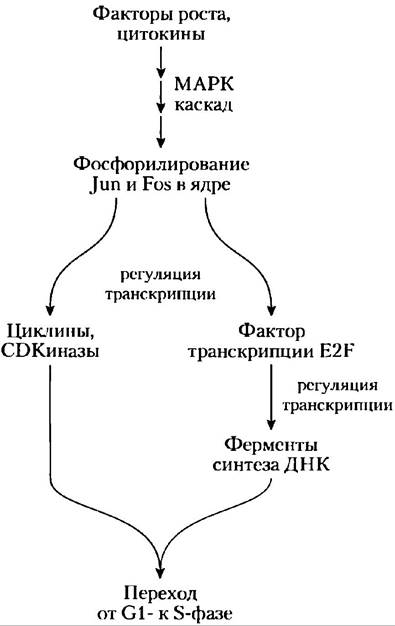
For example, cyclin D, cyclin E, CDK2, and CDK4 are synthesized only when the specific Transcription factor E2F is present in The Nucleus to activate the Transcription of the corresponding genes. The synthesis of E2F, in turn, is regulated by extracellular signals such as growth factors and cytokines (cell division Inducers)—substances essential for the division of mammalian cells in culture. These growth factors induce the Synthesis of specific nuclear transcription factors required for producing enzymes needed for DNA Synthesis. Growth factors trigger the phosphorylation of the Nuclear Proteins Jun and Fos, which are transcription factors that promote the synthesis of products from A large number of genes, including cyclins, CDKs, and E2F. In turn, E2F regulates The production of several enzymes required for deoxynucleotide and DNA synthesis, thereby facilitating the transition of the cell into the S phase (Fig. 12-47).
Fig. 12-47. Regulation of Cell division by growth factors. The pathway from growth factors to cell division proceeds via an enzymatic cascade that activates the MAPK pathway, the phosphorylation of the nuclear transcription factors Jun and Fos, and the activity of the transcription factor E2F, which promotes the synthesis of several enzymes required for DNA synthesis.
CDK inhibition. Finally, specific protein inhibitors bind to specific CDKs and inactivate them. One such protein is p21, which we discuss below.
These four regulatory mechanisms modulate the activity of specific CDKs, which in turn determine whether a cell will divide or differentiate, remain in a permanent state of quiescence, or begin a new cycle after a resting period. The details of cell cycle regulation (the number of different cyclins, kinases, and their combinations) vary from species to species, but the general mechanism has largely been established and conserved throughout the evolution of all eukaryotic cells.
CDKs regulate cell division by phosphorylating essential proteins
We have seen that cells maintain strict control over CDK activity. But how does CDK activity regulate the cell cycle? The list of proteins known to be targets of CDK action continues to grow, and much remains to be learned. Nevertheless, we can envision the General mechanism of CDK regulation by tracing the effects of CDK on The structure of lamins and Myosin and on the activity of the retinoblastoma protein.
The STRUCTURE OF THE nuclear envelope is maintained in part by highly organized networks of Intermediate filaments composed of the protein lamin. The breakdown of the nuclear envelope prior to the Separation of sister chromatids in mitosis is due, in part, to the phosphorylation of lamin by CDKs, which causes lamin filaments to depolymerize.
A second target of the kinase is the ATP-driven Actomyosin contractile system, which constricts the dividing cell, splitting it into two equal parts during cytokinesis. Following cell division, the CDK phosphorylates a small regulatory subunit of myosin, causing myosin to dissociate from Actin filaments and inactivating the contractile machinery. Subsequent dephosphorylation allows the contractile apparatus to reassemble for the next round of cytokinesis.
A third and crucially important substrate of CDK is the retinoblastoma protein, pRb; when DNA damage is detected, this protein participates in a mechanism that halts cell division in the G1 phase (Fig. 12-48). Named after the retinal tumor cell line in which it was discovered, pRb functions in most, if not all, cells, regulating cell division in response to a multitude of stimuli. Unphosphorylated pRb binds to the transcription factor E2F, and upon binding to pRb, E2F is unable to stimulate the transcription of a group of genes required for DNA synthesis (genes for DNA polymerase α, Ribonucleotide reductase, and other proteins; Chapter 25). In this state, the cell cycle cannot progress from the G1 phase to the S phase, which commits the cell to mitosis and division. The mechanism blocking pRb-E2F is turned off when pRb is phosphorylated by cyclin E-CDK2, which appears in response to a cell division signal.
When the protein kinases ATM and ATR detect DNA damage, such as a single-strand break, they activate p53, which acts as a transcription factor stimulating the Synthesis of the p21 protein (Fig. 12-48). This protein inhibits the protein kinase activity of cyclin E-CDK2. In the presence of p21, the pRb protein remains unphosphorylated and bound to E2F, transcription factor activity is blocked, and the cell cycle halts in the G1 phase. This gives the cell time to repair the damaged DNA before entering the S phase, thereby preventing the catastrophic transfer of a damaged genome to one or both daughter cells. If the damage is too severe to be efficiently repaired, the same mechanism triggers the apoptosis pathway described below, leading to cell death and preventing the potential development of cancer.
Fig. 12-48. Regulation of the G1-to-S phase transition by pRb phosphorylation. The transcription factor E2F stimulates the transcription of genes for several enzymes required for DNA synthesis. The retinoblastoma protein pRb can bind the E2F factor (bottom left), inactivating it and preventing the transcription of such genes. Phosphorylation of pRb by CDK2 prevents the binding and inactivation of E2F, allowing genes to continue being transcribed and cells to divide. Cellular DNA damage (top left) triggers a series of events leading to the inactivation of CDK2, resulting in the cessation of cell division. When the MRN protein detects DNA damage, it activates two protein kinases, ATM and ATR, which phosphorylate and activate the transcription factor p53. Active p53 stimulates the synthesis of the p21 protein, a CDK2 inhibitor. Inhibition of CDK2 halts the phosphorylation of pRb, which consequently continues to bind and inhibit E2F. With E2F inactivated, genes necessary for cell division are not transcribed, and cell division stops. Following DNA Repair, these events unfold in reverse, and the cell resumes division.

Summary of Section 12.11 Cell Cycle Regulation by Protein Kinases
■ Progression through the cell cycle is regulated by cyclin-dependent kinases (CDKs), which act at specific checkpoints in the cycle by phosphorylating key proteins and modulating their activity. The catalytic subunit of a CDK is inactive unless it is bound to a regulatory cyclin subunit.
■ The activity of the cyclin-CDK complex varies throughout the cell cycle via differential CDK synthesis, specific cyclin degradation, phosphorylation and dephosphorylation of critical residues in the CDK, and the binding of inhibitory proteins to specific cyclin-CDK complexes.
Last update: 06/08/2026
Editorial and Educational Adaptation: This material has been compiled based on the primary/original source text. The project team performed an editorial review, corrected technical inaccuracies, structured sections, and adapted the content for an educational format.
What was processed:
- elimination of formatting defects (OCR errors, structural breaks, corrupted characters);
- editorial organization of content;
- standardization of terminology in accordance with academic sources;
- verification of factual statements against the original source text.
All mentions of the author, publication year, and origin of the primary text have been preserved in accordance with the source.