Review of Medical Physiology - William F. Ganong 2002
Introduction
General Principles and Cellular Basis of Human Physiology
Functional Morphology of the Cell
The application of modern CELLULAR AND MOLECULAR biology Methods has driven a series of revolutionary shifts in our understanding of Cell Structure and function, as well as in The Study of embryonic and postembryonic development at THE CELLULAR LEVEL. Although postembryonic development and the finer details of cell biology lie beyond The Scope of this study, a foundational knowledge of cell biology is essential for understanding the body's Organ Systems and how they function. The cellular specialization of various Organs is remarkably diverse, and no single cell can be considered typical of the whole Organism. Nevertheless, certain specialized structures (Organelles) are common to most Cells. These structures are illustrated in Figs. 1–4. Many of them can be isolated using ultracentrifugation combined with other technologies. Following cell homogenization and ultracentrifugation of the resulting suspension, nuclei sediment first, followed by Mitochondria. High-speed centrifugation, which generates gravitational forces of 100,000g or greater, yields a fraction composed of granules known as microsomes due to their sedimentation properties. This fraction includes organelles such as Ribosomes and Peroxisomes.
Cell Membrane
The membrane surrounding The Cell is a remarkable structure. Composed of Lipids and Proteins, it is semipermeable, meaning it allows certain substances to pass through while blocking others. However, the permeability of the membrane is adjustable, as it contains numerous Gated Ion Channels and other transport proteins capable of regulating the volume of substances moving across it. This membrane is commonly referred to as The Plasma Membrane. The Cell Nucleus is enclosed by a membrane of a similar type, and cellular organelles are also bounded by or composed of it.
Class="center">
Fig. 1–4. Schematic representation of a hypothetical cell in the center, as viewed under a Light Microscope. Various organelles are shown surrounding it (after Bloom and Fawcett. Reproduced with permission from Junqueira LC, Carneiro J, Kelley RO: Basic Histology. 9th ed. McGraw-Hill, 1998).
Although the chemical COMPOSITION AND PROPERTIES of membranes vary considerably depending on their Location, certain common features can be identified. In particular, the average thickness of a membrane is 7.5 nm (75 Å). Biomembranes consist of proteins and lipids. The Chemistry of Proteins and lipids is described in Chapter 17. The principal lipids are Phospholipids, such as phosphatidylcholine and phosphatidylethanolamine. A phospholipid molecule resembles a clothespin in shape (Fig. 1–5). Its upper end contains a phosphate group and is relatively Water-soluble (polar, hydrophilic). The tail ends are relatively insoluble (nonpolar, hydrophobic). The hydrophilic ends of the biomembrane molecules face the aqueous environment bathing the exterior of the cells and the watery Cytoplasm, whereas the hydrophobic ends converge in the anhydrous interior of the membrane. In prokaryotes (cells lacking a nucleus, such as Bacteria), membranes are relatively simple, whereas in eukaryotes (cells possessing a nucleus), they contain a variety of glycosphingolipids, sphingomyelins, and Cholesterol.
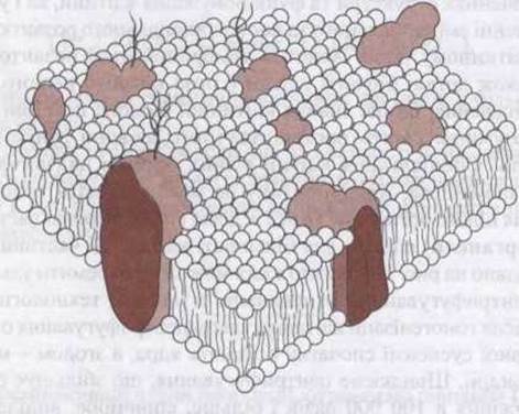
Fig. 1–5. Biological membrane. Each phospholipid molecule has two fatty acid chains (wavy lines) attached to a phosphate HEAD (spherical structure). Proteins are depicted as shaded, irregularly shaped entities. Many of these are integral proteins that span the membrane, whereas peripheral proteins are attached to the inner (not shown) and outer surfaces of the membrane, sometimes via glycosylphosphatidylinositol (GPI) anchors (left).
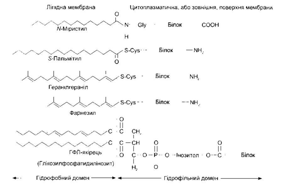
Fig. 1–6. Attachment of proteins to Membrane Lipids. Some proteins are anchored via their amino terminals, others via their carboxyl terminals, and many via glycosylphosphatidylinositol (GPI) anchors (reproduced with permission from Fuller GM, Shields D: Molecular Basis of Medical Cell Biology. McGraw-Hill, 1998).
Membranes contain A wide variety of proteins. They exist as separate units; many span the membrane (integral proteins), while others (peripheral proteins) cluster on its inner and outer surfaces (see Fig. 1–5). The protein content depends on the function of the membrane, but on average, proteins account for about 50% of the membrane's mass, with roughly one protein molecule for every 50 much smaller phospholipid molecules. Membrane Proteins perform diverse functions. Some act as adhesion molecules, attaching cells to neighboring cells or to basement membranes. Others function as pumps, actively transporting ions across the membrane. Still others act as carriers, facilitating the diffusion of substances down their electrochemical gradients, or function as ion channels that, when activated, allow ions to pass through the membrane. The roles of pumps, carriers, and other channels in transmembrane transport are described below.
Another group of proteins acts as receptors that bind Neurotransmitters and Hormones, triggering physiological changes inside the cell. Proteins also function as Enzymes, catalyzing reactions at the membrane surfaces. In addition, some Glycoproteins contribute to antibody maturation and distinguish host cells from foreign ones as "self" versus "non-self" (see Chapter 27).
The hydrophobic portions of proteins are typically located within the interior of the membrane, whereas the hydrophilic portions reside on the surfaces. Peripheral proteins are attached to the membrane surface in various ways. One such mechanism is attachment via glycosylphosphatidylinositol groups. Proteins anchored by these glycosylphosphatidylinositol (GPI) linkages (Fig. 1–6) include enzymes such as alkaline phosphatase, various Antigens, A number of Cell Adhesion molecules, and three proteins that protect cells against Complement-mediated lysis (see Chapter 27). Over 40 GPI-linked cell surface proteins have been described to date. Other proteins are lipidated, meaning they have specialized lipids covalently attached to them (see Fig. 1–6). They may be myristoylated, palmitoylated, or prenylated, meaning they are attached to geranylgeranyl or farnesyl groups.
The structure of proteins, particularly the ENZYMES OF BIOLOGICAL membranes, varies not only among different cells but even within the same cell. For instance, cell membranes and mitochondrial membranes contain different sets of enzymes. In epithelial cells, the enzymes on the apical (mucosal) surface of the plasma membrane differ from those on the lateral surfaces; in other words, the cells are polarized. Membranes are dynamic structures, and their components are continuously renewed at various levels. Some proteins are anchored to the Cytoskeleton, whereas others can move laterally. For example, receptors migrate within the membrane and cluster in regions of endocytosis (see below). Most cell membranes are associated with a thin, amorphous layer containing fibrils that collectively form the basement membrane or basal lamina. The basement membrane (or Extracellular matrix, broadly speaking) consists of numerous proteins that hold cells together, regulate their development, and determine their growth. These proteins include collagens, laminins (see below), Fibronectin, and Proteoglycans.
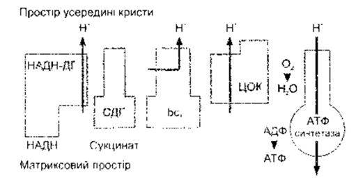
Fig. 1–7. Principal enzymes of The inner mitochondrial membrane involved in Oxidative Phosphorylation; NADH-DH, NADH dehydrogenase; SDH, succinate dehydrogenase; bc1, cytochrome bc1 complex; COX, cytochrome c oxidase (modified from Saraste M: Oxidative phosphorylation at the fin de siècle. Science 1999;283:1488).
Mitochondria
The Morphology of mitochondria varies somewhat among cell types, but generally, each mitochondrion is an elongated, oval-shaped structure (see Fig. 1–4). It consists of an outer and an inner membrane. The inner membrane is folded, forming structures called cristae. The space between the two membranes is known as the intermembrane space, and the space enclosed by the inner membrane is the mitochondrial matrix. Mitochondria are the primary source of cellular energy. These organelles are most numerous and best developed in regions of cells where energy-consuming processes take place. Chemical processes within cells are described in detail in Chapter 17. The outer membrane of each mitochondrion is embedded with enzymes responsible for Biological Oxidation, providing the raw Materials for Chemical Reactions that occur inside the mitochondrion. Within the mitochondrial matrix are enzymes that convert carbohydrate, protein, and lipid metabolites into CO2 and water via The Citric Acid Cycle (Krebs cycle). This enzyme complex comprises four main components (Fig. 1–7): NADH dehydrogenase, succinate dehydrogenase, cytochrome bc1 complex, and cytochrome c oxidase. During the processes catalyzed by these enzymes, protons (H+) are pumped from the matrix into the intermembrane space, establishing a proton gradient. Protons diffusing back down this gradient drive the synthesis of adenosine triphosphate (ATP) via ATP synthase. ATP is an energy-rich triphosphate that serves as the primary energy source for metabolic processes not only in animals (see Chapter 17) but also in bacteria and plants, which synthesize it through analogous mechanisms. The Coupling of oxidation to ATP formation in mitochondria is termed oxidative phosphorylation.
ATP synthase is a unique enzyme composed of multiple subunits, featuring a base embedded in the inner mitochondrial membrane, a stalk, and a spherical head located in the mitochondrial matrix. Much of the stalk and base actually rotates during ATP generation.
There is little doubt that mitochondria were once autonomous microorganisms that established a symbiotic relationship and became permanently incorporated into ancestral Eukaryotic cells. As a consequence of this evolutionary origin, mitochondria possess their own genome, which contains far less DNA than the nuclear genome (see below), with 99% of mitochondrial proteins being products of nuclear genes. Nevertheless, Mitochondrial DNA plays a crucial role in oxidative phosphorylation. Specifically, mitochondrial DNA is a double-stranded, circular molecule containing 16,569 Base Pairs (compared to over a billion in nuclear DNA). It encodes 13 protein subunits that combine with proteins encoded by nuclear genes to form the four enzyme complexes, as well as two Ribosomal RNAs and 22 Transfer RNAs (see below) required for Protein Synthesis by intra-mitochondrial ribosomes.
Mitochondria are inherited exclusively maternally, as they are contributed to the zygote by the egg cell rather than the sperm. Mitochondria lack an efficient DNA Repair system, and the mutation rate of mitochondrial DNA is more than 10 times higher than that of nuclear DNA. Furthermore, a significant number of relatively rare disorders have been shown to be caused by Mutations in mitochondrial DNA. These conditions typically affect Tissues with high metabolic demands, leading to impaired ATP production.
The Cell Cytoplasm contains large, amorphous, membrane-bound structures known as lysosomes. These structures are more acidic than the rest of the cytoplasm and may contain fragments of other cellular components. Certain granules found in Blood granulocytes are also lysosomes. Each lysosome contains a variety of hydrolytic enzymes (Table 1–3) that would destroy most cellular components if they were not sequestered from the cytoplasm by the lysosomal membrane. Lysosomes function as the Digestive System of the cell. Exogenous substances, as well as bacteria engulfed by the cell, enter via membrane-bound vacuoles. Such vacuoles (phagocytic vacuoles) can fuse with lysosomes, allowing the Contents of the vacuole and the lysosome to mix within a common membrane. Some products of the Digestion of engulfed material are absorbed across the vacuolar wall, while the indigestible residues are expelled from the cell by exocytosis (see below). Lysosomes also engulf worn-out cellular components within the cell, forming autophagic vacuoles. When a cell dies, lysosomal enzymes mediate the autolysis of its remains.
Table 1–3. Enzymes contained in lysosomes and their intracellular substrates
Enzyme |
Substrate |
RNA |
|
Deoxyribonuclease |
DNA |
Phosphatase |
Phosphate esters |
Glycosidase |
Complex CARBOHYDRATES, |
Arylsulfatase |
Sulfate esters |
Collagenase |
Proteins |
Cathepsin |
Proteins |
It is well known that in Gout, phagocytes engulf uric acid crystals, which triggers the extracellular release of lysosomal enzymes that promote the inflammatory response in the joints. When a lysosomal enzyme is congenitally deficient, the cytoplasm becomes filled with material that would normally be degraded by these enzymes. This results in one of the lysosomal storage diseases. For example, a deficiency in cathepsin K+ causes pycnodysostosis, whereas a deficiency in glycosylasparaginase leads to aspartylglucosaminuria. Over 25 such disorders have been described. Although rare, they are associated with well-known conditions such as Tay-Sachs disease.
Peroxisomes
Peroxisomes are located in the microsomal fraction of the cell. They are approximately 0.5 µm in diameter and bounded by a membrane. This membrane contains a set of specific proteins responsible for transporting substances into and out of the peroxisomal matrix. The matrix contains over 40 enzymes which, upon interacting with extraperoxisomal enzymes, catalyze a variety of anabolic and catabolic reactions. Catabolic reactions include The breakdown of long-chain Fatty acids. Peroxisomes deserve special attention because a mutation in one of the genes encoding a peroxisomal membrane transporter causes X-linked adrenoleukodystrophy, a fatal condition in childhood. In this disease, the ß-Oxidation of long-chain fatty acids is impaired. An autosomal mutation causing even more severe defects in Transport Across the peroxisomal membrane leads to Zellweger syndrome, which results in early childhood death.
The number of peroxisomes increases in response to peroxisome proliferators, which act on peroxisome proliferator-activated receptors (PPARs) in The Nucleus. These receptors belong to the superfamily of hormone-sensitive nuclear METABOLISM/31.html">Transcription factors, which include steroid Hormone Receptors (see below). Three distinct isoforms are encoded by three already identified genes: PPARα, PPARγ, and PPARδ. The drug troglitazone binds to PPARγ and increases cellular sensitivity to Insulin (see Chapter 19). Activation of PPARγ also stimulates Cell Differentiation into adipocytes and produces other widespread metabolic effects. A dominant-negative mutation in the PPARγ genes increases insulin resistance, leading to Diabetes Mellitus and Hypertension.
Cytoskeleton
All cells possess a cytoskeleton—a system of organelles that not only maintains Cell Structure but also enables the cell to change shape and move. The cytoskeleton consists of microtubules, Intermediate filaments, and microfilaments, along with anchoring and cross-linking proteins. In addition, proteins and organelles move along microtubules and microfilaments from one part of the cell to another, propelled forward by Molecular Motors. Microtubules (Figs. 1–8, 1–9) are long, hollow structures with walls 5 nm thick surrounding a lumen 15 nm in diameter. They are composed of Two Types of spherical protein subunits: α- and ß-tubulin. A third subunit, γ-tubulin, plays a role in The formation of the centrosome from microtubules (see below). The α and ß subunits form heterodimers (see Fig. 1–8), which polymerize to form long tubes consisting of a series of rings, each containing 13 subunits. Microtubules also contain other proteins that participate in their assembly. The self-assembly of microtubules is promoted by warmth, whereas cold leads to their disassembly. The end where self-assembly predominates is called the plus end, and the end where dissociation prevails is called the minus end. Both processes occur simultaneously in vitro.
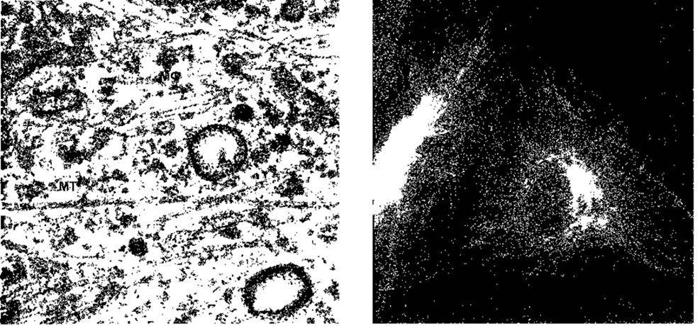
Fig. 1-8. Left: Electron micrograph of fibroblast cytoplasm showing microfilaments (MF) and microtubules (MT) (reproduced with permission from Junqueira LC, Carneiro J, Kelley RO: Basic Histology, 9th ed. The McGraw-Hill Companies, Inc., 1998). Right: Localization of microtubules in fibroblasts. Cells were stained with a fluorescent antibody against tubulin to visualize microtubules (reproduced with permission from Connolly J et al.: Immunofluorescent staining of cytoplasmic and spindle microtubules in mouse fibroblasts with antibody to τ protein. Proc Natl Acad Sci USA 1977;74:2437).
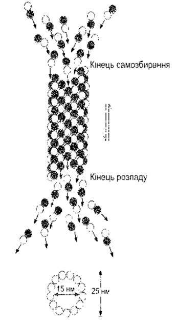
Fig. 1-9. Self-assembly and disassembly of a microtubule resulting from the aggregation and disaggregation of dimers composed of α- and ß-tubulin (reproduced with permission from Sloboda RD: The Role of microtubules in cell structure and Cell Division. Am Sci 1980;68:290).
Due to constant self-assembly and dissociation, microtubules are a highly dynamic component of the cytoskeleton. They mediate The transport of vesicles and organelles—such as secretory granules and mitochondria—from one part of the cell to another, and they form the mitotic spindle that segregates Chromosomes during mitosis (see below). Microtubules can transport cargo in both directions; indeed, a single microtubule has been observed transporting two structures in opposite directions.
Microtubule self-assembly is inhibited by colchicine and vinblastine. The anticancer drug paclitaxel (Taxol) immobilizes microtubules, paralyzing organelles and disrupting mitotic spindle formation, which leads to Cancer cell death.
In addition to microtubules, cells contain intermediate filaments, which are 8–14 nm in diameter and composed of diverse subunits. Some of these link the nuclear membrane to The cell membrane. These filaments form a flexible cellular framework and help the cell withstand external pressure. In the absence of intermediate filaments, cells rupture much more easily. In humans, intermediate filament anomalies are frequently accompanied by Skin blistering. Microfilaments (see Fig. 1–8) are long, robust fibers 4–6 nm in diameter. They are composed of Actin and a protein that, upon interacting with Myosin, drives Muscle contraction (see Chapter 3). Actin, like its mRNA (see below), is present in all cell types. It is the most abundant protein in mammalian cells, accounting for 15% of total cellular protein. Actin structure is remarkably stable; for instance, the Amino Acid Sequence of Yeast and rabbit actin is 88% identical. Actin molecules (G-actin) polymerize in vivo to form F-actin—long, thread-like chains known as microfilaments. They also readily depolymerize in vivo, with polymerization predominantly occurring at one end (the plus end, as in microtubules) and depolymerization at the opposite end (the minus end). Microfilaments attach to various elements of the cytoskeleton (Fig. 1–10). Many microfilaments anchor to the tips of microvilli in intestinal epithelial cells. They are present in large quantities in lamellipodia, the extensions projected by cells as they migrate across surfaces. Actin filaments interact with integrin receptors to form focal adhesion complexes (focal adhesions, see below), which serve as traction sites for surface motility as the cell extends.
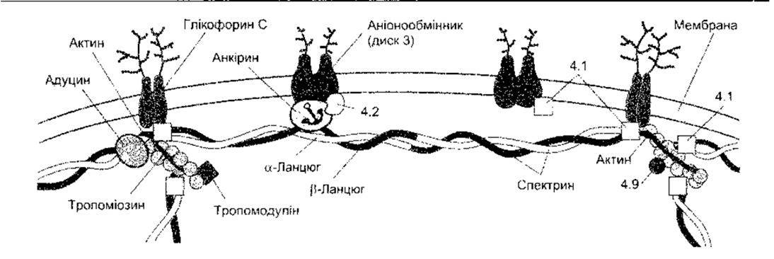
Fig. 1-10. Attachment of the cytoskeleton to The erythrocyte membrane. Various proteins linking actin filaments to the membrane are shown. Some are designated by numbers (4.1, 4.2, 4.9) and others by names (reproduced with permission from Luna EJ, Hitt AL: Cytoskeleton-plasma membrane interactions. Science 1992;258:955).
Molecular Motors
Molecular motors are ATPases of 100–500 kDa that bind to cellular cargo (such as proteins, organelles, and other components) and transport them along microtubules and microfilaments throughout the cell. Their globular heads form cross-bridges with the filament and hydrolyze ATP into energy, which drives the flexing of these bridges and powers the movement of the ATPase molecules. The types of molecular motors are summarized in Table 1–4, and Examples of each type are illustrated in Fig. 1–11. It is now known that each type constitutes a large superfamily comprising numerous forms across the animal kingdom.
The conventional form of kinesin is a two-headed molecule (see Fig. 1–9) that transports particles and membranes toward the plus end of microtubules. This occurs as follows: one head attaches to the microtubule and then bends its neck to allow the other head to bind.
Table 1-4. Molecular motors
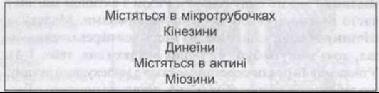
This mechanism produces near-continuous movement. Some kinesins are involved in Mitosis and Meiosis, while others perform different functions, including transporting cargo toward the minus end of microtubules.
Dyneins have two heads, with their necks partially embedded in a protein complex (see Fig. 1–11). Cytoplasmic dynein shares a similar structure and moves particles and membranes toward the minus end of microtubules. Axonemal dynein is responsible for the beating of flagella and cilia (see below).
Myosins form cross-bridges with Actin filaments and slide along them, generating force strokes. This produces movements such as those involved in intestinal villus contraction, cell migration, and the contraction of all human skeletal and other Muscles. The myosin superfamily is divided into 15 classes. Myosin-I and myosin-II are depicted in Fig. 1–11. Myosin-I binds to actin in cell membranes, whereas myosin-II (described in detail in Chapter 3) is found in Skeletal Muscle. All currently known myosins move toward the plus end of actin polymers, and no myosins moving in the opposite direction have been described to date. Various Forms of myosin bind to actin. A myosin molecule consists of globular heads with ATPase activity and tails of varying lengths (see Fig. 1–10). Myosin-I molecules have a single head and frequently interact with cell membranes in conjunction with actin. Myosin-II molecules have two heads and are located predominantly in muscles, although they can also be found elsewhere (see Table 1–4). The formation and movement of cross-bridges with actin molecules, which underlie muscle contraction, are described in detail in Chapter 3.
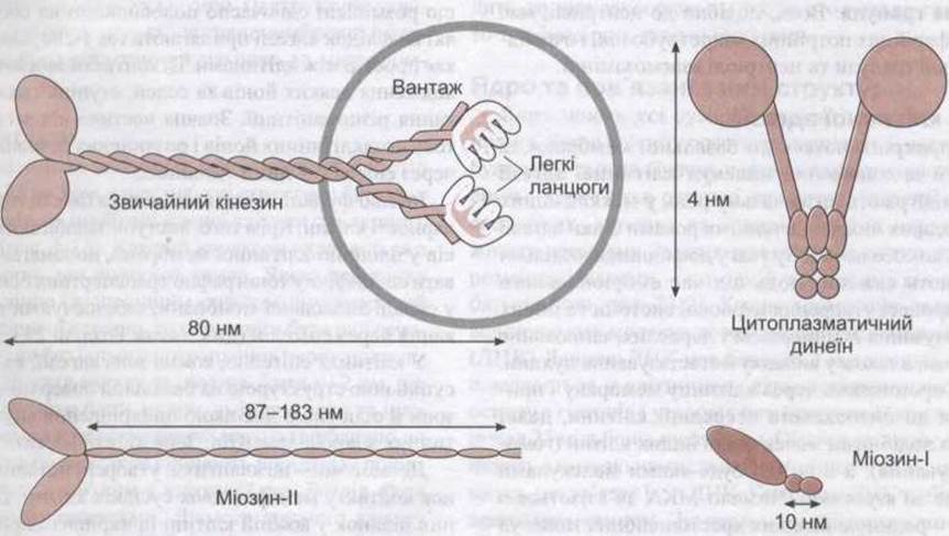
Fig. 1-11. Examples of Three types of molecular motors. A conventional kinesin molecule attaches to cargo (a membrane-bound organelle). All motors feature motile heads that hydrolyze ATP and use the released energy to flex their heads at the neck region (reproduced with permission from Luna EJ, Hitt AL: Cytoskeleton-plasma membrane interactions. Science 1992;258:955).
Centrosomes
Near the nucleus in the cytoplasm of eukaryotic animal cells lies the centrosome. It consists of two centrioles surrounded by amorphous pericentriolar material. Centrioles are short cylinders positioned near the nucleus at right angles to each other. In the walls of each centriole, microtubules extend longitudinally in groups of three (see Figs. 1–4). There are nine such triplets, arranged at regular intervals.
Centrosomes serve as microtubule-organizing centers (MTOCs) containing y-tubulin. Microtubules self-assemble from y-tubulin within the pericentriolar material. During cell division, the centrosome duplicates into two, and these pairs migrate to form the poles of the mitotic spindle, which is composed of microtubules. In multinucleated cells, centrosomes are located adjacent to each nucleus.
Cilia
Cellular projections come in various forms. True cilia are motile appendages used by unicellular organisms for locomotion in water and by Multicellular Organisms to move mucus and other substances across The surface of various epithelia. They resemble centrioles in having nine pairs of microtubules in their wall. In addition, they contain a central pair of microtubules, and each of the nine peripheral structures consists of a doublet rather than a triplet of microtubules. Each cilium is anchored to a structure known as the basal body. Similar to a centriole, the basal body possesses nine peripheral triplet microtubules, demonstrating that basal bodies and centrioles are functionally interchangeable.
Cell Adhesion Molecules
Cells attach to the basement membrane and to one another via cell adhesion molecules (CAMs), which play a critical role in the intercellular interactions described below. In recent years, adhesion proteins have attracted intense research interest due to their vital roles in embryonic development, the formation of The Nervous system and other tissues, the maintenance of tissue integrity in adults, Inflammation and Wound healing, and tumor metastasis. Many of these molecules span the Cell Membrane and anchor to the intracellular cytoskeleton; some bind to similar molecules on other cells (homophilic binding), while others bind to different molecules (heterophilic binding). Numerous CAMs bind to laminins, a family of large, cross-shaped molecules with multiple receptor domains within the extracellular matrix.
The nomenclature of CAMs is somewhat chaotic, partly due to their rapid discovery and partly because of the widespread use of acronyms across various fields of modern biology. Nevertheless, CAMs can be classified into four major families: Integrins, heterodimers that bind to various receptors; members of the immunoglobulin (Ig) superfamily of adhesion molecules, some of which bind heterophilically and others homophilically; cadherins, which are Ca2+-dependent molecules mediating cell-cell adhesion via homophilic reactions; and selectins, which feature lectin-like domains that bind carbohydrates. The functions of CAMs in granulocytes and platelets are discussed in Chapter 27, and their role in inflammation and wound healing in Chapter 33.
CAMs not only anchor cells to their neighbors but also mediate cell-to-cell signaling. Cells that lose contact with the extracellular matrix via integrins exhibit a higher rate of apoptosis (see below) than attached cells, whereas the interaction between integrins and the cytoskeleton is essential for cell motility.
Two MAIN TYPES OF intercellular junctions are distinguished in tissues: junctions that anchor cells to one another and to the surrounding extracellular matrix, and junctions that allow the direct passage of ions and other molecules between cells. Anchoring junctions, which provide mechanical strength and stability to tissues, include tight junctions (also known as zonula occludens). Desmosomes and adherens junctions (Figs. 1–12) hold cells together, whereas hemidesmosomes and focal adhesions anchor cells to the basal lamina. Tight junctions between epithelial cells are also essential for transepithelial ion transport. Junctions that permit molecular exchange are termed Gap Junctions.
Tight junctions generally encircle the apical margins of epithelial cells, such as those lining the intestine, renal tubules, and Blood Vessels. They consist of a network of sealing strands contributed by membranes of adjacent cells, which appose so closely that the intercellular space is virtually obliterated. These junctions permit the passage of certain ions and solutes, with varying degrees of permeability. A significant portion of the total extracellular flux of ions and solutes crosses the epithelium through these pathways.
Another key function of tight junctions is the maintenance of cell polarity. Additionally, the junctional strands restrict the lateral diffusion of proteins within the plane of the cell membrane, helping to preserve the specific topographical distribution of transport proteins and channels in the apical membrane, which is essential for Transepithelial Transport (see also Chapters 25 and 38).
In epithelial cells, each adherens junction typically forms a continuous band located just basal to the tight junction and serves as a primary anchoring site for intracellular microfilaments. Adherens junctions contain cadherins.
Desmosomes are patch-like structures formed by localized thickenings of the apposed membranes of adjacent cells. Intermediate filaments attach to these dense plaques in each cell, running either parallel to the membrane or radiating outward. The intercellular space between the membranes contains a filamentous material rich in cadherins and other proteins.
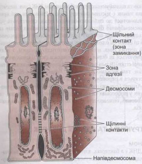
Fig. 1–12. Intercellular junctions in the mucosa of the Small Intestine. Various types of desmosomes are not shown in detail.
Hemidesmosomes resemble half-desmosomes; they anchor cells to the basement membrane and connect intracellularly to intermediate filaments. However, they contain integrins rather than cadherins. Focal adhesions also anchor cells to their basal lamina. As noted above, these are labile structures linked to intracellular actin filaments that play a crucial role in cell migration. These structures form dense plaques where transmembrane integrins tether the extracellular matrix to microfilaments.
Gap Junctions
At gap junction sites, the intercellular space narrows from 25 to 3 nm, and hexagonal arrays of Protein Complexes—connexons—in the membranes of adjacent cells align with one another (Fig. 1–13). Each connexon is composed of six subunits surrounding a central channel. When this channel aligns with a corresponding channel formed by a connexon in the neighboring cell, a pathway is created allowing direct molecular traffic between cells without entering the extracellular fluid. The channel diameter is approximately 2 nm, permitting the passage of ions, sugars, Amino Acids, and other solutes with molecular weights up to roughly 1000. Thus, gap junctions facilitate the rapid cell-to-cell spread of electrical potentials (see Chapter 4) and the exchange of signaling molecules. The diameter of each channel is regulated by intracellular Ca2+; an increase in Ca2+ concentration causes the channel-forming subunits to shift closer together, thereby narrowing the channel. Channel conductance and diameter can also be modulated by changes in pH and Membrane Potential.
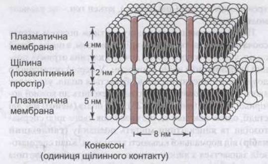
Fig. 1–13. Gap junction. Note that each connexon consists of six subunits, and connexons in the membrane of one cell align with those in the membrane of an adjacent cell to form a channel through which substances can pass from one cell to another without entering the ECF (reproduced with permission from Kandel ER, Schwartz JH, Jessell TM [editors]: Principles of Neural Science, 4th ed. McGraw-Hill, 2000).
In rodents, connexon isoforms are encoded by at least 13 different genes. Humans possess a comparable number of isoform genes. The tissue-specific expression patterns of these isoforms vary widely. In mice, the oocyte is coupled to granulosa cells via gap junctions, and knockout mice lacking specific connexon types exhibit defects in ovulation and complete meiosis. Mutations in connexons are linked to several human disorders, including X-linked Charcot-Marie-Tooth disease, peripheral neuropathy, and heterotaxy syndromes, which are accompanied by various developmental abnormalities, including the failure to establish normal left-right Asymmetry. The exact mechanism by which connexon pathology leads to these diseases remains unclear, although other genes are likely implicated in heterotaxy.
The Nucleus and Associated Structures
The nucleus is present in all eukaryotic cells capable of division. If a cell is bisected, the enucleated portion will die. A major component of the nucleus is the chromosomes—structural units that carry the complete set of hereditary, species-specific, and individual characteristics of a living organism. Except for Germ Cells, chromosomes exist in pairs, with one chromosome contributed by each parent (see Fig. 23–2). Each chromosome consists of giant molecules of deoxyribonucleic acid (DNA). Although a DNA molecule is nearly 2 m long, it fits compactly within the cell nucleus by coiling around a core of histone proteins to form nucleosomes. Each nucleus contains over 25 million nucleosomes. The structure of Chromatin is often compared to "beads on a string," where the beads represent nucleosomes and the connecting thread represents DNA. The complex of DNA and associated proteins is termed chromatin. During cell division, the coiling of DNA around Histones is relaxed, likely mediated by histone Acetylation, allowing chromosome pairs to become visible as distinct entities, whereas during interphase only diffuse chromatin networks can be distinguished within the nucleus. The primary units of heredity residing within chromosomes are genes (see below), with each Gene representing a discrete segment of a DNA molecule.
During normal cell division by mitosis, chromosomes are first duplicated and then divided, resulting in each daughter cell receiving a complete (diploid) set of chromosomes. During the final maturation of germ cells, a division takes place in which only half of the chromosomes pass to each daughter cell (see Chapter 23). This division (meiosis) has two stages, but crucially, it ensures that mature spermatozoa and oocytes contain half the normal number of chromosomes (a haploid set). When a spermatozoon fuses with an oocyte, the newly formed cell (zygote) restores the diploid set of chromosomes, receiving one half from each parent. The chromosomes undergo recombination, which results in the mixing of maternal and paternal genes.
The nucleus of most cells contains a nucleolus (see Figs. 1-4), a ribonucleic acid (RNA)-rich cluster of granules. Some cells have several such structures within their nucleolus. Nucleoli are most prominent and numerous in growing cells. They serve as the site of synthesis for ribosomes, the cytoplasmic structures in which proteins are synthesized (see below).
Internally, the nucleus features a filamentous Skeleton attached to the nuclear membrane, or envelope (see Figs. 1-4), which surrounds the nucleus. This membrane is double-layered, and the space between its two layers is called the perinuclear cisterna. While the membrane is permeable only to ions and small molecules, it contains nuclear pore complexes. Each complex consists of about 100 proteins organized into a tunnel shape through which proteins and mRNA are transported. Many transport pathways are concentrated here; specific proteins known as importers and exporters have been isolated and characterized. The Ran protein plays an organizational role. Most contemporary research is devoted to the detailed interpretation of nuclear and retrograde transport mechanisms.
The endoplasmic reticulum is a labyrinth of interconnected tubules located within the cytoplasm of the cell (see Figs. 1-4). The walls of these tubules are formed by a membrane. In the rough, or granular, endoplasmic reticulum, granules known as ribosomes are attached to the cytoplasmic face of the membrane. Conversely, the smooth, or agranular, endoplasmic reticulum lacks these granules. Free ribosomes are also found in the cytoplasm. The granular endoplasmic reticulum is responsible for the synthesis of proteins and primary polypeptide chain regions involving the formation of Disulfide Bonds. The agranular endoplasmic reticulum serves as the site of steroid synthesis in steroid-secreting cells and functions in detoxification within other cells. As the sarcoplasmic reticulum (see Chapter 3), it plays a vital role in skeletal and cardiac muscle.
Ribosomes
In eukaryotic cells, ribosomes measure between 22 and 32 nm in length. Each ribosome consists of large and small subunits, designated on The basis of their sedimentation in an ultracentrifuge as the 60S and 40S subunits, respectively. Ribosomes are complex structures containing numerous distinct proteins and at least three ribosomal RNAs (see below). They also serve as the site of protein synthesis. Ribosomes attached to the endoplasmic reticulum synthesize all transmembrane proteins, the majority of secreted proteins, and most proteins destined for the Golgi apparatus, lysosomes, and endosomes. All of these proteins feature a hydrophobic signal peptide at one end (see below). The polypeptide chains that form these proteins are synthesized within the endoplasmic reticulum. Free ribosomes synthesize cytoplasmic proteins, such as Hemoglobin (see Chapter 27), as well as proteins localized to peroxisomes and mitochondria.
The Golgi apparatus, which plays a role in the Processing of proteins synthesized in ribosomes and packaged into secretory granules, vesicles, and endosomes, is described below in the context of protein Synthesis and Secretion.
Last update: 10/08/2026
Editorial and Educational Adaptation: This material has been compiled based on the primary/original source text. The project team performed an editorial review, corrected technical inaccuracies, structured sections, and adapted the content for an educational format.
What was processed:
- elimination of formatting defects (OCR errors, structural breaks, corrupted characters);
- editorial organization of content;
- standardization of terminology in accordance with academic sources;
- verification of factual statements against the original source text.
All mentions of the author, publication year, and origin of the primary text have been preserved in accordance with the source.