Review of Medical Physiology - William F. Ganong 2002
Introduction
General Principles and Cellular Basis of Human Physiology
General Principles
Body Composition
The Cells that make up all organisms—except for the simplest multicellular aquatic and terrestrial animals—are bathed in extracellular fluid (ECF), which is bounded by the outer integument. From this fluid, cells obtain O2 and nutrients, and into it they release Metabolic waste products. Although the ECF is more dilute than modern seawater, its composition closely resembles that of the primordial ocean from which life is hypothesized to have originated.
In animals with a closed Circulatory system, ECF is divided into two main types: interstitial fluid and intravascular Blood Plasma. Plasma and cellular blood elements, primarily red Blood Cells, fill The Vascular System and together constitute the total blood volume. Interstitial fluid lies outside the vascular bed and bathes the cells. Special fluids, combined together as transcellular fluids, are described below. The ECF volume accounts for one-third of the total body Water (TBW), with the remainder being intracellular fluid (ICF).
Human Body Organization
In an average young adult male, 18% of body weight consists of Proteins and related substances, 7% minerals, and 15% fats. The remaining 60% is water. The body's fluid compartments are illustrated in Fig. 1-1.
Intracellular fluid accounts for approximately 40% of body weight, and extracellular fluid for 20%. Nearly 25% of the ECF is located within the vascular system (plasma makes up 5% of body weight) and 75% in the extravascular space (interstitial fluid accounts for 15% of body weight). Total blood volume is about 8% of body weight.
Measurement of Body Fluid Volumes
The volume of each type of fluid in The Human Body can be determined only theoretically. Upon administering a substance that remains confined to a single compartment, one calculates the volume of distribution in which the test substance has spread (the volume of distribution of the administered substance). This volume equals The amount of the substance administered (minus any amount lost through METABOLISM or excretion during mixing) divided by its concentration in the sample. For example: 150 mg of sucrose is administered to an individual with a mass of 70 kg. The plasma sucrose level is 0.01 mg/ml. It is known that 10 mg may be excreted or metabolized during the mixing period. The volume of distribution for sucrose
Class="center">

Fig. 1-1. Body fluid compartments. Arrows indicate the direction of fluid movement. Transcellular fluids, which represent a very small percentage of total Body Fluids, are not shown
Since 14,000 mg corresponds to the volume of distribution of sucrose, this space is also referred to as the sucrose space.
The volume of distribution can be calculated for any substance introduced into the body, provided its concentration in body fluid and the amount lost via excretion and metabolism can be accurately determined.
Although the principle underlying this measurement is simple, A number of confounding factors must be taken into account. The administered substance must be non-toxic, distribute evenly throughout the volume being measured, and not independently affect the distribution of water or other substances in the body. Furthermore, it must remain stable during mixing, or the extent of any alteration must be known. Finally, the substance must be relatively easy to measure.
Plasma Volume, Total Blood Volume, and Red Blood Cell Volume
Plasma volume is measured using Dyes that bind to Plasma Proteins, such as Evans blue (T-1824). It can also be measured by injecting radioiodinated albumin. Corresponding aliquots of the injected solution and post-injection plasma samples are analyzed using a scintillation counter. The average plasma volume is 3,500 mL (5% of body weight in a 70 kg human).
Once the plasma volume and the hematocrit (i.e., the percentage of a blood sample volume occupied by cells) are known, the total blood volume can be calculated using the following formula:
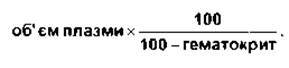
Example. The hematocrit is 38%, and the plasma volume is 3,500 mL. Then the total blood volume

Red blood cell volume (the total volume of circulating erythrocytes in the human body) is determined by subtracting the plasma volume from the total blood volume. It can also be determined by injecting labeled red blood cells and, after mixing, calculating the fraction of labeled erythrocytes.
A commonly used label is 51Cr, a radioactive chromium isotope that binds to red blood cells and is measured in a specific volume of blood. Iron and phosphorus isotopes (59Fe and 32P) as well as antigenic markers are also utilized in practice.
Extracellular Fluid Volume
Extracellular fluid volume is difficult to measure because the boundaries of this space are poorly defined, and few substances both mix rapidly within this space and remain strictly extracellular. Lymph cannot be separated from the extracellular fluid, so they are measured together. Many substances enter the CEREBROSPINAL FLUID (CSF) slowly due to the presence of the blood-Brain barrier (see Chapter 32). Equilibration of substance concentrations in synovial fluid, aqueous humor, and the ECF of avascular Tissues—such as Cytology/practical/45.html">Dense Connective Tissue, Cartilage, and parts of bone—occurs sluggishly. Substances that distribute throughout the ECF may also be detected in glandular secretions and the Contents of the gastrointestinal tract. Because these fluids are sequestered from the rest of the ECF, they—along with the CSF, intraocular fluids, and certain others—are termed transcellular fluids. Their volume is relatively negligible.
The ECF volume can be measured most accurately using inulin, a polysaccharide with a Molecular Weight of 5,200. Mannitol and sucrose are also employed to measure ECF volume. The conventionally accepted ECF volume is 20% of body weight, or 14 L in a 70 kg human (3.5 L plasma; 10.5 L interstitial fluid).
Interstitial Fluid Volume
The space occupied by the interstitial fluid cannot be measured directly because this fluid is difficult to sample, and substances that equilibrate within it also equilibrate within the plasma. The volume of the interstitial fluid is calculated by subtracting the plasma volume from the ECF volume. The ratio of ECF volume to intracellular fluid volume is greater in infants and children than in adults. However, It is important to note that the total ECF volume in children is smaller, which is why dehydration develops much faster in them and often presents in more acute forms than in adults.
Intracellular Fluid Volume
The intracellular fluid volume also cannot be measured directly. It is calculated by subtracting the ECF volume from the TBW. TBW is measured using the same dilution principle applied to determine other restricted fluid compartments of the body. Most commonly, deuterium oxide ($D_2O$ - heavy water) is used for this purpose. In its properties, $D_2O$ differs little from $H_2O$, yet in body water measurement experiments, it yields more accurate results due to THE PRINCIPLE OF concentration equilibration. Tritiated water and aminopurine are also used for this purpose. The water content in lean* body mass is constant at 71-72 mL per 100 g of tissue; since fat is virtually devoid of water, the ratio of TBW to body mass varies inversely with body fat content. TBW is slightly lower in women than in men, and in both sexes, these values decrease with age (Table 1-1).
Units of Concentration for Dissolved Substances
When analyzing the effects of various physiologically important substances and their interactions, the number of molecules, electrical charges, or solute particles per unit volume of a given body fluid is often more important than simply the mass of the substance per unit volume. Consequently, concentration is frequently expressed in moles, equivalents, or osmoles.
Mole
A mole represents the gram-molecular mass of a substance, that is, the molecular mass of a substance in grams. One mole contains approximately $6 \times 10^{23}$ molecules. A millimole (mmol) is 1/1000 of a mole, and a micromole (µmol) is 1/1,000,000 of a mole. Thus, 1 mole of NaCl has a mass of 23 + 35.5 = 58.5 g, and 1 mmol has a mass of 58.5 mg. The mole is the standardized unit of measurement for the amount of substance in the SI system (see Appendix).
* Lean body mass - body mass excluding adipose tissue.
Table 1-1. Total body water volume in humans (as a percentage of body mass) according to AGE AND SEX
Age |
Men |
Women |
10-18 |
59% |
57% |
18-40 |
61% |
51% |
40-60 |
55% |
47% |
Over 60 |
52% |
46% |
The molecular mass of a substance is the ratio of the mass of a molecule of the substance to the mass of 1/12 of a carbon-12 atom. Since molecular mass is a ratio, it is dimensionless. The unit of mass equal to 1/12 of the mass of a carbon-12 atom is called the dalton** (Da); and 1000 Da = 1 kilodalton (kDa). The kilodalton, often denoted by the letter K, is a convenient unit for measuring protein mass. For instance, one can speak of a 64 K protein, or state that the molecular mass of the protein is 64,000 Da. However, because molecular mass is a ratio and is dimensionless, stating that the molecular mass of a protein is 64 kDa is technically incorrect.
Equivalents
THE CONCEPT OF electrical equivalence is important in physiology because many critical solutes in the human body exist as charged particles. One equivalent (Eq) is 1 mole of an ionized substance divided by its valence. One mole of NaCl dissociates into 1 Eq of $Na^+$ and 1 Eq of $Cl^-$. One equivalent of $Na^+$ = 23 g/mol = 23 g; and 1 equivalent of $Ca^{2+}$ = 40 g/2 = 20 g. A milliequivalent (mEq) is equal to 1/1000 of an equivalent.
Electrical equivalence is not always identical to chemical equivalence. A gram-equivalent is the mass of a substance chemically equivalent to 8,000 g of oxygen. The normality (N) of a solution is the number of gram-equivalents per liter; a 1 N solution of Hydrochloric acid contains (1 + 35.5) g/L = 36.5 g/L.
pH
Maintaining a constant hydrogen ion concentration in human aqueous environments is vital. The pH of a solution is defined as the negative logarithm of $[H^+]$. For example, the pH of water at 25°C, in which the amounts of $H^+$ and $OH^-$ ions are equal, is 7.0 (Fig. 1-2). For each unit change in pH below 7.0, the $[H^+]$ increases tenfold; for each unit increase in pH above 7.0, the $[H^+]$ decreases tenfold.
** In some countries, this unit is referred to as the atomic mass unit (amu).

Fig. 1-2. pH values (reproduced with permission from Alberts B et al: Molecular Biology of The Cell. Garland, 1983).
Buffers
Intracellular and extracellular pH are normally maintained at constant levels. For instance, the pH of ECF is 7.40, and in a healthy individual, this value typically does not deviate by more than ±0.05 pH units. In the body, pH is stabilized by the buffering capacity of fluids. A buffer is a physiological system capable of binding or releasing $H^+$ in solution, thereby keeping the pH of the solution relatively constant despite significant additions of acid or alkali. One such buffer is the system formed by carbonic acid, which partially dissociates into $H^+$ and bicarbonate: $H_2CO_3 \rightleftharpoons H^+ + HCO_3^-$. If $H^+$ is added to a carbonic acid solution, the equilibrium shifts to the left, and most of the $H^+$ is removed from the solution. When $OH^-$ is added, $H^+$ and $OH^-$ combine, resulting in a decrease in $H^+$ concentration. However, this decrease in $H^+$ concentration is minimized by the further dissociation of $H_2CO_3$. Other buffer systems include blood proteins and intracellular proteins. The Quantitative Aspects of buffering, as well as the respiratory and renal regulatory mechanisms that manage buffers to maintain a stable ECF pH of 7.40, are described in Chapter 39.
Diffusion
Diffusion is the process by which a gas or a substance whose particles are in motion spreads out to fill all available space. Particles (molecules or atoms) of a solute dissolved in a solvent are in constant random motion. They are equally likely to move toward or away from regions of high concentration. However, because there are more particles in a region of high concentration, the net movement of particles is toward regions of lower concentration; in other words, a net flux of solute particles occurs from areas of high concentration to areas of low concentration. The time required to achieve equilibrium by diffusion is proportional to the square of the diffusion distance. The rate of diffusion from one Location to another is directly proportional to the cross-sectional area of the volume through which diffusion occurs and to the concentration gradient (or chemical gradient), which is the difference in concentration of the diffusing substance divided by the diffusion path length (Fick's law of diffusion).
Thus,

where J is the rate of diffusion; D is the diffusion coefficient; A is the area; and ∆c/∆x is the concentration gradient. The minus sign indicates the direction of diffusion. When molecules move from a region of higher concentration to a lower one, ∆c/∆x is negative, and multiplying it by DA yields a positive value. Although the permeability of the media through which diffusion occurs varies within the body, diffusion remains the primary factor governing the distribution of water and solutes.
Osmosis
When a substance is dissolved in water, the concentration of water molecules in the resulting solution is lower than that in pure water, because adding a solute increases the volume beyond that of the pure water alone. If this solution is placed on one side of a membrane that is permeable to water but impermeable to the solute, and an equal volume of pure water is placed on the other side, water molecules will diffuse into the solution, thereby reducing the concentration gradient (Fig. 1-3). The net movement of solvent molecules into a region containing a higher concentration of a solute to which the membrane is impermeable is called osmosis. This process plays a vital role in physiological Functions. The tendency of solvent molecules to move into a region of higher solute concentration can be counteracted by applying pressure to the more concentrated solution. The pressure required to prevent the movement of the solvent is known as osmotic pressure.
Like vapor pressure lowering, freezing point depression, and boiling point elevation, osmotic pressure depends primarily on the number of particles in a solution rather than their chemical nature; this is a fundamental colligative property of solutions. In an ideal solution, the osmotic pressure P relates to Temperature and volume in the same manner as gas pressure:

where n is the number of particles; R is the gas constant; T is the absolute temperature; and V is the volume. Evidently, at a constant T, osmotic pressure is directly proportional to the number of particles per unit volume of solution. Therefore, the concentration of osmotically active particles is conventionally expressed in osmols. One osmol equals the molecular weight of a substance in grams divided by the number of particles released by each molecule in solution. A milliosmol (mOsm) is 1/1000 of an osmol.
When the solute is a nonelectrolyte, such as glucose, the osmotic pressure is a function of the number of glucose molecules present. However, if the solute ionizes and forms an ideal solution, each ion acts as an osmotically active particle. For instance, NaCl dissociates into Na+ and Cl- ions; thus, each mole in solution yields 2 osmols. One mole of Na2SO4 dissociates into two Na+ and one SO42-, yielding 3 osmols. Body fluids, however, are not ideal solutions; although strong electrolytes dissociate completely, the number of particles exerting osmotic pressure is reduced due to interionic interactions. Consequently, the actual effective concentration (activity) of body fluids, rather than the number of electrolyte equivalents in solution, determines its osmotic effect. This is why, for example, 1 mmol of NaCl in 1 L of body fluid accounts for less than 2 mOsm of osmotically active particles. The more concentrated the solution, the greater the deviation from ideal behavior. The osmolal concentration of a substance in a fluid is determined by the degree of freezing point depression, given that a one-molsol (1 osmol/kg) solution has a freezing point 1.86°C lower than that of pure water. The number of milliosmols per liter of solution equals the freezing point depression divided by 0.00186. Osmolarity refers to the number of osmols per liter of solution (e.g., plasma), whereas osmolality refers to the number of osmols per kilogram of solvent. Consequently, osmolarity is affected by solute volume and temperature, whereas osmolality is independent of these factors. Because osmotically active substances in the body are dissolved in water, and the density of water is virtually unity, osmolal concentrations can be expressed as osmols per liter (osmol/L) of water. In the following discussions, we will primarily use osmolal (rather than osmolar) concentrations, expressing osmolality in milliosmols per liter (of water).

Fig. 1-3. Schematic representation of osmosis. Water molecules are depicted as small white rings, and solute molecules as large black circles. In the left diagram, water is placed on one side of a membrane that is permeable to water and impermeable to solute, while an equal volume of solution is placed on the other side. Water molecules move down their concentration gradient into the solution, and as shown in the right diagram, the volume of the solution increases. The arrow on the right indicates that osmotic pressure is the pressure that must be applied to prevent the Movement of water molecules.
Although a homogeneous solution contains osmotically active particles and is said to possess an osmotic pressure, it can only exert that pressure when it is in contact with another solution separated by a membrane that is permeable to the solvent and impermeable to the solute.
Plasma Osmolality: Tonicity
The freezing point of human plasma averages -0.54°C, which corresponds to an osmolal concentration of 290 mOsm/L and an Osmotic Pressure of 7.3 atmospheres. One might expect the osmolality to be higher because the sum of all cationic and anionic equivalents in plasma exceeds 300. However, the value is lower because plasma is not an ideal solution, and interionic interactions reduce the number of particles exerting osmotic pressure. Except in cases where equilibrium has not yet been reestablished following an acute change in composition, all fluid compartments of the human body are in osmotic equilibrium or very close to it.
The term tonicity is used to describe the osmolality of a solution relative to plasma. Solutions with the same osmolality as plasma are termed isotonic; those with a higher osmolality are hypertonic, and those with a lower osmolality are hypotonic. All solutions that are initially isoosmotic with plasma (i.e., having the same osmotic pressure or freezing point depression) would remain so were it not for the fact that some solutes diffuse into cells while others undergo metabolism. Thus, a 0.9% NaCl solution is isotonic because there is no net movement of osmotically active particles into cells, nor are the particles metabolized. On the other hand, a 5% glucose solution is also initially isotonic when administered intravenously; however, glucose is metabolized, with the net effect equivalent to infusing a hypotonic solution.
The impact of various plasma components on total osmolality is significant. Nearly 20 of the 290 mOsm per liter of plasma are contributed by Na+ and its associated anions, primarily Cl- and HCO3-. THE CONTRIBUTION OF other cations and anions is relatively small. Although the concentration of plasma proteins expressed in grams per liter is considerable, they typically account for less than 2 mOsm/L. This is due to their exceptionally high molecular weight. The principal nonelectrolytes in plasma are glucose and urea, which are in steady-state equilibrium with cells, each generally contributing about 5 mOsm/L to osmolality, though this can increase markedly in hyperglycemia and uremia.
Total plasma osmolality is crucial for assessing dehydration, overhydration, and other fluid-electrolyte disorders. Hyperosmolality can lead to coma (hyperosmolar coma; see Chapter 19). Despite The Influence of numerous solutes on osmotic pressure and the deviation of plasma from an ideal solution, the approximate plasma osmolality can be readily calculated with an error of only a few milliosmols per liter using the following formula:

where standard clinical units are converted into millimoles of solute per liter, and BUN stands for blood urea nitrogen. This formula is also clinically significant because an unexpectedly high concentration of other solutes can be detected through it. If the measured plasma osmolality (determined by freezing point depression) significantly exceeds the value calculated by the formula, it strongly suggests the presence of foreign substances, such as ethanol or mannitol (sometimes administered to osmotically reduce cellular edema), or toxic agents like Ethylene glycol or methanol (common components of antifreeze).
Cell Volume Regulation
Unlike plant cells, which possess rigid walls, animal cell membranes are flexible. Living cells swell when exposed to extracellular hypotonicity and shrink when exposed to extracellular hypertonicity. Cell Swelling triggers the activation of cell membrane channels, leading to an enhanced efflux of K+, Cl-, organic anions, and minor amounts of organic solutes known as organic osmolytes.
Water follows these osmotically active particles out of the cell, thereby restoring normal cell volume. Ion Channels and other membrane transport proteins are discussed in detail below.
Nonionic Diffusion
Certain weak acids and bases are sufficiently lipid-soluble in their undissociated form to cross cell membranes, whereas their ionized forms penetrate membranes poorly. Consequently, if undissociated molecules diffuse from one side of a membrane to the other and subsequently dissociate, a net directional movement of the substance from one side to the other is established. This phenomenon is termed nonionic diffusion. It occurs notably in the gastrointestinal tract (see Chapter 25) and the Kidneys (see Chapter 38).
Donnan Effect
When a nondiffusible ion is present on one side of a membrane, the distribution of other, diffusible ions is predictably altered. For example, the negative charge of a nondiffusible anion retards the diffusion of cations while accelerating the diffusion of anions. Consider the following example:

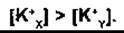
where the membrane (m) between compartments X and Y is impermeable to protein, yet freely permeable to K+ and Cl-. Let us assume that the concentrations of anions and cats on both sides are initially equal; Cl diffuses, resulting in a decrease of its concentration gradient from Y to X, while K+ moves along with the negatively charged Cl, maintaining the electroneutrality of side Y and, consequently, the state of equilibrium:
Next

that is, there are more osmotically active particles on side X than on side Y.
Donnan and Gibbs proved that in the presence of a non-diffusible ion, diffusible ions distribute themselves such that at equilibrium the ratios of their concentrations are equal:

By multiplying the terms of the proportion, we obtain the Gibbs-Donnan equation:

It is valid for any pair of cations and anions with the same valence.
The Donnan ion distribution effect influences physiological processes in various ways. First, given the presence of proteins within cells (Protein), the number of osmotically active particles there is greater than in the interstitial fluid. Since the cells of living organisms have elastic walls, they would swell or even rupture due to osmosis if the Na+-K+-ATPase did not pump ions out of them (see below). Therefore, normal cell volume and pressure depend on the Na+-K+-ATPase. Second, As a result of equilibrium, the distribution of ions penetrating the membrane (m) is asymmetric, leading to an electrical potential difference across the membrane, the magnitude of which can be calculated using the Nernst equation (see below). In our example, side X will be negatively charged relative to Y. Note that in any macroscopic region of the solution, the number of positive and negative charges is equal. However, in the situation described, charges are distributed along the membrane with a concentration gradient for Cl that is precisely balanced by an opposing electrical gradient; the same applies to K+. Third, since plasma contains more proteins than interstitial fluid, the Donnan effect occurs during movement across the Capillary Wall (see below).
Forces Acting on Ions
The forces acting across The cell membrane on each ion can be described mathematically. In the extracellular fluid, the concentration of chloride ions is higher than inside the cells, so these ions naturally diffuse into the cell along their concentration gradient. The net intracellular charge is negative relative to the net extracellular charge, which drives chloride ions out of the cell along the electrical gradient. Equilibrium is reached when the influx and efflux of Cl are equal. The Membrane Potential at which this equilibrium is stable is called the equilibrium potential. Its value can be calculated from the Nernst equation

where ECl is the equilibrium potential for Cl; R is the gas constant; T is the absolute temperature; F is the Faraday constant (the number of coulombs per mole of charge); ZCl is the valence of Cl (I); [Cl]o is the extracellular concentration of Cl; [Cl]i is the intracellular concentration of Cl.
After converting the common logarithm to a base-10 logarithm and substituting numerical values for some of the constants, we obtain

Note that when simplifying the expression, the concentration ratio is inversely proportional because the valence of Cl (-1) has been factored out of the expression.
The equilibrium potential ECl, calculated from the values given in Table 1-2, is -70 mV, which is identical to the value of the membrane potential. Therefore, to explain the distribution of Cl across the membrane, one must account for the forces expressed in the chemical and electrical gradients.
An identical equilibrium potential can also be calculated for K+ ions:

where EK is the equilibrium potential for K+; ZK is the valence of K+ (I); [K+]o is the extracellular concentration of K+; [K+]i is the intracellular concentration of K+.
In this case, the concentration gradient is directed outward, while the electrical gradient is directed inward. In mammalian spinal motor Neurons, EK is equal to -90 mV (see Table 1-2). Since the membrane potential is -70 mV, neurons contain slightly more K+ than would be predicted by the Electrical and Chemical gradients alone.
Unlike the concentration ratios of K+ and Cl-, the chemical gradient of Na+ is directed inward—toward the region of its lower concentration—just like the electrical gradient. The equilibrium potential ENa is +60 mV (see Table 1-2). Since neither EK nor ENa equals the membrane potential, one might expect the cell to gradually accumulate Na+ and lose K+ if only passive electrical and chemical forces acted on the membrane. However, intracellular concentrations of Na+ and K+ remain constant because Na+ is actively transported out of the cell against its electrical and concentration gradients. This transport is coupled with the active uptake of K+ into the cell (see below).
The value of the membrane potential at any given time depends, of course, on the distribution of K+ and Cl- and the membrane permeability to each of these ions. The equation that fairly accurately describes this interaction is known as the Goldman constant-field equation:
where E is the membrane potential; R is the gas constant; T is the absolute temperature; F is the Faraday constant; and PK+, PNa+, and PCl represent the membrane permeability to K+, Na+, and Cl-, respectively. Brackets denote concentration, with in and out referring to the intracellular and extracellular spaces. Since PNa is relatively low compared to PK+ in resting cells, Na+ has little effect on the value of E.
As derived from the Goldman Equation, changes in extracellular Na+ concentration do not cause significant shifts in the cellular potential, whereas an increase in extracellular K+ concentration leads to its decrease.
Table 1-2. Concentration of Certain Ions Inside and Outside Mammalian Spinal Motor Neurons
Ion |
Concentration, mmol/L H2O |
Resting Membrane Potential, mV |
|
inside the cell |
outside the cell |
||
Na+ |
15,0 |
150,0 |
+60 |
К+ |
150,0 |
5,5 |
-90 |
Сl- |
9,0 |
125,0 |
-70 |
This phenomenon is characteristic of various cell types. For example, in Skeletal Muscle cells, the resting membrane potential is approximately -90 mV; ECl is -86 mV; EK is -100 mV; and ENa is +55 mV.
Origin of the Membrane Potential
The process of ion translocation across the Cell Membrane and The Nature of the membrane itself help explain the Generation of the membrane potential. As is well known, the concentration gradient of K+ drives the movement of K+ out of the cell through K+ channels, whereas the electrical gradient for K+ acts in the opposite (inward) direction. This establishes a balance where the tendency of K+ to leave the cell is counterbalanced by its tendency to enter it. Consequently, a slight excess of cations accumulates extracellularly and anions intracellularly. This state is maintained by the Na+-K+-ATPase, which pumps K+ back into the cell and keeps intracellular Na+ concentration low. The activity of the Na+-K+ pump also contributes to the ESTABLISHMENT OF THE membrane potential because for every three Na+ ions pumped out of the cell, two K+ ions are pumped in. Thus, the Na+-K+-ATPase also exerts a minor influence on the membrane potential. It should be noted that the ions responsible for the membrane potential constitute a very small fraction of the total ion pool; the overall concentration of positive and negative ions is equal everywhere, except for those located immediately along the membrane. The influx of Na+ does not offset the efflux of K+ because, via K+ channels (see below), the membrane is far more permeable to K+ than to Na+.
Last update: 10/08/2026
Editorial and Educational Adaptation: This material has been compiled based on the primary/original source text. The project team performed an editorial review, corrected technical inaccuracies, structured sections, and adapted the content for an educational format.
What was processed:
- elimination of formatting defects (OCR errors, structural breaks, corrupted characters);
- editorial organization of content;
- standardization of terminology in accordance with academic sources;
- verification of factual statements against the original source text.
All mentions of the author, publication year, and origin of the primary text have been preserved in accordance with the source.