TEXTBOOK OF PEDIATRIC GYNECOLOGY - 2013
Chapter 10. INFLAMMATORY DISEASES OF THE REPRODUCTIVE ORGANS IN ADOLESCENT GIRLS
ESTABLISHMENT OF THE GENITAL TRACT MICROBIOCENOSIS IN GIRLS
The vaginal microecology in a healthy girl is a hormonally dependent system whose state is interrelated with the body's immune characteristics and is determined by the functional state of the Ovaries, the concentration of lactoflora, the pH of the vaginal content, and the state of local Immunity.
The microbial COMPOSITION OF THE Vagina in girls is conventionally divided into two groups:
1) microorganisms that persistently colonize the vagina;
2) potentially hazardous or opportunistic vaginal microorganisms.
According to their type of Respiration and metabolic processes, microorganisms are classified into aerobes, facultative anaerobes, and obligate anaerobes. The vaginal lactoflora comprises 11 strains of lactobacilli, which represent the Gram-positive rod-shaped flora of the genital tract. The quantitatively dominant strain (42.9%) is Döderlein's bacilli, whose role is multifaceted:
— they ensure the colonization resistance of the genital tract;
— they exhibit enzymatic activity that maintains the acidic environment of the vagina and exerts a detrimental effect on pathogenic flora.
Modern studies of the microflora of vaginal contents in girls lead to the following Conclusions:
— Structure/149.html">The problem of vulvovaginitis in girls is multifaceted, complex, and far from a satisfactory solution;
— a significant number of technologies and Treatment regimens tested on adult women and not routinely used by pediatric gynecologists can be applied with caution (e.g., Antibiotics, Hormones, immunomodulators, Vaccines);
— the onset of vulvovaginitis in girls is driven by A number of factors, one of which stems from the age-related instability and Variability of the vaginal microflora during childhood;
— METABOLISM/2.html">THE CONCEPT OF the monoetiological nature of nonspecific vulvovaginitis should be revised: in more than half of the observations, an association of microorganisms is noted, each of which is capable of causing inflammation on its own;
— the presence of opportunistic flora in the vagina does not prove the etiological role of this specific agent in The Development of vulvovaginitis;
— the absence of clinical manifestations of urogenital infection in the presence of opportunistic microorganisms can be regarded either as carriage or as the natural microbial landscape.
Some lactobacilli act as Inducers of local immunity. The coccal flora vegetating in the genital tract of girls belongs to opportunistic flora; at the same time, some coccal strains are part of the normal microflora of the Skin, mucous membranes of the respiratory tract, digestive tract, and genital Organs. Among staphylococci, the most frequent colonizers of the genital tract are: Staphylococcus aureus, Staphylococcus epidermidis, and micrococcus (saprophytic staphylococcus); among streptococci — alpha-hemolytic streptococcus, hemolytic streptococcus, enterococcus (fecal streptococcus), and pneumococcus (Streptococcus pneumoniae), which has a diplococcal shape. The ecological niche of peptostreptococci (anaerobic cocci) is the Oral Cavity, intestines, and genital organs. A biological feature of cocci is that their young forms stain intensely Gram-positive, whereas Aging forms become Gram-negative.
Rod-shaped flora is opportunistic and vegetates in the genital tract.
From the genus Corynebacterium: diphtheroids, Gram-positive bacilli; certain types of diphtheroids are members of the normal flora of the respiratory tract, skin mucous membranes, and the girl's genital tract. Enterobacteria are Gram-negative bacilli: Escherichia coli, Proteus, Enterobacter (Pseudomonas aeruginosa), and Klebsiella, which constitute a significant part of the normal intestinal flora and participate in ensuring its physiological functioning.
Gardnerellae (small Gram-negative bacilli) are facultative anaerobes, biologically close to the genera Haemophilus and Corynebacterium. Gardnerellae are detected in girls aged from 2 months to 15 years without any clinical manifestations.
Leptotrichia are Gram-variable bacilli that may have a coccobacillary shape. They are facultative anaerobes that vegetate in the genital tract and show weak growth on Blood Agar upon cultivation.
Acinetobacter are aerobic, Gram-negative, thick, short rods with a diplococcal shape; in smears, they resemble gonococci and meningococci vegetating on the skin, mucous membranes, and in the respiratory and urogenital tracts.
During the period of sexual quiescence in girls, common causative agents of bacterial vulvovaginitis are staphylococci, streptococci, and enterococci in various associations.
Dozortseva E. A. (1948) established that a girl's vagina is sterile immediately after birth in 52% of cases. Colonization of the genital tract by microflora begins 3–4 hours after birth, predominantly with coccal flora. Coccal flora predominates in girls up to 12 years of age. During the period of sexual quiescence (up to 10 years), the vaginal environment reaction is alkaline or neutral. Lactobacilli are absent during this period. Cultural studies reveal coccal flora, enterobacteria, and corynebacteria.
During the prepubertal period, the secretion of Sex Hormones increases, and the vagina is colonized by lactobacilli, the number of which increases significantly during Puberty. The vaginal environment becomes acidic, and physiological pubertal leukorrhea appears (Fig. 34, Table 21).
Class="center">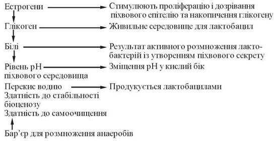
Fig. 34. Factors influencing the vaginal microflora (V. Kask, R. Mandar, 1997)
Table 21. Vaginal biocenosis and local immunity factors in healthy girls
Parameter |
Girls aged 5–8 years |
Girls from 9 years to menarche |
Menstruating girls up to 17 years |
Leukocytes |
1-15 per high-power field |
4-15 per high-power field |
4-10 per high-power field |
Epithelium |
1-3 per high-power field |
3-5 per high-power field |
3-5 per high-power field |
pH |
7.0-8.5 |
5.8-7.2 |
4.0-4.5 |
Total microbial count |
102-105 CFU/ml |
105-106 CFU/ml |
105-107 CFU/ml |
Flora |
Coccal |
Mixed |
Bacillary |
Lactobacilli |
+ 102 CFU/ml |
++ 102-103 CFU/ml |
++++ 105-107 CFU/ml |
Cocci |
++++ 102-105 CFU/ml |
+++ 103-106 CFU/ml |
+ 104-105 CFU/ml |
Hemolytic flora |
++++ 102-105 CFU/ml |
++ 103-104 CFU/ml |
+ 102-103 CFU/ml |
Interferon level, IU/ml |
1.13-5.53; mean — 3.33 |
5.8-17.0; mean — 11.2 |
15.1-45.7; mean — 30.4 |
IgG, mcg/ml |
22.1-46.42; mean — 34.26 |
20.81-36.25; mean — 28.53 |
34.72-75.44; mean — 55.08 |
IgA, mcg/ml |
0.67-1 1.03; mean — 5.85 |
0.02-2.62; mean — 1.32 |
2.86-18.58; mean — 10.72 |
IgM, mcg/ml |
0.79-2.07; mean — 1.43 |
0.22-0.62; mean — 0.42 |
0.12-1.12; mean — 0.62 |
S IgA, mcg/ml |
7.87-18.07; mean — 12.97 |
4.37-11.69; mean — 8.03 |
6.31-24.87; mean — 15.59 |
It is known that the detection of cytomegalovirus at a concentration of 50–200 genomes or Ureaplasma at a microbial titer of less than 104 in the reproductive tract of healthy girls does not require specific antiviral or antibacterial therapy.
With the onset of menarche, specific sex Steroids acquire particular importance: progesterone has a stimulating effect on the development of St. haemolyticus and inhibits the growth of E. coli, whereas estriol suppresses the development of populations of these microorganisms.
There is a certain correlation between a history of vulvovaginitis in childhood and intrauterine infection (chorioamnionitis, embryopathy, etc.) during subsequent reproductive function.
Infection, bacterial carriage, and vulvovaginitis in girls may first occur during childbirth: passing through the birth canal, the girl acquires the maternal microflora, which determines the initial spectrum of both normal and pathological vaginal biocenosis. This spectrum remains relatively stable During the first three years of life or even longer.
Among various factors contributing to the development of vulvovaginitis, the anatomical, morphological, and functional Features of the external genitalia in girls are of great importance. The skin in early childhood has several distinct characteristics: a thin dermis and a loose stratum corneum of the epidermis that is easily susceptible to harmful influences. The Protective Functions of the skin, especially its bactericidal properties, are reduced (the normal skin pH in adults is 4.5–6.0, whereas in children it is 6.7). The development and Maintenance of the inflammatory process are also facilitated by individual Anatomical Features, such as the absence of the posterior fourchette, low placement of the external urethral meatus, cicatricial deformations, incomplete synechiae, and Developmental anomalies of the external genitalia.
Disruptions in the vaginal microflora of girls are caused by certain PHYSIOLOGICAL CHARACTERISTICS OF the pediatric vagina and vulva. The stratified squamous epithelium covering these organs in girls consists of a small number of layers (5–8), its Cells do not keratinize and contain almost no Glycogen; therefore, the girl's vagina lacks the conditions necessary for the existence of lactic acid Bacteria that create an acidic environment in adult women. The girl's vagina is colonized by opportunistic microorganisms (usually Staphylococcus epidermidis) and exhibits an alkaline reaction.
The main physiological protective mechanisms of the vagina are:
a) physiological desquamation and cytolysis of superficial vaginal epithelial cells;
b) nonspecific antimicrobial mechanisms:
— acting at THE CELLULAR LEVEL: phagocytosis mediated by macrophages and polymorphonuclear leukocytes;
— nonspecific humoral factors: the plasma protein transferrin; opsonins, which enhance the phagocytic activity of cells; Lysozyme peptide, which exhibits antimicrobial activity; and lysins released by platelets at the site of inflammation;
c) immune defense mechanisms: lymphocytes, IMMUNOGLOBULINS, cytokines, The Complement System, etc.
A decrease in the reactivity of the child's body, occurring after any illness in girls with a chronic inflammatory process, leads to an imbalance between the vaginal microflora and the child's Organism. With a significant decline in defense mechanisms, opportunistic flora may acquire pathogenic properties. Under these conditions, pathogenic microorganisms easily penetrate the vagina, frequently originating from the oropharynx, intestine, or skin.
Failure to observe hygiene rules promotes the development of pathogenic microflora, which can be introduced into the vagina via hands, clothing, or bathing in contaminated Water bodies.
Some girls experience vesicourethral reflux, in which vaginal contents can be continuously forced into the Urinary Tract, as well as a neurogenic bladder.
M. L. Korshunov (1986), to improve colpobacterioscopic Diagnostics, identified Three types of vaginal smears in girls:
Type I — normal. Leukocyte count at ages 1–3 years is 1–2 per high-power field, at 4–6 years — 1–3 per high-power field, at 7–10 years — 3–5 per high-power field, and at 11–15 years — 7–10 per high-power field. Phagocytosis is not pronounced. In blondes, children with paratrophia, and those suffering from lymphatic diathesis, physiological values may be twice as high. Mucus is present in insignificant amounts up to 7 years of age, in small amounts after 9 years, and starting from 11–12 years, the mucus content in the smear increases along with the enhancement of the secretory function of the genital tract. The number of desquamated vaginal epithelial cells is insignificant up to 7–9 years of age. After 9 years, it increases to 10–12–20 per high-power field due to hormonal desquamation.
The vaginal flora in girls up to 8 years of age is predominantly coccal (in small amounts). Lactobacilli may be detected up to 6 months of age, after which they are absent until 9 years of age and reappear in smears with the onset of puberty. In the prepubertal and pubertal periods, the girl's vaginal flora is mixed, with gram-positive bacillary flora predominating.
Type II — a transitional state from the first smear type to the third. The leukocyte count is 20 or more per high-power field. Desquamated epithelial cells and mucus are present in moderate amounts. Phagocytosis is not pronounced.
Discharges are turbid, with a significant amount of mixed opportunistic flora present in the absence of clinical signs of inflammation.
Type III is characteristic of a clinically pronounced inflammatory process. The leukocyte count exceeds 20 per field of view, with a predominance of neutrophils and macrophages, active phagocytosis, and a pronounced mucous reaction. There is a significant amount of desquamated epithelium and numerous degeneratively altered cells. Lactobacilli are absent. Discharges are mucopurulent or purulent. Culturing of genital secretions is mostly performed to detect aerobic microflora.
Last update: 08/08/2026
Editorial and Educational Adaptation: This material has been compiled based on the primary/original source text. The project team performed an editorial review, corrected technical inaccuracies, structured sections, and adapted the content for an educational format.
What was processed:
- elimination of formatting defects (OCR errors, structural breaks, corrupted characters);
- editorial organization of content;
- standardization of terminology in accordance with academic sources;
- verification of factual statements against the original source text.
All mentions of the author, publication year, and origin of the primary text have been preserved in accordance with the source.