Human Anatomy Part 1 - K. A. Dyubenko, A. K. Kolomiysev, Yu. B. Chaykovsky 2002
Special Part
Axial Skeleton, sceleton axiale - The Skull as a Whole
Age and Sex-Related Features of Skull Structure
The shape, dimensions, and proportions of a newborn's Skull differ significantly from those of an adult skull (Fig. 56). The volume of the newborn's facial cranium accounts for only 13% of the neurocranium volume, whereas in adults, the facial cranium makes up 40% of the neurocranium. This is due to the underdevelopment of the newborn's jaws, particularly the alveolar processes, the absence of Teeth, and the poor Development of the Nasal cavity and Paranasal Sinuses. Other Characteristic Features of the newborn skull include: relatively small facial bones compared to the neurocranium, a greater number of bones than in adults, the presence of six fontanelles (fonticuli), and well-developed frontal and parietal eminences. Starting from the age of 13-14, the facial cranium enlarges considerably.
Fontanelles are remnants of The First stage of skull development (the membranous stage); they are located at the intersections of the sutures where remnants of unossified Connective Tissue persist. Externally, the fontanelles are covered by the Skin and epicranial aponeurosis, while the dura mater adheres to them from the cranial cavity. The fontanelles allow the cranial bones to strengthen and enable the shape and size of the skull to change during childbirth. Another important role of the fontanelles is that, due to their pliability, they cushion fluctuations in intracranial pressure caused by the increasing weight of the Brain.
Class="center">
Fig. 56. Proportional relations between the newborn and adult skull (after A. Andronescu)
The following fontanelles are distinguished: the anterior fontanelle (fonticulus anterior), posterior fontanelle (fonticulus posterior), sphenoidal fontanelle (fonticulus sphenoidalis), and mastoid fontanelle (fonticulus mastoideus) (Fig. 57).
The anterior fontanelle (frontal, fonticulus anterior) is diamond-shaped and located along the midline at the junction of the sagittal, frontal, and coronal sutures. Its dimensions range from 18x20 to 26x30 mm. The anterior fontanelle closes at the age of 1.5-2 years. Delayed closure of the fontanelles indicates developmental disorders or Rickets in the child.
The posterior fontanelle (occipital, fonticulus posterior) is triangular in shape and located at the end of the sagittal suture between the squamous part of the Occipital bone and the parietal bones; it closes by the 2nd month of life.
The sphenoidal fontanelle (fonticulus sphenoidalis) is paired and located between the anteroinferior angle of the Parietal bone, the Frontal bone, the greater wing of the Sphenoid bone, and the squamous part of the Temporal bone; it closes by the 2nd-3rd months of life.
The mastoid fontanelle (fonticulus mastoideus) is paired and located between the posteroinferior angle of the parietal bone, the Base of the petrous part of the temporal bone, and the squamous part of the occipital bone. The sphenoidal and mastoid fontanelles close after birth, but they are often observed only in premature infants. In addition to the listed fontanelles, inconstant fontanelles may occur (such as the Casserian, parietal, and cerebellar fontanelles), which can be sites for The formation of craniocerebral hernias.

Fig. 57. Newborn skull (after Morris):
A - right lateral view; B - superior view
The frontal and occipital fontanelles are of great practical importance in obstetric practice, serving as landmarks for determining THE POSITION OF the fetal HEAD during labor. The venous sinuses of the dura mater run beneath the membranous tissue of the fontanelles. In clinical practice, puncture of the superior sagittal sinus is performed through the frontal fontanelle to administer medications.
Newborns lack fully formed sutures, diploë is insufficiently developed, and the cranial roof is more developed than the base. Cartilaginous layers are located between the individual PARTS OF THE cranial bones, and the paranasal sinuses are not yet formed.
The postnatal growth of the neurocranium and facial cranium occurs unevenly. The linear dimensions of the neurocranium increase by 1.5 times over the entire growth period, whereas the facial cranium increases by 3 times. Changes in the skull are conventionally divided into five age periods (Table 2).
Table 2. Age-related features of skull Structure
|
Periods |
Characteristics |
|
1. From birth to 7 years |
Characterized by active skull growth. The neurocranium enlarges particularly fast in the first months of life: the volume of the cranial cavity doubles in the first 6 months after birth, triples by the age of 2, and reaches 3/4 of the adult skull volume by the age of 5. |
|
2. From 8 to 13-14 years |
After the age of seven, skull growth slows down somewhat, although the nasal cavity, Orbit, and maxillary sinus enlarge significantly. |
|
3. From the onset of Puberty (14-16 years) to 20-25 years, when skeletal growth ceases |
During this period, skull growth is fully completed. The facial cranium grows more intensively than the neurocranium, especially in males. Paranasal sinuses, eminences, prominences, sulci, and the glabella take their final shape. Growth of the cranial vault is completed at 16-18 years, while the base grows in length until 18-20 years, when the spheno-occipital synchondrosis ossifies. |
|
4. From 26 to 45 years |
This is the most stable period, during which the skull remains unchanged. Starting from the age of 20-30, cranial sutures begin to close, and syndesmoses are replaced by synostoses. |
|
5. From suture ossification to old age |
This period is characterized by tooth loss and Atrophy of the alveolar processes of the jaws. As a result, the angle of the Mandible increases, and the chin projects forward. In senile age, Osteoporosis of the cranial bones occurs, making them thin and lightweight. |
Sexual Dimorphism of the skull involves the size and shape of both The Skull as a whole and its individual regions (Fig. 58). The cranial cavity in males is approximately 10% larger than in females. The external relief of the male skull is more pronounced: the zygomatic bones, frontal eminences, supraorbital margins, mastoid processes, and mental eminence project outward significantly. The surface of the female skull is smoother, the bones are thinner, and the facial cranium and supraorbital margins are less developed than in males.
Anomalies of the cranial bones are caused by The complexity of their ontogeny. These include: sutural and fontanelle bones, inconstant sutures, cranioschisis, turricephaly, scaphocephaly, trigonocephaly, as well as microcephaly and macrocephaly. Anomalies of the facial cranium are associated with developmental Disorders of the teeth, palate, and jaws, leading to Asymmetry manifested by unequal sizes of the orbits, frontal sinuses, and maxillary sinuses.
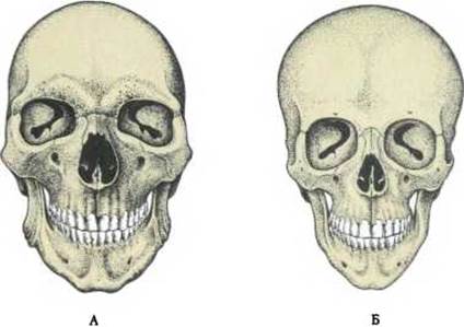
Fig. 58. Male (A) and female (B) skulls (after A. P. Bystrov)
Last update: 08/08/2026
Editorial and Educational Adaptation: This material has been compiled based on the primary/original source text. The project team performed an editorial review, corrected technical inaccuracies, structured sections, and adapted the content for an educational format.
What was processed:
- elimination of formatting defects (OCR errors, structural breaks, corrupted characters);
- editorial organization of content;
- standardization of terminology in accordance with academic sources;
- verification of factual statements against the original source text.
All mentions of the author, publication year, and origin of the primary text have been preserved in accordance with the source.