Molecular Biology: Protein Structure and Function - Stepanov V.M. 2005
Fibrous proteins
Keratins
Keratin is a Structure-forming fibrous protein synthesized by epithelial Cells, forming what are known as Intermediate filaments—strands that contribute to the Organization OF THE Cytoskeleton. Proteins related to keratin are found in the cytoskeleton of other Cell types. Horny structures such as Hair, Nails, claws, beaks, feathers, spines, shells, and the dry outer layer of the Skin are also built from keratin.
As epithelial cells migrate toward the surface, keratin synthesis intensifies significantly, and eventually, keratin fibers fill the entire cell. The Cell's surface structures are displaced to the periphery of the fiber, forming its outer sheath, or cuticle.
In epithelial cells, at least 20 structural genes encode the synthesis of keratins belonging to either type I or type II; type III keratins are synthesized as cytoskeletal proteins in non-epithelial cells. All keratins are evolutionarily related proteins sharing a common structural plan. For instance, human epidermal keratin II contains 562 amino acid residues within a single polypeptide chain, with its N- and C-terminal segments (effectively domains) appearing to lack any dominant regular Secondary structure. In contrast, The sequence of the central part of the polypeptide chain, corresponding to four exons, consists of four contiguous helical domains. Each of these domains features a repeatedly occurring identical structural motif of seven amino acid residues, of which the first and fourth are typically hydrophobic. For example, starting from the 386th residue, the fragment runs as follows (hydrophobic residues are highlighted in bold): Ile—Gin—Arg—Leu—Arg—Ser—Glu—Ile—Asp—His—Val— Lys—Lys—Gm—Cys—Ala—Asn—Leu, etc.
The regular appearance of hydrophobic side chains every three or four amino acid residues in the a-helical regions of keratin leads to The formation of distinct hydrophobic "ridges," the interaction of which stabilizes the superhelix—the so-called protofibril—formed by the twisting of two identically oriented keratin chains around each other (Fig. 14.4). Evidently, the terminal non-helical segments in keratin polypeptide chains, much like in Collagen, perform an "assembly" function, facilitating their proper folding and accelerating superhelix formation. Eleven protofibrils bundle together to form a microfibril, and microfibrils assemble into fibrils.
During the formation of horny structures such as hair, wool, shells, etc., a major role is played by the matrix protein in which the keratin fibrils are embedded. Unlike keratin proper, this non-helical protein is very rich in Cysteine residues which, upon oxidation, form numerous Disulfide Bonds that reinforce The structure of the keratin fibrils and render them virtually insoluble. Thus, the cysteine-rich proteins of human hair and sheep wool, which are very similar in Primary Structure, contain 168 amino acid residues in their polypeptide chain, 36% of which are cysteine, converting into cystine upon oxidation. A characteristic feature is the N-terminal sequence of the sulfur-rich human hair protein, in which cysteine residues occupy every third or fourth position:
Class="center">Met—Gly—Cys—Ser—Gly—Cys—Ser—Gly—Gly—Cys—Gly—Ser—Ser— Cys—Cly—Gly—Cys—Gly—Ser—Arg—Cys—Gly— Gly—Cys—Ser— Ser— Ser—Cys—Cys—
The content of cysteine residues in keratinized horny structures heavily depends on the animal species, diet, and Nature of the epidermal Tissues. For instance, the cysteine content in sheep wool is significantly higher than in horns, while in turtle shells it reaches 20%.
Stretching keratin fibers while simultaneously moistening them leads to a profound reorganization of their secondary structure: the a-helical chains extend and transition into a ß-Structure, facilitated by moisture through the establishment of an interchain Hydrogen bond network. This transition and the resulting stretching of keratin fibers, such as hair, are reversible: upon load removal and humidity reduction, the fibers revert to the a-helical structure and contract in length. This property is utilized in psychrometers—devices used to measure atmospheric humidity.
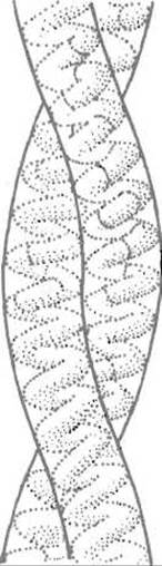
Fig. 14.4. Keratin double helix
The reduction of disulfide bonds, typically achieved by Treatment with an excess of a mercapto compound (thioglycolic acid or mercaptoethanol), removes the cross-links in the cysteine-rich matrix protein and makes the keratin fibers highly flexible and moldable. Subsequent oxidation induces the formation of a different set of disulfide bonds, thereby fixing the new shape of the fiber. This treatment is used in Permanent hair waving and for imparting crease-resistant pleats to wool fabrics made of keratin.
Last update: 13/08/2026
Editorial and Educational Adaptation: This material has been compiled based on the primary/original source text. The project team performed an editorial review, corrected technical inaccuracies, structured sections, and adapted the content for an educational format.
What was processed:
- elimination of formatting defects (OCR errors, structural breaks, corrupted characters);
- editorial organization of content;
- standardization of terminology in accordance with academic sources;
- verification of factual statements against the original source text.
All mentions of the author, publication year, and origin of the primary text have been preserved in accordance with the source.