BIOCHEMISTRY - Textbook - Ostapchenko L. I. - 2012
Chapter 4. MOLECULAR ORGANIZATION AND BIOLOGICAL FUNCTIONS OF MEMBRANES
4.1. Structure of Biological Membranes
The first direct chemical Methods in membrane research were applied by E. Overton (1895-1902). Having established that lipid-soluble substances penetrate Cells faster, he proposed the first model of Cell/30.html">The Plasma Membrane (1902), stating that the membrane is a thin layer of Phospholipids. In 1925, E. Gorter and F. Grendel demonstrated that the area of a lipid monolayer extracted from erythrocytes is twice as large as the total surface area of the erythrocytes. Based on these findings, they hypothesized that Membrane Lipids are arranged as a bimolecular layer 3-4 nm thick. The electrical capacitance of such a Structure is 0,5 · 10-2 F/m2, which corresponds to a dielectric thickness (the lipid layer of the membrane) of 3,5 nm.
Concurrently with these studies, it was found that the surface tension coefficient at The Cell-Water interface is closer to that of the protein-water interface (~0,1 dyn/cm) than the lipid-water interface (~10 dyn/cm). This contradiction was resolved by J. Danielli and H. Davson, who in 1935 proposed a "sandwich" model of the biological Membrane Structure (Fig. 4.1, 1). This membrane is formed by two protein layers with a lipid bilayer sandwiched between them. Later (1956), to reconcile the model with the then-known data on Facilitated Diffusion of low-molecular-weight hydrophilic substances through the membrane, J. Danielli and W. Stein suggested the existence of polar protein pores within the membrane (Fig. 4.1, 2).
With advancements in ultrathin tissue sectioning techniques (1950-1960) for Electron Microscopy, it became evident that the trilaminar structure is characteristic of all membranes studied at the time. Based on these morphological data, J. Robertson put forward the unit membrane concept in 1960 (Fig. 4.1, 3). This model is similar to the Danielli-Davson model, yet it also reflects another membrane feature: its Asymmetry (where protein layers are depicted differently in the diagram). The Development of electron microscopy enabled scientists to "see" the membrane not as a continuous trilaminar line, but as a granular structure containing breaks (protein pores) and appearing to consist of separate globular parts. Furthermore, when membranes were disrupted using detergents (see below), lipoprotein fragments were obtained which, upon detergent removal, spontaneously reassembled into membrane-like structures. These findings cast doubt on Robertson's model and sparked a series of models reflecting METABOLISM/2.html">THE CONCEPT OF globular membrane Organization. Examples include the models by F. Sjöstrand (1963) and J. Lucy (1964), which envisioned the membrane as alternating lipid micelles and protein globules; D. Green and J. Purdue (1966), proposing a mitochondrial membrane model made of complex lipoprotein subunits; and A. Benson (1966), suggesting that lipid hydrocarbon chains are intertwined with Polypeptides and held together by hydrophobic interactions. J. Finean's model (1966) emphasized differences in the distribution of membrane components and made the first attempt to describe the membrane taking specific lipid molecules into account (Fig. 4.1).
The discovery of phase polymorphism in water-lipid systems and the liquid-crystalline state of lipids at physiological temperatures, the concept of lipid bilayer fluidity, and the measurement of lateral diffusion rates in membranes (1968-1971) provided the foundation for S. Singer and G. Nicolson to propose a fundamentally new model of molecular membrane organization (1972), known as the fluid-mosaic model (Fig. 4.1, 10). The main postulates of this membrane model are:
✵ the membrane is a lipid "sea" in which individual Proteins "float";
✵ There are two types of Membrane Proteins: peripheral (bound to the membrane via polar and ionic interactions) and integral (spanning The Lipid Bilayer and interacting with lipids through their hydrophobic domains);
✵ membrane molecules are in constant lateral motion, etc. Fig. 4.1, 10 illustrates the fluid-mosaic model of the plasma membrane, taking into account the attachment of its structural components to the Cytoskeleton and glycocalyx.
Class="center">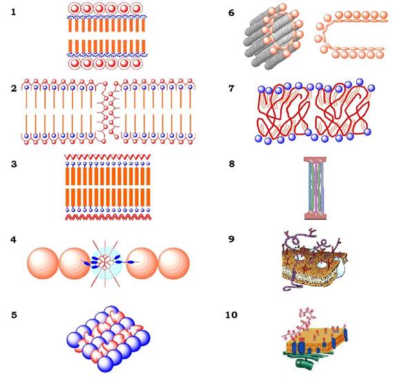
Fig. 4.1. Models of biological membrane structure:
1 - Danielli-Davson; 2 - Stein-Danielli; 3 - Robertson; 4 - Lucy; 5 - Sjöstrand; 6 - Green and Purdue; 7 - Benson; 8 - Finean; 9 - Singer-Nicolson (fluid-mosaic); 10 - fluid-mosaic model incorporating membrane connections with the cytoskeleton and glycocalyx
Last update: 06/08/2026
Editorial and Educational Adaptation: This material has been compiled based on the primary/original source text. The project team performed an editorial review, corrected technical inaccuracies, structured sections, and adapted the content for an educational format.
What was processed:
- elimination of formatting defects (OCR errors, structural breaks, corrupted characters);
- editorial organization of content;
- standardization of terminology in accordance with academic sources;
- verification of factual statements against the original source text.
All mentions of the author, publication year, and origin of the primary text have been preserved in accordance with the source.