Introduction to Molecular Biology: From Cells to Atoms - Anthony Rees, Michael Sternberg 2002
Proteins
Secondary structure of proteins
Class="center">Introduction/introduction.files/image018.jpg" width="523"/>
Fig. 9.1.
The Secondary Structure of a polypeptide chain segment refers to the conformation of the backbone of this fragment, disregarding the conformation of its side groups. According to an earlier definition, which is sometimes still used, secondary structure comprises those segments of the polypeptide chain that participate in forming a regular Hydrogen bond network. Certain Conformations yield regular, repeating structures stabilized by Hydrogen Bonds between the >N—H and >C=O groups of the polypeptide backbone (e.g., the a-helix, ß-sheet, and Collagen helix).
A regular or helical secondary structure is formed when the phi (φ) and psi (ψ) torsion angles are identical for all residues. In this case, the backbone twists by the exact same angle at each Ca atom. Any helix can be viewed as the result of winding a chain around the lateral surface of an imaginary cylinder. A helix is characterized by the number of repeating units (residues) per turn (denoted as n) and the distance between adjacent residues along the helical axis (d). The pitch of the helix (p) is equal to the product of n and d. Helices can be either right-handed or left-handed. Stating that a helix is right-handed means that if you point the outstretched thumb of your right hand along the axis of the helix, your remaining fingers curled into a fist perpendicular to the thumb will point in the direction of the chain's advance.
In 1951, Pauling and Corey suggested that Two Types of regular structure should occur in Proteins: the a-helix (n = 3.6) and the ß-Structure (n = 2). This hypothesis was based, firstly, on known data concerning the dimensions and planar STRUCTURE OF THE peptide group, and secondly, on the fact that optimal conditions for forming the N—H···O hydrogen bond are achieved when all three atoms—N, H, and O—lie on a straight line. It was precisely the ability of the polypeptide chain to adopt the conformation of an a-helix and a ß-structure that successfully explained the results of X-Ray Diffraction experiments on fibers of the fibrous protein keratin (Chapter 11), which exist in the a- and ß-forms, respectively (hence the names a- and ß-structures). The a-helix and ß-structure are the most energetically favorable conformations because both are stabilized by hydrogen bonds (Chapter 7) between the >N—H and >C=O backbone groups. Furthermore, both the a-helix and ß-structure are additionally stabilized by the tight packing of backbone atoms, which fit together like pieces of a jigsaw puzzle.
In a right-handed a-helix, φ = -57° and ψ = -47°. The number of residues per turn (n) is 3.6, the distance between adjacent residues along the helical axis (d) is 0.15 nm, and the pitch of the helix (p) is 0.54 nm (= 3.6 × 0.15 nm). Each >C=O group forms a hydrogen bond with
the fourth >N—H group down the chain. The regularity of the structure means that all >N—H and >C=O groups, except those located at the ends of the helix, are able to form hydrogen bonds. In terms of shape, the a-helix resembles a twig in which the stem represents the backbone and the protruding side branches represent the side chains (R groups).
A ß-sheet is formed from two or more ß-structural segments of a polypeptide chain, referred to as ß-strands. In each ß-strand, the polypeptide chain is almost fully extended; consequently, the >N—H and >C=O groups are oriented approximately perpendicular to the direction of the strand and can form hydrogen bonds with adjacent strands. As a result, several ß-strands combine to form a structure that, to a first approximation, appears flat and sheet-like. However, because the planes of the peptide groups in each ß-strand tilt alternately in opposite directions relative to the strand axis, the flat ß-sheet acquires a pleated conformation. Moving along one of the ß-strands that make up a ß-sheet, the side groups project alternately from one side and then the other of the sheet.
Parallel and antiparallel ß-structures. There are two different patterns of hydrogen bonding between strands within a ß-sheet. Each pattern corresponds to specific values of the φ and ψ angles. In a parallel ß-sheet (φ = -119°, ψ = 113°), adjacent ß-strands run in the same direction (Fig. 9.2), whereas in an antiparallel ß-sheet (φ = -139°, ψ = 135°), they run in opposite directions (Fig. 9.1).
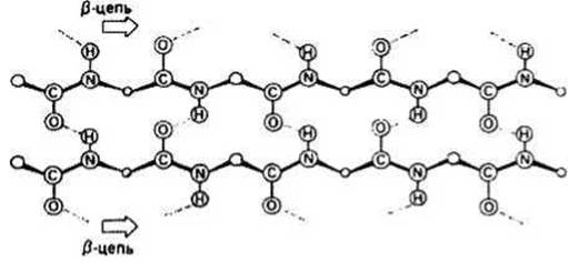
Fig. 9.2. Parallel ß-sheet.
The collagen helix represents a third type of regular secondary structure, with torsion angles of φ = -60° and ψ = 140°. This conformation is adopted by the major portion of the polypeptide chain in the fibrous protein collagen (Chapter 11).
Most Globular proteins contain a-helical and/or ß-structural regions. a-Helices typically consist of 6–24 residues, and their lengths correspondingly vary from 0.9 to 2.4 nm. The φ and ψ angles in a helix always deviate slightly from standard values. Occasionally, several residues adopt a 310-helical conformation, which differs from the a-helix. A 310-helix has 3 residues per turn, and its Hydrogen bonds are formed between residues n and n — 3 along the chain. ß-Sheet regions generally consist of 3–10 residues and span lengths from 1.0 to 3.3 nm. A typical ß-sheet contains from 2 to 10 ß-strands. In some ß-sheets, all strands are aligned parallel to one another, while in others, all adjacent ß-strands are antiparallel. Additionally, both types of arrangement can sometimes be found within the same sheet. Just as with the a-helix, the actual conformation of a ß-sheet may deviate somewhat from the standard geometry.
A ß-turn is another type of secondary structure found in many globular proteins at sites where the direction of the polypeptide chain reverses. This structure often acts as a connecting link between two antiparallel ß-strands within a ß-sheet. Fig. 9.3 illustrates an example of a four-residue ß-turn stabilized by a single hydrogen bond between the >C=O group of residue 1 and the >N—H group of residue 4. Other types of ß-turns are also found in proteins. Unlike the a-helix, collagen helix, and ß-sheet, the φ and ψ angles in a ß-turn vary from one residue to another. ß-Turns are typically located On the surface of protein globules.
It is possible to predict which regions of a globular protein molecule will form regular secondary structures based on their Amino Acid Sequence with an accuracy of about 70%. Certain residues (such as Glu, Met, Ala, and Leu) occur frequently in a-helices, whereas others (Gly and Pro) are found much less often. As for Proline (Pro), this residue can only reside within the first three positions of an a-helix; in any other position, the covalent bond between the proline side chain and the backbone nitrogen atom prevents the >N—H group from forming a hydrogen bond. Some residues show a strong preference for ß-strands (e.g., Val, Ile, Tyr, and Phe), whereas Asp and Glu rarely adopt this conformation. Such statistical data regarding the occurrence of various residues in different secondary structure types are utilized to predict the locations of a-helical and ß-strand regions within a protein.
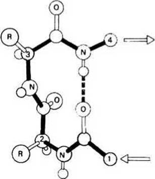
Fig. 9.3. ß-Turn.
Last update: 13/08/2026
Editorial and Educational Adaptation: This material has been compiled based on the primary/original source text. The project team performed an editorial review, corrected technical inaccuracies, structured sections, and adapted the content for an educational format.
What was processed:
- elimination of formatting defects (OCR errors, structural breaks, corrupted characters);
- editorial organization of content;
- standardization of terminology in accordance with academic sources;
- verification of factual statements against the original source text.
All mentions of the author, publication year, and origin of the primary text have been preserved in accordance with the source.