Introduction to Molecular Biology: From Cells to Atoms - Anthony Rees, Michael Sternberg 2002
Proteins
Peptide Conformation
Class="center">Introduction/introduction.files/image015.jpg" width="635"/>
Fig. 8.1
Molecular Conformations refer to spatial arrangements of a molecule's constituent atoms that can be interconverted by rotation around single covalent bonds. In contrast, configurations are spatial arrangements whose interconversion requires the breaking and formation of covalent bonds. For example, the D- and L-isomers of amino acid residues (Chap. 6) represent different configurations, whereas the α-Helix and Collagen helix (see below) represent different conformations of the polypeptide chain.
A peptide bond is the linkage between the >C=O group of one residue and the >N—H group of the next. Contrary to expectations, this C—N bond is not purely single, but possesses partial double-bond character (C—N) due to Resonance. Consequently, the bond order of the adjacent C=O double bond is correspondingly reduced. This shift in bond order occurs because one electron pair of the C=O bond is delocalized among the O, C, and N atoms. As a result of this electron redistribution, the O, C, N, and H atoms (the peptide group) and the two covalently attached Cα atoms all lie in a single plane, known as the amide plane, and rotation around the C—N bond becomes restricted. The coplanarity of the peptide group was first proposed by Pauling and Corey. They observed that the length of the peptide bond (0.132 nm) is intermediate between the lengths of a single C—N bond (0.149 nm) and a double C=N bond (0.127 nm).
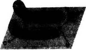
Fig. 8.2.
Trans-conformation of the peptide group. The Cα atoms flanking a peptide group on either side are typically in a trans-conformation relative to each other. The alternative cis-conformation is energetically unfavorable due to severe steric hindrance arising from the close approach of bulky groups attached to the Cα atoms.
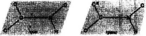
Fig. 8.3.
The Cα atom is linked by single bonds to the nitrogen atom of one peptide group and the carbonyl carbon atom of the next. Relatively free rotation is possible around these single bonds.
The φ and ψ angles. Rotation around the N—Cα and Cα—C bonds is described by the torsion angles φ (phi) and ψ (psi), respectively. The φ angle defines the positions of all atoms lying in the amide plane preceding the Cα atom, whereas the ψ angle determines the positions of all atoms in the amide plane following the Cα atom. Certain pairs of φ and ψ values are forbidden due to steric constraints, which occur when two atoms approach each other closer than the sum of their Structure/103.html">Van der Waals radii. For example, at φ = 0° and ψ = 180°, steric clashes arise between two oxygen atoms.
The Ramachandran conformation map, or (φ, ψ) plot, illustrates which pairs of φ and ψ values are allowed. This map is named after the Indian scientist who first calculated the permissible values for these angles. Each point on the map corresponds to a specific (φ, ψ) pair and indicates whether the spatial arrangement of the two flanking amide planes and the side chain attached to the Cα atom is energetically favorable. The (φ, ψ) plot shown in Fig. 8.1 is typical for most amino acid side chains, with the exception of Glycine and Proline. In fully allowed conformations, unfavorable atomic contacts are absent. Forbidden angles (such as φ = 0°, ψ = 180°) correspond to conformations where certain atoms are separated by a distance less than the sum of their contact radii. It should be noted that because atoms are not rigid spheres, they can approach one another to a distance slightly less than the sum of their contact radii. Although such close contacts are unfavorable, they can occur nonetheless, and the corresponding conformations are referred to as partially allowed (e.g., φ = −180°, ψ = 180°). If every fully allowed (φ, ψ) pair is represented on the map by a dark brown dot, the resulting dark brown regions correspond to the fully allowed areas of φ and ψ angle space. Partially allowed regions are marked in light brown. It must be emphasized that (φ, ψ) plots indicate the allowed pairs of angles for a specific residue. The side chain of glycine, consisting of a single hydrogen atom, is smaller than that of any other residue; consequently, the Ramachandran map for glycine features more extensive allowed regions than those of other residues. Conversely, in proline, the constraints imposed by the covalent bond between the side-chain carbon atom and the backbone nitrogen atom lead to a significant reduction in the size of the allowed areas.
Regular or Helical structures. When the φ and ψ values remain identical for several consecutive residues along the chain, the main chain undergoes a uniform rotation at each Cα atom within that segment. This gives rise to a regular Secondary structure. Such structures are frequently found in both globular and Fibrous Proteins. Of particular importance are parallel and antiparallel β-structures (Chap. 9), the α-helix (Chap. 9), and the collagen helix (Chap. 11). The values of φ and ψ corresponding to these types of secondary structure are presented in Fig. 8.1.
Last update: 13/08/2026
Editorial and Educational Adaptation: This material has been compiled based on the primary/original source text. The project team performed an editorial review, corrected technical inaccuracies, structured sections, and adapted the content for an educational format.
What was processed:
- elimination of formatting defects (OCR errors, structural breaks, corrupted characters);
- editorial organization of content;
- standardization of terminology in accordance with academic sources;
- verification of factual statements against the original source text.
All mentions of the author, publication year, and origin of the primary text have been preserved in accordance with the source.