Biological Membranes - A. N. Ogurtsov 2012
Structure and Functions of Biomembranes
Membrane Proteins
Structure of Transmembrane Domains
Transmembrane a-helices. Hundreds of specific protein motifs (or repeats) have been identified in Water-soluble Proteins. In contrast, the number of "standard" motifs found in integral Membrane Proteins is quite small. The transmembrane a-helix is the most common among them.
Integral proteins containing transmembrane a-helical domains are anchored in the membrane through hydrophobic interactions with specific Lipids, as well as potential ionic interactions with the polar HEAD groups of Phospholipids.
A clear example of such proteins is Glycophorin A, the major protein of the erythrocyte Cell/33.html">Plasma Membrane. Its transmembrane domain consists of a single a-helix (Figure 36).
Class="center">
Figure 36 - Diagram of the glycophorin A dimer
Typically, a transmembrane a-helix consists of 20-25 hydrophobic Amino Acids. In glycophorin A, the transmembrane a-helix is composed of 23 amino acids. The length of such a helix is approximately 3.75 nm, which is in good agreement with data on the thickness of The Lipid Bilayer.
In the a-helix, hydrophobic amino acid side chains are oriented outward and interact with the hydrophobic lipid environment, whereas the carbonyl (C=O) and imino (NH) groups, which form intra-helical Hydrogen Bonds, are located inside the a-helix and are shielded from the lipid medium by amino acid residues. It is energetically favorable for two glycophorin A molecules to form a dimer through The formation of a coiled-coil motif by their transmembrane helices.
Such protein dimerization via coiled-coil formation is a widespread phenomenon. For instance, many Membrane Receptors are activated only upon dimerization.
There is an extensive "family" (over 150) of integral membrane proteins characterized by the presence of seven transmembrane a-helices. These "seven-helix" multi-pass proteins include G protein-coupled receptors, which play a pivotal role in cellular signaling.
The General Principles of protein architecture in this family are illustrated by The Structure of Bacteriorhodopsin (Figure 37), which Functions as a photoreceptor in certain Bacteria.
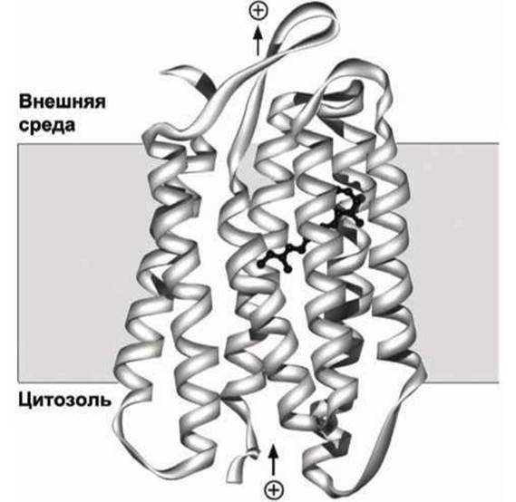
Figure 37 - Diagram of bacteriorhodopsin. The retinal molecule is shown in black
A retinal molecule is covalently attached to one of the transmembrane a-helices. Upon absorption of a light quantum, the retinal molecule undergoes a conformational change (Figure 38), which triggers a conformational transition in the protein molecule, resulting in the translocation of a single proton from the Cytosol across the membrane into the extracellular space.

Figure 38 - Conformations of the retinal molecule: a - all-trans-retinal, b - 13-cis-retinal
Bacteriorhodopsin functions as a proton pump, utilizing light energy to generate a proton concentration gradient across the membrane. This proton gradient is subsequently utilized for ATP synthesis by membrane F0F1 complexes (ATP synthases). Experimental Determination of the atomic STRUCTURE OF THE transmembrane domain has revealed that virtually all amino acids at the periphery of the bacteriorhodopsin transmembrane domain are indeed hydrophobic, interacting with the hydrocarbon interior of the lipid bilayer.
Another important class of integral membrane proteins is Ion Channels, the STRUCTURE AND FUNCTIONS of which will be discussed in detail in Chapter 13.
Ion channels are predominantly tetrameric proteins. Each of the four subunits contains at least two transmembrane a-helices, and together, the a-helices of all subunits form a selective aqueous pore that allows the passage of only specific types of ions.
The amino acids lining this channel are polar and hydrophilic, whereas the peripheral amino acids of the ion channel's transmembrane domain are hydrophobic, much like in bacteriorhodopsin.
In many ion channels, external factors such as Ligand binding, Changes in membrane potential, or mechanical stress induce a shift in the relative arrangement of the a-helices, which serves to regulate ion flux across the membrane.
Transmembrane ß-structures. The class of integral proteins known as porins differs radically from the transmembrane proteins discussed above (Figures 39, 40).

Figure 39 - Diagram of a porin OmpX subunit from the E. coli Cell wall
Several types of porins have been discovered both in the outer cell wall of Gram-negative bacteria, such as E. coli, and in the outer membranes of Mitochondria and METABOLISM/14.html">Chloroplasts. The Cell wall protects enteric bacteria from hazardous environmental factors (e.g., Antibiotics, Bile salts, proteases), yet it remains permeable to small water-soluble molecules, letting nutrients in and Metabolic waste products out. Porins in the E. coli cell wall (Figure 39 facilitate the inward passage of Disaccharides and phosphates.
The polypeptide sequences of porins consist predominantly of polar Amino Acids and lack continuous hydrophobic segments characteristic of integral proteins with transmembrane a-helices.
Porins are trimeric proteins consisting of three identical subunits. Each subunit is formed by sixteen ß-strands that create a cylindrical structure with a central channel (Figure 39). Unlike typical water-soluble Globular proteins, a porin features a hydrophobic outer surface and a hydrophilic inner core; in this sense, porins represent "inside-out" globular proteins. In the porin monomer, hydrophobic amino acid residues of each ß-strand face outward, collectively forming a hydrophobic belt that encircles the protein monomer along its perimeter.
The amino acid residues oriented toward the interior of the channel are predominantly hydrophilic, lining the pore through which small, water-soluble molecules can cross the membrane. Two chains of aromatic (carbon ring-containing) amino acids shown in Figure 39, along with aliphatic (non-cyclic) amino acids within the ß-strands, help properly orient the porin within the membrane.
Figure 40 illustrates a schematic diagram of another bacterial porin found in the outer membrane of the bacterium Rhodopseudomonas blastica. This porin is constructed from sixteen ß-sheet secondary structural segments of the polypeptide protein chain, which form a cylindrical pore acting as a passive channel in the bacterial outer membrane. The pore runs along the axis of this cylinder.

Figure 40 - Structure of the bacterial porin from Rhodopseudomonas blastica: a - porin integrated into the biomembrane, b - spatial arrangement of the sixteen ß-sheet secondary structural segments of the porin protein chain
Animal cell Plasma Membranes contain water channels known as Aquaporins. Like most integral membrane proteins, aquaporins feature multiple transmembrane a-helices. Therefore, despite their name, aquaporins differ from porins both structurally and functionally, as they selectively allow only water molecules to pass through.
Last update: 13/08/2026
Editorial and Educational Adaptation: This material has been compiled based on the primary/original source text. The project team performed an editorial review, corrected technical inaccuracies, structured sections, and adapted the content for an educational format.
What was processed:
- elimination of formatting defects (OCR errors, structural breaks, corrupted characters);
- editorial organization of content;
- standardization of terminology in accordance with academic sources;
- verification of factual statements against the original source text.
All mentions of the author, publication year, and origin of the primary text have been preserved in accordance with the source.