Biological Membranes - A. N. Ogurtsov 2012
Structure and Functions of Biomembranes
Membrane Proteins
Protein-Membrane and Protein-Cytoskeleton Interactions
Lipid-linked Proteins and hydrocarbon anchors. In Eukaryotic Cells, specific covalently attached Lipids anchor certain proteins to one of the surfaces of Cell/30.html">The Plasma Membrane or to specific intracellular membranes. In such proteins attached via a lipophilic anchor, the lipid hydrocarbon chain is embedded in The Lipid Bilayer, whereas the protein itself does not span the membrane.
Membrane anchors include Fatty acids (acyl residues of palmitic (C16) or myristic (C14) acids) or Isoprenoids (prenyl residues of farnesol (C15) or geranylgeraniol (C10)). In addition, glycosylated phosphatidylinositol (glycosylphosphatidylinositol, GPI) is utilized (Figure 41).
Class="center">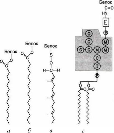
Figure 41 - Lipophilic anchors: a - myristoyl; b - palmitoyl; c - farnesyl; d - GPI anchor. Gray circles designate: I - Inositol; M - mannose; C - N-acetylglucosamine; G - galactose; E - ethanolamine
Depending on the type of lipophilic anchors employed, lipid-linked proteins are divided into three groups.
1. Cytosolic Proteins of the first group attach a fatty acid acyl group (e.g., myristate or palmitate) to the Glycine (Gly) residue at the N-terminus of the protein chain, which in turn embeds into the lipid monolayer on the cytosolic side of the plasma membrane (Figure 42, acylation). The binding of first-group proteins can play a crucial role in their function. For instance, the v-Src protein, a mutant form of Tyrosine kinase, becomes an oncogenic protein and can transform a cell only if its N-terminus is myristylated (acylated).
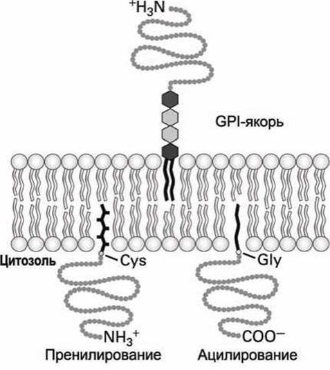
Figure 42 - Binding of Proteins to the membrane via lipid anchors
2. Cytosolic proteins of the second group are anchored to the cytosolic side of the membrane via an unsaturated fatty acid acyl group (an isoprenoid chain) attached to a Cysteine (Cys) residue at or near the C-terminus of the protein (Figure 39, prenylation).
In these proteins, farnesol (C15) or geranylgeraniol (C20) is linked via a thioether bond to the thiol (-SH) group of cysteine. These prenyl anchors are synthesized from isoprene.
In some cases, to strengthen the attachment of the protein to the membrane, a second geranylgeranyl or palmitate group is attached to an adjacent cysteine residue of the protein. Such a dual anchor, for example, retains the signaling GTPase protein Ras on the cytosolic side of the plasma membrane. Another example of utilizing a dual anchor is the binding of the GTPase protein Rab—which is involved in membrane fusion—to the cytosolic side of intracellular vesicle membranes.
3. Certain cell surface proteins and heavily glycosylated Extracellular matrix Proteoglycans bind to the exoplasmic face of the plasma membrane via a third type of lipophilic anchor: glycosylphosphatidylinositol (the GPI anchor) (Figure 42). The exact Structure OF THE GPI anchor varies widely across different cell types, but it invariably contains phosphatidylinositol (PI), whose two fatty acid tails embed into the membrane, phosphoethanolamine, which covalently attaches the anchor to the C-terminus of the protein, and several sugar residues. An example of GPI anchor Organization is shown in Figure 41.
A series of experiments has demonstrated that the presence of a GPI anchor is both necessary and sufficient for protein binding to the membrane. For instance, the enzyme phospholipase C (Figure 28) cleaves the phosphate-glycerol bond in both Phospholipids and GPI anchors. Treating cells with phospholipase C leads to the release of such GPI-anchored proteins as Thy-1 and PLAP from The Cell surface (Figure 33).
As noted above, PLAP is concentrated in more ordered membrane microdomains, or lipid rafts, enriched in Sphingolipids and Cholesterol (Figure 33). Although PLAP and other GPI-anchored proteins reside on the opposite side of the membrane relative to acyl-anchored proteins, both types of Membrane Proteins concentrate in lipid rafts. By contrast, prenylated proteins are not found in lipid rafts.
Asymmetry of integral proteins and Glycolipids. Lipid-anchored proteins provide one example of membrane proteins that are asymmetrically localized relative to the two sides (cytosolic and exoplasmic) of The cell membrane.
Each class of transmembrane proteins also exhibits a specific orientation with respect to the membrane surfaces: certain PARTS OF THE protein molecule are always oriented toward the Cytosol, whereas others are always directed toward the exoplasmic space. Such asymmetry in membrane orientation determines the distinct properties of a membrane protein on opposite sides of the membrane.
To date, not a single instance of flip-flop for integral proteins has been recorded. Therefore, the asymmetry of a membrane protein established immediately upon its synthesis on Endoplasmic reticulum membranes persists throughout the entire lifetime of that protein.
Many proteins contain carbohydrate chains covalently linked to Serine, Threonine, or asparagine residues. Such Glycoproteins are oriented such that their carbohydrate chains are always located on the exoplasmic side (Figures 35 and 36).
Similarly, glycolipids—in which carbohydrate chains are attached to glycerol or sphingosine backbones—are always situated with their carbohydrate chains on the exoplasmic face of the membrane, protruding outward into the extracellular environment.
Glycoproteins and glycolipids are particularly abundant in the plasma membrane of eukaryotic cells. However, they are absent from The inner mitochondrial membrane, chloroplast lamellae, and certain other intracellular membranes.
Because the carbohydrate chains of plasma membrane glycoproteins and glycolipids reside in the exoplasmic space, they can interact with extracellular matrix components, Lectins (phytoagglutinins), growth factors, Antibodies, and other extracellular entities.
Interaction with the Cytoskeleton. It has been demonstrated experimentally (see, for example, Figure 21) that many integral and lipid-linked proteins actively participate in rapid lateral diffusion along the membrane. Depending on the cell type, 30% to 90% of all integral proteins diffuse freely within the plasma membrane.
The rate of lateral diffusion for proteins in a pure lipid bilayer is comparable to that of lipid diffusion. However, the diffusion rate of proteins in the plasma membrane of a living cell is typically 10 to 30 times lower than the diffusion rate of the same proteins reconstituted in an artificial liposome.
This reduction in the diffusion rate of membrane proteins in living cells is attributed to their interactions with the underlying submembranous cytoskeleton. Some proteins are permanently bound to this cytoskeletal network, rendering them completely immobile within the membrane. As for mobile membrane proteins, their movement is hindered by the continuous process of breaking and reforming weak non-covalent bonds with the underlying cytoskeleton as the proteins diffuse along the membrane.
Last update: 13/08/2026
Editorial and Educational Adaptation: This material has been compiled based on the primary/original source text. The project team performed an editorial review, corrected technical inaccuracies, structured sections, and adapted the content for an educational format.
What was processed:
- elimination of formatting defects (OCR errors, structural breaks, corrupted characters);
- editorial organization of content;
- standardization of terminology in accordance with academic sources;
- verification of factual statements against the original source text.
All mentions of the author, publication year, and origin of the primary text have been preserved in accordance with the source.