Molecular Biology of the Cell - Volume 1 - Alberts B., Bray D., Lewis J., Raff M., Roberts K., Watson J. 1994
Molecular Organization of Cells
Energy Conversion: Mitochondria and Chloroplasts
Mitochondrial and Chloroplast Genomes
As Cells grow and divide, new Organelles must form within their Cytoplasm. Even in non-dividing cells, a continuous turnover of organelles takes place, with new ones replacing those that degrade. This requires the regulated synthesis of necessary Proteins and Lipids, followed by the precise delivery of each component to the correct site within the organelle. Chapter 8 already discussed the import of specific proteins and lipids—synthesized outside the organelles—into Cell/35.html">Mitochondria and METABOLISM/14.html">Chloroplasts; here, we focus on THE CONTRIBUTION OF these organelles to their own Biosynthesis.
Two distinct genetic systems participate in The biosynthesis of mitochondrial and chloroplast proteins. Although the majority of these proteins are encoded by nuclear DNA and imported into the organelle after being synthesized on cytosolic Ribosomes, certain proteins are encoded by the organelle's own DNA and synthesized on ribosomes located inside the organelle itself. Protein traffic appears to be strictly unidirectional—from the Cytosol into the organelles; at least, no proteins are known to be exported from mitochondria or chloroplasts into the cytosol.
Class="center">
Fig. 7-64. General scheme of Protein Synthesis for proteins localized in mitochondria and chloroplasts. Bold arrows indicate the sites of action of inhibitors that specifically block protein synthesis either in the mitochondria or in the cytosol.
The participation of the two genetic systems in The formation of mitochondria and chloroplasts is tightly coordinated (Section 7.5.12). However, this coordination is not absolute, and isolated organelles continue to synthesize DNA, RNA, and proteins in vitro for some time, which makes it possible to determine which proteins are encoded by the organelle's own DNA versus nuclear DNA. Another approach involves studying the effects of specific inhibitors on intact cells. For example, cycloheximide inhibits Protein synthesis in the cytosol but does not affect protein synthesis in mitochondria and chloroplasts. Conversely, certain Other Antibiotics, such as chloramphenicol, tetracycline, and erythromycin, suppress protein synthesis in energy-transducing organelles without having any noticeable effect on cytosolic protein synthesis (Fig. 7-64). Such inhibitors are widely used to study mitochondrial and chloroplast Functions.
7-33
7.5.1. The number of mitochondria and chloroplasts in a cell is maintained by their division [46]
Mitochondria and chloroplasts never arise de novo; they are always formed by the division of pre-existing organelles. Observations of living cells show that mitochondria not only divide but can also fuse with one another. On average, however, each organelle must double its mass and then divide in half once per cell generation. Electron micrographs suggest that mitochondrial division begins with the formation of a circumferential constriction in the inner membrane, much like the process seen in many bacterial cells (Figs. 7-65 and 7-66); thus, mitochondrial division appears to be a controlled process rather than a random splitting in two.
In most cells, energy-converting organelles divide throughout interphase; thus, each divides independently of the others and of The Cell as a whole. Similarly, organellar DNA Replication occurs not only during the period of nuclear DNA Synthesis (S phase) but throughout other Phases of the Cell Cycle as well. Although individual DNA molecules apparently replicate at random (so that some may double several times in a given cell cycle while others do not at all), their total number doubles every cell cycle, maintaining a constant amount of this DNA within the cell.
The number of energy-transducing organelles can be regulated According to the cell's energy demands; for instance, a significant increase (5- to 10-fold) in the number of mitochondria is observed in Skeletal Muscle after prolonged, repetitive contraction. Moreover, in some cases, organelle division is directly controlled by the cell: for example, the chloroplasts of certain Algae containing only one or a few such organelles divide immediately before cytokinesis, in the exact plane where the subsequent Cell Division will occur (Fig. 7-67). However, the molecular mechanisms regulating these processes remain poorly understood.
7.5.2. In most cases, chloroplast and Mitochondrial Genomes consist of circular DNA molecules [47]
Organellar DNA molecules are relatively simple, small, and (with the exception of mitochondrial genomes in certain Algae and Protozoa) closed circles. Chloroplast Genome sizes are similar across all organisms studied, whereas mitochondrial genomes in plants are much larger than those in animals (Table 7-2). In many organelles, the DNA molecules are comparable in size to viral DNAs. For example, The Genome of mammalian mitochondria is a circular DNA molecule containing about 16,500 Base Pairs (more than 10,000 times smaller than the nuclear genome). In animals as diverse as Drosophila and sea urchins, Mitochondrial DNA sizes are nearly identical (Fig. 7-68). In plants, however, the circular Mitochondrial Genome is 150 to 2,500 times larger, depending on the species. The largest of these mitochondrial DNA molecules is about half the size of a bacterial circular DNA.
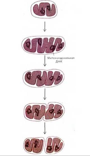
Fig. 7-65. Diagram of mitochondrial division. The sequence of events shown is inferred from static images, such as the micrograph in Fig. 7-66.
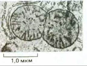
Fig. 7-66. Electron micrograph of a dividing mitochondrion from a Liver cell. (Courtesy of Daniel S. Friend.)

Fig. 7-67. In the primitive filamentous alga Klebsormidium, chloroplast division occurs at a specific time in early mitosis. The cell contains a single chloroplast, and its plane of division coincides with the plane of subsequent cell Cleavage. (From J. D. Pickett-Heaps, Cytobios, 1972, 6, 167-183.)
All mitochondria and chloroplasts contain multiple copies of their genomic DNA (Table 7-3). These DNA molecules are typically organized into discrete clusters within the mitochondrial matrix and chloroplast stroma, where they are believed to be attached to the inner membrane. Although the exact packaging mechanism of this DNA is unknown, its Structure resembles a bacterial genome rather than eukaryotic Chromatin. For example, like Bacteria, they lack Histones entirely.
In mammalian cells, mitochondrial DNA accounts for less than 1% of total cellular DNA. However, in other cells (such as higher plant leaves or very large amphibian eggs), the fraction of DNA belonging to energy-transducing organelles can be much higher (Table 7-3); in these cells, a major share of total cellular RNA and Protein synthesis also takes place within the organelles.
Table 7-2. Sizes of organellar genomes1)
|
DNA Type |
Size in kilobase pairs |
|
|
Chloroplast DNA |
||
|
Higher plants |
120-200 |
|
|
Chlamydomonas (green alga) |
180 |
|
|
Mitochondrial DNA |
||
|
Animals (including Flatworms, insects, and mammals) |
16-19 |
|
|
Higher plants |
150-2500 |
|
|
Schizosaccharomyces pombe (Yeast) |
17 |
|
|
Aspergillus nidulans |
32 |
|
|
Neurospora crassa |
60 |
|
|
Saccharomyces cerevisiae (yeast) |
78 |
|
|
Chlamydomonas (green alga) |
16 (linear molecule) |
|
|
Protozoa |
||
|
Trypanosoma brucei |
22 |
|
|
Paramecium |
40 (linear molecule) |
1) These genomes consist of circular DNA molecules unless otherwise stated.

Fig. 7-68. Electron micrograph of a replicating circular DNA molecule from a mammalian mitochondrion. Only the region between the two arrowheads has replicated so far (strands highlighted in white). (Courtesy of David Clayton.)
Table 7-3. Relative amount of organelle DNA in some Cells and Tissues
|
Organisms |
Tissue or cell type |
Number of DNA molecules per 1 organelle |
Number of organelles per cell |
Share of organelle DNA in total cellular DNA, % |
|
Mitochondrial DNA |
||||
|
Rat |
Liver |
5-10 |
1000 |
1 |
|
Mouse |
L-cell culture |
5-10 |
100 |
<1 |
|
Yeast * |
Vegetative |
2-50 |
1-50 |
15 |
|
Frog |
cells Egg cell |
5-10 |
107 |
99 |
|
Chloroplast DNA |
||||
|
Chlamydomonas |
Vegetative cells |
80 |
1 |
7 |
|
Corn |
Leaves |
20-40 |
20-40 |
15 |
7.5.3. Mitochondria and Chloroplasts Possess a Complete Genetic System [48]
Despite the small number of proteins encoded by mitochondrial and chloroplast genes, these organelles replicate and transcribe their DNA and carry out protein synthesis. These processes take place in the mitochondrial matrix and chloroplast stroma. Although the proteins involved in all these processes are organelle-specific, the majority of them are encoded by the nuclear genome (Section 7.5.17). This is all the more surprising given that the entire protein-synthesis machinery in organelles resembles that of bacteria rather than eukaryotes. In chloroplasts, this similarity is particularly striking:
1. Chloroplast ribosomes closely resemble E. coli ribosomes both in their sensitivity to various antibiotics (chloramphenicol, streptomycin, erythromycin, tetracycline, etc.) and in their structure. Not only are The nucleotide sequences of chloroplast and E. coli Ribosomal RNAs strikingly similar, but chloroplast ribosomes are also capable of using bacterial tRNAs in protein synthesis. In all these respects, chloroplast ribosomes differ from those located in The plant cell cytosol.
2. Protein synthesis in chloroplasts begins with N-formylmethionine, just as in bacteria, rather than with Methionine as in the cytosol of eukaryotic
cells.
3. Unlike nuclear DNA, chloroplast DNA can be transcribed using E. coli RNA polymerase to yield chloroplast mRNAs that are efficiently translated by the E. coli protein-synthesizing system.
Although mitochondrial genetic systems are much less similar to those of modern bacteria than are chloroplast systems, mitochondrial ribosomes are also sensitive to antibacterial antibiotics, and mitochondrial protein synthesis begins with N-formylmethionine.
7.5.4. The Chloroplast Genome of Higher Plants Contains about 120 Genes [49]
Chloroplast genes have been most thoroughly studied in plants and green algae, in which these organelles are very similar. The chloroplast genome is a circular DNA molecule; its complete nucleotide sequence has now been determined in tobacco and a liverwort. The obtained data indicate that the chloroplast genes of these very distantly related higher plants are practically identical. In addition to four ribosomal RNAs, these genomes encode about 20 ribosomal proteins, several subunits of chloroplast RNA polymerase, a few proteins that are part of Photosystems I and II, ATP synthase subunits, parts of Electron Transport Chain enzyme complexes, one of the two subunits of ribulose bisphosphate carboxylase, and 30 tRNAs (Fig. 7-69). Furthermore, the DNA sequence apparently encodes at least 40 more proteins of unknown function. Surprisingly, all known chloroplast-encoded proteins are components of large enzyme complexes that also contain one or more nuclear-encoded subunits. The possible reasons for this will be discussed later (Section 7.5.17).
The similarity between the chloroplast and bacterial genomes is striking. Essential regulatory sequences, such as promoters and Transcription terminators, are virtually identical in both genomes. Chloroplast-encoded Proteins are very similar to bacterial ones, and certain groups of genes with related functions (e.g., those encoding ribosomal proteins) are organized similarly in the genomes of chloroplasts, E. coli, and cyanobacteria.

Fig. 7-69. Organization OF THE chloroplast genome in a liverwort. The complete nucleotide sequence of this genome has been determined. The organization of chloroplast genomes is very similar in all higher plants; the size of the circular DNA molecule varies from species to species depending on the proportion of DNA surrounding the genes encoding the 16S and 23S ribosomal RNAs that is present in two copies.
Tracing the evolutionary pathway from bacteria to chloroplasts will require detailed comparisons of homologous nucleotide sequences, but several Conclusions can already be drawn:
1) higher plant chloroplasts originated from photosynthetic bacteria;
2) the chloroplast genome has remained almost unchanged for at least several hundred million years (roughly the time when the evolutionary lineages of liverworts and tobacco diverged);
3) many of the ancestral bacterial genes can now be identified in the nuclear genome, into which they were transferred and have been preserved to the present day. For instance, although higher plant chloroplast ribosomal proteins are related to bacterial proteins and the ribosomes themselves resemble bacterial ones, two-thirds of the approximately 60 proteins of chloroplast ribosomes are encoded in the Cell Nucleus.
7-34
7.5.5. The Mitochondrial Genome Has Several Striking Features [50]
The chloroplast genome was not the first organellar genome to be completely sequenced. The first was the human mitochondrial genome: its relatively small size made it an especially attractive object for molecular geneticists armed with the latest DNA Sequencing techniques (see Section 4.6.6), and in 1981 the complete sequence of this genome, consisting of 16,569 nucleotide pairs, was published. By comparing it with known tRNA nucleotide sequences and partial Amino acid sequences of proteins encoded by Mitochondrial Genes, the locations of all these genes on the circular DNA molecule were mapped (Fig. 7-70). Compared to nuclear, chloroplast, and bacterial genomes, the human mitochondrial genome possesses several remarkable features:
1) here, unlike in other genomes, virtually every nucleotide is part of a coding sequence either for a protein or for one of the rRNAs or tRNAs. Because these coding sequences abut one another directly, very little space is left for regulatory DNA sequences;
2) whereas the cytosol contains at least 31 tRNAs for various Amino Acids and chloroplasts contain 30 tRNAs, mitochondria use only 22 tRNAs to carry out protein synthesis. In mitochondria, the conventional codon-anticodon pairing rules are observed less strictly, and many tRNA molecules are capable of recognizing any of the four NUCLEOTIDES at the third (wobble) position (Section 5.1.6). This "two-out-of-three" reading mechanism enables a single tRNA to bind to any of four different codons, making it possible to get by with fewer tRNAs during protein synthesis;
3) comparison of the nucleotide sequences of mitochondrial genes with The amino acid sequences of proteins has revealed perhaps the most striking feature: The Genetic Code in mitochondria is modified, and the meanings of four out of the 64 codons differ from those in other genomes (Table 7-4).
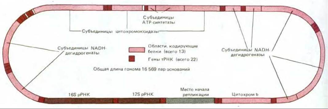
Fig. 7-70. Organization of the human mitochondrial genome, determined by complete DNA sequencing. The genome contains two rRNA genes, 22 tRNA genes, and 13 protein-coding regions. The complete mitochondrial DNA sequences of the cow and mouse have also been determined; they contain the same genes and exhibit a similar organization.
Table 7-4. Differences between the "universal" code and mitochondrial genetic codes*
|
Codon |
"Universal" code |
Mitochondrial codes |
|||
|
Mammals |
Drosophila |
Yeast |
Plants |
||
|
UGA |
STOP |
Trp |
Trp |
Trp |
STOP |
|
AUA |
Ile |
Met |
Met |
Met |
Ile |
|
CUA AGA |
Leu |
Leu |
Leu |
Thr |
Leu |
|
AGG |
Arg |
STOP |
Ser |
Arg |
Arg |
* Codon meanings that differ from the "universal" code are highlighted in italics and color.
The near-universal identity of the genetic code across all organisms serves as compelling evidence that all cells evolved from a common ancestor. How, then, can we explain the distinct deviations found in the mitochondrial genetic code? Recent data on the variations in mitochondrial genetic codes among different organisms have shed light on this question. For instance, the triplet UGA, which functions as a stop codon in the universal code, encodes Tryptophan in the mitochondria of mammals, fungi, and protozoa, whereas it serves as a stop codon in plant mitochondria. Similarly, the triplet AGG, which typically encodes Arginine, acts as a "stop" signal in mammalian mitochondria and encodes Serine in Drosophila (Table 7-4). Such exceptions indicate that occasional alterations can occur in the mitochondrial genetic code. The emergence and fixation of these random changes in codon meaning are likely permitted by the exceptionally small number of proteins encoded by the mitochondrial genome; in a large genome, such modifications would disrupt the function of many proteins and, consequently, lead to cell death.
7.5.6. Animal mitochondria possess the simplest known genetic systems [52]
A comparison of DNA sequences among various organisms reveals that The rate of nucleotide substitution in the mitochondrial genome during evolution is roughly 10 times higher than that in The Nucleus. This is presumably due to a lower fidelity of mitochondrial DNA replication, repair, or both. Because all RNAs and proteins in an animal cell are produced via the replication and expression of a DNA sequence consisting of only about 16,500 nucleotides, the probability of error per read nucleotide during DNA Replication and Repair, RNA polymerase-mediated transcription, and Translation at the mitochondrial ribosome can remain relatively high without impairing the organelle. This likely explains why the mechanisms underlying these processes are relatively simple compared to those employed by the cell for the same purposes outside the organelles. For example, it has been hypothesized (though not yet definitively proven) that the presence of only 22 tRNAs and the unusually small size of rRNAs (less than two-thirds of their size in E. coli) reduce The fidelity of protein synthesis in mitochondria.
Owing to the relatively Rapid Evolution of mitochondrial genes, sequence comparisons of their DNA can be particularly useful for dating relatively recent events, such as the stages of primate evolution (see Section 1.2.2).
7-35
7.5.7. Why do plants have such a large mitochondrial genome? [52]
The mitochondrial genome in plants is significantly larger than that in animal cells, with mitochondrial DNA content varying widely—roughly from 150,000 to 2.5 million base pairs. Nevertheless, the plant mitochondrial genome apparently encodes little more proteins than that of animals. Furthermore, within a single plant family (such as the Cucurbitaceae), mitochondrial genome sizes can vary sevenfold; meanwhile, the linear mitochondrial genome of the green alga Chlamydomonas is comparable in size to that of animal cells, measuring 16,000 base pairs.
Very little is known about the nucleotide sequences of higher plant mitochondrial DNA, but the large (78,000 bp) mitochondrial genome of the yeast Saccharomyces cerevisiae has been almost completely sequenced, revealing that only about one-third of it codes for proteins. These findings suggest that much of the "excess" DNA in yeast mitochondria (and potentially in higher plants as well) may not be essential for the Organism.
7.5.8. Some organelle genes contain introns [53]
In both thoroughly studied mitochondrial systems—human and yeast—the Processing of precursor RNAs plays a crucial role. In human cells, both strands of mitochondrial DNA are transcribed at equal rates from a single promoter on each strand, yielding two distinct giant RNA molecules, each representing a complete copy of One DNA strand. Thus, transcription is entirely symmetrical. The RNA molecule transcribed from one of the DNA strands—designated the heavy strand (H-strand) due to its high density in a CsCl gradient—is cleaved by Nucleases to generate two rRNA molecules, the majority of mitochondrial tRNAs, and about a dozen poly(A)-containing RNAs. In contrast, Processing of the RNA transcribed from the light strand (L-strand) produces only eight tRNAs and one small poly(A)-containing RNA. The remaining 90% of this transcript apparently carries no useful information (being complementary to the coding sequence synthesized on the opposite strand) and is degraded. The poly(A)-containing RNAs are believed to represent mitochondrial mRNAs; they lack a 5' cap, and their 3' poly(A) region contains about 55 nucleotides. This polynucleotide tail is added post-transcriptionally through the action of mitochondrial poly(A) polymerase.
Unlike humans, certain plants and fungi (including Yeasts) harbor introns within their mitochondrial genes, which must be excised from the transcript followed by splicing (Section 3.2.7). In plants, introns have also been found in approximately 20 chloroplast genes. Many introns in organelle genes contain related nucleotide sequences that can self- excise from RNA transcripts via an RNA-catalyzed reaction (Section 9.4.14), although proteins typically assist in this "self-splicing". The discovery of introns in organelle genes was unexpected from the perspective of the endosymbiotic theory of energy-converting organelle origins, given that introns are absent from The genes of bacteria, the presumptive ancestors of mitochondria and chloroplasts.
In yeast, introns may be present in the mitochondrial Gene of one strain yet absent from the same gene in another strain. Such "facultative" introns are apparently capable of entering and leaving genes much like Transposons. Conversely, in certain yeast mitochondrial genes, introns occupy the exact same positions as those in the mitochondria of Aspergillus and Neurospora, indicating that they were inherited from a common ancestor of these three fungi. Introns are likely of ancient origin; although lost in many bacteria, they have been conserved in the genomes of those organelles where The regulation of RNA splicing helps control Gene Expression (Section 10.5.5).
7-36
7.5.9. Non-Mendelian (cytoplasmic) inheritance of mitochondrial genes distinguishes them from nuclear genes [54]
For several reasons, most experimental studies on the mechanisms of mitochondrial biogenesis are performed using cultures of Saccharomyces carlsbergensis (brewer's yeast) and S. cerevisiae (baker's yeast). First, when grown on glucose, these yeasts exhibit The unique ability to survive solely via Glycolysis, thus functioning independently of active mitochondria, i.e., without Oxidative Phosphorylation. This makes it possible to work with cells whose mitochondrial and nuclear DNAs carry Mutations that would otherwise impair normal mitochondrial development—mutations that are lethal in virtually all other organisms. Second, yeasts are simple, Unicellular Eukaryotes that are easily cultivated and biochemically analyzed. Finally, yeasts—which typically reproduce asexually by budding (asymmetric mitosis)—also undergo a sexual cycle. During sexual reproduction, two haploid cells fuse to form a diploid zygote, which then either divides mitotically or undergoes Meiosis to yield haploid cells once again. The ability to control the alternation of asexual and Sexual reproduction in the laboratory (Section 13.2) greatly facilitates genetic analysis. Such analysis makes it possible to identify genes responsible for mitochondrial function and determine whether they reside in nuclear or mitochondrial DNA, since mutations in mitochondrial genes do not obey the Mendelian laws that govern nuclear gene inheritance.

Fig. 7-71. Difference in the inheritance patterns of mitochondrial and nuclear genes in yeast. Two of the four yeast cells resulting from meiosis inherit a given nuclear gene from one haploid parent cell, and the other two inherit it from the other parent (Mendelian inheritance). By contrast, through gradual mitotic segregation of mitochondria during vegetative growth (see text), it may well happen that all four meiotic cells receive mitochondrial genes from only one of the two haploid parents (non-Mendelian or CYTOPLASMIC INHERITANCE). In this example, a mitochondrial gene mutation confers resistance to chloramphenicol—an inhibitor of protein synthesis in energy-converting organelles and bacteria (Section 5.1.15).
Fig. 7-71 illustrates an example of non-Mendelian (cytoplasmic) inheritance of mitochondrial genes in the progeny of haploid yeast cells. The mutant gene confers resistance to chloramphenicol upon the mitochondrial protein-synthesizing system; yeast cells carrying this mutation can be readily identified by cultivating them in the presence of chloramphenicol on a carbon source such as glycerol, which cannot be utilized for glycolysis. Under conditions where glycolysis is blocked, ATP must be supplied exclusively by functionally active mitochondria; therefore, only cells possessing chloramphenicol-resistant mitochondria are able to grow on such medium. When a chloramphenicol-resistant haploid cell fuses with a wild-type haploid cell sensitive to the antibiotic, a diploid zygote is formed containing a mixture of both mutant and wild-type mitochondria. However, if a diploid daughter cell buds off from the zygote via mitosis, it receives only a small fraction of the mitochondrial population. After several mitotic cycles, all mitochondria in a given newly formed cell may become uniform—either entirely mutant or entirely wild-type. Consequently, all progeny of such a cell will possess genetically identical mitochondria. This stochastic process, which generates diploid offspring containing only a single type of mitochondrial DNA, is termed mitotic segregation. When a diploid cell carrying a single mitochondrial type undergoes meiosis, all four daughter haploid cells inherit identical mitochondrial genes. This inheritance pattern is termed non-Mendelian or cytoplasmic, in contrast to the Mendelian inheritance of nuclear genes (Fig. 7-71); it indicates that the gene in question resides outside the nuclear Chromosomes, presumably within cytoplasmic organelles.
7.5.10. In many organisms, organelle genes are maternally inherited [55]
For certain organisms, including humans, the consequences of cytoplasmic gene transmission are far more significant than for yeast. In yeast, the two fusing haploid cells are of equal size and contribute equivalent amounts of mitochondrial DNA to the zygote (Fig. 7-71). Thus, in yeast, the mitochondrial genome is inherited from both parents, with each making an equal genetic contribution to the progeny (although, as we have seen, after several generations individual descendants often end up containing mitochondria from only one parental type). By contrast, in higher animals, the egg cell contributes vastly more cytoplasm to the zygote than the sperm, and in some animals sperm may contribute no cytoplasm at all. Consequently, one would expect the mitochondrial genome in higher animals to be transmitted exclusively from a single parent—specifically, via the maternal Lineage. This has been confirmed in experiments with laboratory animal strains differing in their mitochondrial DNA types. When animals carrying type A mitochondrial DNA are crossed with animals carrying type B, the resulting offspring contain mitochondrial DNA exclusively of the maternal type. Similarly, tracking the distribution of various mitochondrial DNA sequences in large human families demonstrates that human mitochondrial DNA is also inherited maternally.
In about two-thirds of higher plants, the chloroplasts of the male parent (contained within pollen grains) fail to enter the zygote; thus, chloroplast DNA, much like mitochondrial DNA, is inherited maternally. In other plants, defective chloroplasts cause variegation: mitotic segregation during Plant GROWTH AND DEVELOPMENT sorts out the mixture of normal and defective chloroplasts, leading to the formation of leaves with alternating green and white patches—the green patches contain normal chloroplasts, whereas the white patches contain defective ones.
7.5.11. As studies of "petite" yeast mutants demonstrate, the cell nucleus plays a crucial role in mitochondrial biogenesis
Genetic studies in yeast have played a pivotal role in analyzing mitochondrial biogenesis. A striking example is The Study of mutants with extensive deletions in mitochondrial DNA, which leads to a complete cessation of protein synthesis within mitochondria. It is hardly surprising, therefore, that such mutants lack "respiring" mitochondria. A rare but important group of these mutants completely lacks mitochondrial DNA. Because such mutants form unusually small colonies when grown on low-glucose media, all mutants with defective mitochondria are referred to as cytoplasmic petite mutants.
Fig. 7-72. Cytology/cytology/93.html">ELECTRON MICROGRAPHS OF thin sections of yeast cells, showing The structure of normal mitochondria (A) and mitochondria from a petite mutant, which lacks all proteins encoded by the mitochondrial genome (B). In the latter case, the organelle consists exclusively of proteins encoded by the nuclear genome. (Courtesy of Barbara Stevens.)
Although petite mutants lack mitochondrial PROTEIN SYNTHESIS AND thus cannot form ATP-synthesizing mitochondria, they nevertheless possess mitochondria with a normal outer membrane, but with poorly developed inner membrane cristae (Fig. 7-72). Such mitochondria contain virtually all mitochondrial proteins encoded by the nuclear genome and imported into the organelle from the cytosol, including DNA and RNA polymerases, all Enzymes of The Citric Acid Cycle, and the majority of inner Membrane Proteins. This clearly demonstrates the predominant role of the nuclear genome in mitochondrial biogenesis. Furthermore, it is evident that organelles capable of binary fission can reproduce indefinitely in the cytoplasm of proliferating Eukaryotic cells, even in the complete absence of their own genome. Many biologists believe that Peroxisomes normally reproduce in the same manner (Section 8.5.2).
As for chloroplasts, the closest analogues to yeast mitochondrial petite mutants are mutants of unicellular algae such as *Euglena*. Cells that lack chloroplast protein synthesis still contain chloroplasts and are fully viable in the presence of oxidizable substrates. However, if The Development of mature chloroplasts in higher plants is blocked due to the absence of light (Section 20.4.1), defects in their DNA, or its complete absence, such plants die as soon as their nutrient reserves are exhausted.
7-37
7.5.12. The formation of mitochondria and chloroplasts is regulated by nuclear-encoded proteins [57]
The genetic systems of the nucleus and organelles must coordinate their contributions to the construction of mitochondria and chloroplasts. Overall control is undoubtedly exerted by the nucleus, since in mutants with blocked organellar protein synthesis, mitochondria and chloroplasts are formed in normal amounts, albeit with impaired function. In some of these functionally defective organelles, DNA and partial RNA Synthesis continue, implying that all proteins required for these processes are encoded by nuclear genes.
The nucleus must regulate the number of mitochondria and chloroplasts to match cellular demand; it must also control The amount of proteins synthesized on ribosomes within the organelles to maintain a proper balance between nuclear and organellar contributions to mitochondrial and chloroplast biogenesis. Although these regulatory aspects are crucial for understanding Introduction/5.html">Eukaryotic Cell Homeostasis, our knowledge in this area remains insufficient.
Nuclear regulation of mitochondrial protein synthesis has been intensively studied using yeast mutants. In *Saccharomyces cerevisiae*, numerous mutants have been isolated with alterations in the nuclear (as well as mitochondrial — see Section 7.5.9) genome that are unable to form respiring mitochondria. Each of these nuclear petite mutants carries a single defective protein encoded by nuclear DNA and required for mitochondrial function. By growing yeast cultures in media containing labeled amino acids and cycloheximide (which inhibits the synthesis of nuclear-encoded proteins), one can determine The Effect of each such nuclear mutation on the expression of mitochondrial genes. As it turned out, mutations in nuclear genes encoding mitochondrial proteins directly associated with respiratory function (such as a subunit of ATP synthase or an enzyme of The Citric Acid cycle) have no effect on mitochondrial protein synthesis, as expected. Conversely, mutations in nuclear genes encoding mitochondrial ribosomal proteins or subunits of mitochondrial RNA polymerase block the synthesis of all proteins within the mitochondria.
Of greatest relevance to regulatory processes is a third group of nuclear petite mutants, which lack or have altered one or more gene products encoded by mitochondrial DNA. More than 50 such genes have been identified in the yeast nucleus, and some of them, required for the expression of specific mitochondrial genes, have already been cloned and characterized. Some of these genes encode proteins that apparently act directly on a specific mRNA molecule, enhancing either its stability or the efficiency of its translation in mitochondrial protein synthesis. The products of other genes are involved in mitochondrial RNA splicing and are therefore necessary for the expression of mitochondrial genes containing introns. Both types of nuclear genes are thought to participate in regulating the functions of mitochondrially encoded proteins according to the metabolic needs of the cell, though the exact mechanisms of this regulation remain unknown.
Although the nucleus plays the primary role, evidence suggests that the interaction between the nuclear and mitochondrial genetic systems is bidirectional. For example, if mitochondrial Protein synthesis is blocked in an intact cell, there is an upregulation of imported enzymes involved in the synthesis of mitochondrial DNA, RNA, and proteins, as if the cell were attempting to overcome the blockade. The Nature of the signal sent from the mitochondria to the nucleus remains to be elucidated.
7.5.13. Energy-transducing organelles contain tissue-specific proteins [58]
The cell also regulates mitochondrial functions through more conventional mechanisms. In mammals, The Urea Cycle serves as the principal metabolic pathway for processing nitrogenous waste products. The resulting urea is excreted in urine. Nuclear-encoded enzymes catalyze several steps of this cycle within the mitochondrial matrix. Urea is produced only in specific Organs, such as the liver, and the urea cycle enzymes are synthesized and imported into mitochondria exclusively in these organs. In addition, the respiratory enzyme complexes embedded in The inner mitochondrial membrane of mammals contain several tissue-specific subunits that are encoded by the nucleus and presumably act as regulators of electron transport. For instance, in some individuals with an inherited muscle disorder, a specific cytochrome c oxidase subunit is defective; because this subunit is specific to skeletal muscle, The Heart muscle fibers in these patients function normally, allowing them to survive. As expected, tissue-specific differences are also characteristic of nuclear-encoded chloroplast proteins.
Let us now examine how specific cytoplasmic proteins are imported into mitochondria and chloroplasts; this topic is discussed in greater detail in Chapter 8.
7.5.14. The import of proteins into mitochondria and chloroplasts requires energy [59]
Most of the proteins found in mitochondria and chloroplasts are imported into these organelles from the cytosol (Section 8.4). This raises two questions: how does the cell direct proteins to the correct organelle, and how do these proteins cross its membranes?
A partial answer came from studies on The transport of the small subunit (S) of ribulose-1,5-bisphosphate carboxylase into the chloroplast stroma. When mRNA isolated from the cytoplasm of the unicellular alga *Chlamydomonas* or from pea leaves is translated in an in vitro protein-synthesizing system, one of the many products is a precursor to the S protein, termed pro-S, which is larger than S by 50 amino acid residues. When pro-S is incubated with intact chloroplasts, it penetrates the organelles and is converted into the mature S protein by an endopeptidase. This S protein then associates with the large subunit of ribulose-1,5-bisphosphate carboxylase synthesized on chloroplast ribosomes, forming an active enzyme within the stroma. The import of pro-S into the chloroplast, as expected for processes of this type, requires energy provided by ATP Hydrolysis (Section 8.4.7).
Protein transport into mitochondria operates in a similar manner. If purified yeast mitochondria are incubated with a cell extract containing newly synthesized radioactive yeast proteins, one can observe that nuclear-encoded mitochondrial proteins are selectively incorporated into mitochondria—in exactly the same way as in an intact cell. Furthermore, Proteins of the outer and inner membranes, matrix, and intermembrane space all find their way to their proper mitochondrial compartments (see Fig. 8-30).
Protein Transport across mitochondrial and chloroplast membranes apparently occurs at specialized contact sites where the inner and outer membranes are closely apposed (Fig. 7-73). Proteins arrive at these sites in the form of precursors containing a specific signal peptide. For the transported protein to be translocated into the organelle at such a site, its polypeptide chain must unfold (see Section 8.4.4).
7.5.15. Chloroplasts synthesize most of their own lipids, whereas mitochondria largely obtain theirs from the cytosol [60]
In addition to Nucleic Acids and Proteins, the construction of new mitochondria and chloroplasts requires lipids. All lipids necessary for chloroplasts are typically synthesized within the organelles themselves. In spinach leaves, for example, the synthesis of all cellular Fatty acids occurs within the chloroplasts, and only the introduction of double bonds into their molecules takes place elsewhere. Even the major chloroplast Glycolipids are synthesized endogenously.
In contrast, mitochondria obtain the majority of their lipids from external sources. In animal cells, the Phospholipids phosphatidylcholine and phosphatidylserine are synthesized in the Endoplasmic reticulum and subsequently transferred to the outer mitochondrial membrane. It is believed (though not yet definitively proven) that specialized transfer proteins mediate this process (Section 8.6.15), after which the lipids are incorporated into the inner membrane—presumably at membrane contact sites. Aside from decarboxylating imported phosphatidylserine to phosphatidylethanolamine, mitochondria themselves catalyze The conversion of imported lipids into cardiolipin. Cardiolipin is a "double" phospholipid containing four fatty acid chains; this lipid is located primarily in the inner mitochondrial membrane, where it accounts for about 20% of the total lipid content.

Fig. 7-73. Contact sites. A. Schematic drawing of a small portion of a mitochondrion or chloroplast showing a membrane contact region. Such regions are thought to be involved in the selective import of proteins into the organelle. Contact sites (also referred to as contact zones) have recently been isolated, and their specific protein components are currently under investigation. Proteins encoded in the cell nucleus and synthesized in the cytosol are translocated across these zones. B. Electron micrograph of a small region of a pea chloroplast in which the contact region (indicated by arrows) is labeled with antibody-gold conjugates, which are believed to bind to an integral membrane protein involved in protein transport. (From D. Pain, J. S. Kanwar, G. Blobel, Nature, 331: 232-237, 1988.)
7-38
7.5.16. Mitochondria and Chloroplasts Likely Evolved from Endosymbiotic Bacteria [61]
As discussed in Chapter 1, the "prokaryotic" Nature of the organelle genetic system—most prominently seen in chloroplasts—suggests that mitochondria and chloroplasts originated from bacteria once engulfed via endocytosis. According to this endosymbiotic theory, early eukaryotic cells were anaerobic organisms lacking mitochondria and chloroplasts, which subsequently established a stable Symbiosis with bacteria and co-opted their oxidative phosphorylation machinery for their own metabolic needs (Fig. 7-74). The event leading to The Emergence of mitochondria is estimated to have occurred 1.5 billion years ago, following a significant rise in atmospheric oxygen and prior to the divergence of animal and plant lineages (see Fig. 7-61). Plant and algal chloroplasts presumably arose later through a secondary endosymbiotic event, when ancestral cells engulfed oxygen-producing photosynthetic bacteria. It is generally assumed that at least three independent events of this kind took place, as this accounts for the differences in pigments and other characteristics observed among modern Higher Plants and green, brown, and red algae (see Fig. 7-62).

Fig. 7-74. Proposed evolutionary origin of mitochondria (highlighted in color). While it is sometimes suggested that all mitochondria share a single common ancestor, mitochondria from evolutionarily distant groups such as trypanosomes and euglenoids (see Fig. 1-16) may have arisen via independent endosymbioses. Microsporidia (Microsporidia, Protozoa)—contemporary anaerobic unicellular eukaryotes inhabiting the intestines of many animals—completely lack mitochondria. Because rRNA nucleotide sequence analysis reveals that these microorganisms are highly divergent from all other known eukaryotes, it is hypothesized that the ancestors of Microsporidia were also anaerobes, closely resembling the eukaryotic host that originally engulfed the mitochondrial ancestor (Section 1.2.6).
Since the majority of genes encoding proteins in modern mitochondria and chloroplasts reside within the nuclear genome, it is widely believed that a substantial portion of the original organellar genes was transferred to nuclear DNA during eukaryotic evolution. This hypothesis helps explain why certain nuclear genes encoding mitochondrial proteins share striking similarities with bacterial genes. For example, in chickens, the N-terminal Amino Acid Sequence of the mitochondrial enzyme superoxide dismutase bears a much closer resemblance to the corresponding segment of bacterial superoxide dismutase than to the N-terminal region of the same enzyme isolated from the cytosol of the same eukaryotic cells. Further evidence for such gene transfers during evolution comes from the discovery of noncoding DNA sequences within the nuclear genome that appear to be of recent mitochondrial origin; these sequences were evidently integrated into the nuclear genome as "junk" DNA.
What type of bacterium gave rise to mitochondria? Complete amino acid sequencing and three-dimensional X-ray crystallographic analysis of cytochrome $c$ types from various bacteria have revealed striking similarities among these proteins, as well as with cytochrome $c$ of the Respiratory Chain in PLANT AND ANIMAL mitochondria. Based on these and other biochemical data, the evolutionary tree shown in Fig. 7-62 was proposed. Mitochondria appear to have descended from a specialized group of purple photosynthetic bacteria that lost their photosynthetic capability while retaining the respiratory chain. However, it remains uncertain whether all mitochondria (and chloroplasts) originated from a single endosymbiotic event. Although protozoan mitochondria exhibit distinct prokaryotic features, some diverge sufficiently from plant and animal mitochondria to suggest independent origins.
7.5.17. Why Do Mitochondria and Chloroplasts Maintain Their Own Genetic Systems? [62]
Why do mitochondria and chloroplasts require their own genetic systems, whereas other organelles such as peroxisomes and Lysosomes do not? This is far from a trivial question, as maintaining a separate genetic apparatus is metabolically costly for the cell: it requires the nuclear genome to encode over 90 specialized proteins, including numerous ribosomal proteins, Aminoacyl-tRNA synthetases, DNA and RNA polymerases, and RNA Processing and modification enzymes (Fig. 7-75). Most studied mitochondrial and chloroplast proteins differ significantly in amino acid sequence from their counterparts elsewhere in the cell, suggesting that very few organellar proteins are shared with other cellular compartments. Consequently, maintaining the genetic system for each type of energy-transducing organelle demands at least 90 extra genes in the nuclear genome. The evolutionary rationale for this apparent "wastefulness" remains unclear, and hopes that the mystery would be solved by sequencing mitochondrial DNA have not been realized. It is difficult to envision why proteins produced within mitochondria must necessarily be synthesized locally rather than in the cytosol.
It was once hypothesized that certain proteins synthesized inside the organelle are too hydrophobic to traverse its membrane from the cytosol. However, subsequent evidence has shown this explanation to be unlikely. In many cases, even highly hydrophobic subunits are synthesized in the cytosol. Furthermore, although individual protein subunits of various mitochondrial enzyme complexes are remarkably conserved throughout evolution, their sites of synthesis can vary. The differing genomic locations of genes encoding functionally equivalent protein subunits across various organisms are difficult to reconcile with any hypothesis postulating clear evolutionary advantages for the genetic systems retained in modern mitochondria and chloroplasts.

Fig. 7-75. Proteins synthesized in the cytosol and subsequently imported into the mitochondrion not only constitute the majority of the organelle's protein mass but also play essential roles in the mitochondrial protein-synthesis machinery itself. Of the components making up this machinery, the mitochondrion itself synthesizes only mRNA, rRNA, and tRNA.
It is possible that the genetic systems of these organelles represent an evolutionary dead end. Within the framework of the Endosymbiotic Hypothesis, this implies that The transfer of endosymbiont genes to the host nuclear genome stalled before completion; in the case of mitochondria, this halt may have resulted from relatively recent alterations in the mitochondrial genetic code. Such changes would likely render any remaining mitochondrial genes functionally inactive if they were transferred to the nucleus.
Summary
The growth and division of mitochondria and chloroplasts are governed by two distinct genetic systems: the organelle's own genome and the nuclear genome. The vast majority of organellar proteins are encoded by nuclear DNA, synthesized in the cytosol, and subsequently imported into the organelle. However, certain mitochondrial and chloroplast proteins, along with all of their RNAs, are encoded by the organelle's own DNA and synthesized locally. The human mitochondrial genome contains approximately 16,500 nucleotide pairs and encodes 2 rRNAs, 22 tRNAs, and 13 distinct polypeptide chains. The chloroplast genome is roughly 10 times larger than the human mitochondrial genome and contains about 120 genes. Nonetheless, the nucleus plays the dominant role in the biogenesis of both organelle types, as evidenced by the fact that even mutants lacking a functional organellar genome still produce partially functional organelles in normal numbers.
Chloroplast ribosomes closely resemble bacterial ribosomes, whereas mitochondrial ribosomes diverge somewhat more from the latter, making the evolutionary origin of mitochondria more difficult to trace. Nevertheless, protein sequence similarities suggest that both types of organelles evolved from bacteria that established a stable symbiotic relationship (as endosymbionts) with primitive eukaryotic cells; mitochondria are thought to have originated from purple bacteria, and chloroplasts (at a later date) from cyanobacteria or closely related organisms. Although many genes from these ancient bacteria are still utilized for organelle-specific protein synthesis, the majority of them have somehow been integrated into the nuclear genome, where they encode enzymes that resemble bacterial counterparts, are synthesized on cytosolic ribosomes, and are subsequently imported into the organelle.
General
Becker W.M. The World of the Cell, pp. 117-284. Menlo Park CA, Benjamin-Cummings, 1986.
Ernster L. ed. Bioenergetics. New York, Elsevier, 1984.
Harold F. M. The Vital Force: A Study of Bioenergetics. New York, W. H. Freeman, 1986.
Lehninger A.L. Principles of Biochemistry, Chapters 16, 17, 23. New York, Worth, 1982.
Nicholls D. G. Bioenergetics: An Introduction to the Chemiosmotic Theory. New York, Academic Press, 1982.
Stryer L. Biochemistry, 3rd ed., Chapters 16, 17 and 22. New York, W. H. Freeman, 1988.
References
1. Ernster L., Schatz G. Mitochondria: a historical review. J. Cell Biol., 91, 227s-255s, 1981.
Fawcett D. W. The Cell, 2nd ed., pp. 410-485. Philadelphia, Saunders, 1981.
Harold F. M. The Vital Force: A Study of Bioenergetics, Chapter 7. New York, W.H. Freeman, 1986.
Tzagoloff A. Mitochondria. New York, Plenum, 1982.
2. DePierre J. W., Ernster L. Enzyme topology of intracellular membranes. Annu. Rev. Biochem., 46, 201-261, 1977.
Srere P. A. The STRUCTURE OF THE mitochondrial inner membrane-matrix compartment. Trends Biochem. Sci., 7, 375-378, 1982.
3. Krstic R. V. ULTRASTRUCTURE OF THE Mammalian Cell, pp. 28-57. New York, Springer-Verlag, 1979.
Pollak J. K., Sutton R. The differentiation of animal mitochondria during development. Trends Biochem. Sci., 5, 23-27, 1980.
4. Geddes R. Glycogen: a metabolic viewpoint. Biosci. Rep., 6, 415-428, 1986.
McGilvery R. W. Biochemistry: A Functional Approach, 3rd ed. Philadelphia, Saunders, 1983.
Newsholme E.A., Start C. Regulation in Metabolism. New York, Wiley, 1973.
5. Baldwin J.E., Krebs H. The evolution of metabolic cycles. Nature, 291, 381-382, 1981.
Krebs H. A. The history of The Tricarboxylic Acid Cycle. Perspect Biol. Med., 14, 154-170, 1970.
Reed I. J., Damuni Z., Merryfleld M. L. Regulation of mammalian Pyruvate and branched-chain a-keto acid dehydrogenase complexes by phosphorylation-dephosphorylation. Curr. Top. Cell Regul., 27, 41-49, 1985. Williamson J. R., H. Cooper R. H. Regulation of the citric acid cycle in mammalian systems. FEBS Lett., Suppl. 117, K73-K85, 1980.
6. Mitchell P. Coupling of phosphorylation to electron and hydrogen transfer by a chemi-osmotic type of mechanism. Nature, 191, 144-148, 1961.
Racker E. From Pasteur to Mitchell: a hundred years of bioenergetics. Fed. Proc., 39, 210-215, 1980.
7. Hatefi Y. The Mitochondrial Electron Transport and oxidative phosphorylation system. Annu. Rev. Biochem., 54, 1015-1070. 1985.
8. Nicholls D. G. Bioenergetics: An Introduction to the Chemiosmotic Theory, Chapter 3. New York, Academic Press, 1982.
Wood W. В., Wilson J. H., Benbow R. M., Hood L. E. Biochemistry: A Problems Approach, 2nd ed. Menlo Park CA, Benjamin-Cummings 1981. (See problems, Chapter 9, 12 and 14).
9. Al-Awgati A. Proton-translocating ATPase. Annu. Rev. Cell Biol, 2, 179-199, 1986.
Hinkle P. C, McCarty R.E. How cells make ATP. Sci. Am., 283(3), 104-123, 1978.
10. Durand R., Briand Y., Touraille S., Alziari S. Molecular approaches to phosphate transport in mitochondria. Trends Biochem. Sci., 6, 211-214, 1981.
Klingenberg M. The ADP, ATP shuttle of the mitochondrion. Trends Biochem. Sci., 4, 249-252, 1979.
LaNoue K. F., Schoolwerth A. C. Metabolite transport in mitochondria. Annu. Rev. Biochem., 48, 871-922, 1979.
11. Eisenberg D., Crothers D, Physical Chemistry with Applications to the Life Sciences, Chapters 4 and 5. Menlo Park CA, Bemjamin-Cummings, 1979.
12. Hatefi Y. The mitochondrial Electron Transport and oxidative phosphorylation system. Annu. Rev. Biochem., 54, 1015-1069, 1985.
Wikstrom M., Saraste M. The mitochondrial respiratory chain. In: Bioenergetics (L. Ernster ed.), pp. 49-94. New York, Elsevier, 1984.
13. Racher E. A New Look at Mechanisms in Bioenergetics. New York, Academic Press, 1976. (A personal account of the concepts and history).
14. Awzel L. M., McKinney M., Narayanan P., Pedersen P. L. Structure of the mitochondrial F1 ATPase at 9-А resolution. Proc. Natl. Acad. Sci. USA, 79, 5852-5856, 1982.
Futai M., Kanazawa H. Structure and function of protontranslocating adenosine triphosphatase (F0F1): biochemical approaches. Microbiol. Rev., 47, 285-312, 1983.
Racker E., Stoeckenius W. Reconstruction of purple membrane vesicles catalyzing light-driven proton uptake and adenosine triphosphate formation. J. Biol. Chem., 249, 662-663, 1974.
Schneider E., Altendorf K. The proton-translocating portion (F0) of the E. coli. ATP synthase. Trends.Biochem. Sci., 9, 51-53, 1984.
15. Hammes G. G. Mechanism of ATP synthesis and coupled proton transport: studies with purified chloroplast coupling factor. Trends Biochem. Sci., 8, 131-134, 1983.
Ogawa S., Lee Т. М. The relation between the internal phosphorylation potential and the proton motive force in mitochondria during ATP synthesis and hydrolysis. J. Biol. Chem., 259, 10004-10011, 1984.
Pederson P. L., Carafoli E. Ion motive ATPases. II. Energy coupling and work output. Trends Biochem. Sci., 12, 186-189, 1987.
Senior A. E. ATP synthesis by oxidative phosphorylation. Physiol. Rev., 68, 177-231, 1988.
16. Fillingame R. H. The proton-translocating pumps of oxidative phosphorylation. Annu. Rev. Biochem., 49, 1079-1113, 1980.
17. Chance В., Williams G.R. A method for the localization of sites for oxidative phosphorylation. Nature, 176, 250-254, 1955.
Dickerson R. E. The structure and history of an ancient protein. Sci. Am., 226(4), 58-72, 1972. (The conformation and evolution of cytochrome c).
Keilin D. The History of Cell Respiration and Cytochromes. Cambridge U. K., Cambridge University Press, 1966.
Spiro T.G. ed. Iron-Sulfur Proteins. New York, Wiley-Interscience, 1982.
18. Capaldi R.A., Darley-Usmar V., Fuller S., Millet F. Structural and functional Features of the interaction of cytochrome с with complex III and cytochrome с oxidase. FEBS Lett., 138, 1-7, 1982.
Casey R. P. Membrane reconstruction of the energy-conserving enzymes of oxidative phosphorylation. Biochim. Biophys. Acta, 768, 319-347, 1984.
Leonard K., Haiker H., Weiss H. Three-dimensional structure of NADH: ubiquinone reductase (complex I) from Neurospora mitochondria determined by Electron Microscopy of membrane crystals. J. Mol. Biol., 194, 277-286, 1987.
Weiss H., Linke P., Haiker H., Leonard K. Structure and function of the mitochondrial ubiquinol: cytochrome с reductase and NADH: ubiquinone reductase. Biochem. Sci. Trans., 15, 100-102, 1987.
19. Hackenbrock C.R. Lateral diffusion and electron transfer in the mitochondrial inner membrane. Trends Biochem. Soc. Trans., 15, 151-154, 1981.
20. Dutton P. L., Wilson D. F. Redox potentiometry in mitochondrial and photosynthetic bioenergetics. Biochim. Biophys. Acta, 346, 165-212, 1974.
Hamamoto Т., Carrasco N., Matsushita K., Kabak H. R., Montal M. Direct measurement of the electrogenic activity of D-type cytochrome oxidase from E. coli reconstituted into planar lipid bilayers. Proc. Natl. Acad. Sci. USA, 82, 2570-2573, 1985.
Lehninger A. L. Bioenergetics: The Molecular Basis of Biological Energy Transformations, 2nd ed. Menlo Park CA, Benjamin-Cummings, 1971.
21. Prince R.C. The proton pump of cytochrome oxidase. Trends Biochem. Sci., 13, 159-160, 1988.
Slater Е. С. The Q Cycle, an ubiquitous mechanism of electron transfer. Trends Biochem. Sci., 8, 239-242, 1983.
22. Hanstein W. G. Uncoupling of oxidative phosphorylation. Trends Biochem. Sci., 1, 65-67, 1976.
23. Brand M. D., Murphy M. P. Control of electron flux through the respiratory chain in mitochondria and cells. Biol. Rev. Cambridge Philosophic. Soc., 62, 141-193, 1987.
Erecinska A., Wilson D. F. Regulation of cellular Energy Metabolism. J. Membr. Biol., 70, 1-14, 1982.
Racker E. A New Look at Mechanisms in Bioenergetics. New York. Academic Press, 1976.
24. Klinyerberg M. Principles of carrier catalysis elucidated by comparing two similar membrane translocators from mitochondria, the ADP/ATP carrier and the uncoupling protein. Ann. N. Y. Acad. Sci., 456, 279-288, 1985.
Nicholls D.G., RialE. Brown fat mitochondria. Trends Biochem. Sci., 9, 489-491, 1984.
25. Gottskalk G. Bacterial Metabolism, 2nd ed. New York, Springer-Verlag, 1986.
MacNab R.M. The bacterial flagellar motor. Trends Biochem. Sci., 9, 185-189, 1984.
Neidhardt F. C. et al, eds. Escherichia coli and Salmonella typhimurium: CELLULAR AND MOLECULAR Biology. Washington DC, American Society for Microbiology, 1987.
Skulachev V.P. Sodium bioenergetics. Trends Biochem. Sci., 9, 483-485, 1984.
Thauer R., Jungermann K., Decker K. Energy conservation in chemotrophic anaerobic bacteria. Bacteriol. Rev., 41, 100-180, 1987.
26. Bogorad L. Chloroplasts. J. Cell. Biol., 91, 256s-270s, 1981. (A historical review).
Clayton R. K. Photosynthesis: Physical Mechanisms and Chemical Patterns. Cambridge U.K., Cambridge University Press 1980 (Excellent general Treatment).
Haliwell B. Chloroplast Metabolism - The Structure and Function of Chloroplasts in Green Leaf Cells. Oxford U.K., Clarendon, 1981.
Hoober J.K. Chloroplasts. New York, Plenum, 1984.
27. Cramer W.A., Widger W.R., Herrmann R.G., Trebst A. Topography and function of thylakoid membrane proteins. Trends Biochem. Sci., 10, 125-129, 1985.
Miller K.R. The photosynthetic membrane. Sci. Am., 241(4), 102-113, 1979.
28. Akazawa Т., Takabe Т., Kobayashi H. Molecular evolution of ribulose-l,5-bisphos-phate carboxylase/oxygenase (RuBisCO). Trends Biochem. Sci., 9, 380-383, 1984.
Barber J. Structure of key enzyme refined. Nature, 325, 663-664, 1987.
Lorimer G.H. The carboxylation and oxygenation of ribulose-1,5-biphosphate: the primary events in photosynthesis and Photorespiration. Annu. Rev. Plant Physiol., 32, 349-383, 1981.
29. Bassham J.A. The path of carbon in photosynthesis. Sci. Am., 206(6), 88-100, 1962.
Preiss J. Starch, sucrose Biosynthesis and the partition of carbon in plants are regulated by orthophosphate and triose-phosphates. Trends Biochem. Sci., 9, 24-27, 1984.
30. Bjorkman J., Berry J. High-efficiency photosynthesis. Sci. Am. 229(4), 80-93, 1973. (C4 plants).
Cholelet R. The biochemistry of photorespiration. Trends Biochem. Sci., 2,155-159, 1977.
Edwards G., Walker D. С3, С4 Mechanisms, and Cellular and Environmental Regulation of Photosynthesis. Berkeley, University of California Press, 1983.
Heber U., Krause G.H. What is The Physiological Role of photorespiration? Trends Biochem. Sci., 5, 32-34, 1980.
31. Clayton R. К. Photosynthesis: Physical Mechanisms and Chemical Patterns. Cambridge U.K., Cambridge University Press 1980.
Parson W. W. Photosynthesis and other reactions involving light. In: Biochemistry (G. Zubay ed.), 2nd ed., pp. 564-597. New York, Macmillan, 1988.
32. Barber J. Photosynthetic reaction centres: a common link. Trends Biochem. Sci., 12, 321-326, 1987.
Govindjee, Govindjee R. The absorption of light in photosynthesis. Sci. Am 231(6)68-82, 1974.
Li J. Light-harvesting chlorophyll a/b-proteins three-dimensional structure of a reconstituted membrane lattice in negative stain. Proc. Natl. Acad. Sci. USA, 82, 386-390, 1985.
Zuber J. Structure of light-harvesting antenna complexes of photosynthetic bacteria, cyanobacteria, and red algae. Trends Biochem. Sci., 11, 414-419, 1986.
33. Deisenhofer J., Epp J., Miki K., Huber R., Michel H. Structure of the protein subunits in the photosynthetic reaction centre of Rhodopseudomonas viridis at ЗА resolution. Nature, 318, 618-624, 1985.
Deiaenhofer J., Michel H., Huber R. The structural basis of photosynthetic light reactions in bacteria. Trends Biochem. Sci., 10, 243-248, 1985. Knaff D. B. Reaction centers of photosynthetic bacteria. Trends Biochem. Sci., 13, 157-158, 1988.
Michel H., Epp D., Deisenhofer J, Pigment-Protein Interactions in the photosynthetic reaction center from Rhodopseudomonas viridis. EMBO J., 5, 2445-2451, 1986. (Three-dimensional structure by X-Ray Diffraction).
34. Govindjee ed. Photosynthesis: Energy Conversion by Plants and Bacteria. New York, Academic Press, 1982. (Two volumes of review articles at an advanced level.)
Nugent J. H. A. Photosynthetic electron transport in plants and bacteria. Trends Biochem. Sci., 9, 354-357, 1984.
Scolnik P. A., Mans B. L. Genetic research with photosynthetic bacteria. Annu. Rev. Microbiol., 41, 703-726, 1987.
35. Blankenship R.E., Prince R.C. Excited-state Redox Potentials and The Z scheme of photosynthesis. Trends Biochem. Sci., 10, 382-383, 1985. Prince R. C. Manganese at the Active Site of the chloroplast oxygen-evolving complex. Trends Biochem. Sci., 11, 491-492, 1986.
36. Anderson J. M. Photoregulation of the composition, function and structure of thylakoid membranes. Annu. Rev. Plant Physiol., 37, 93-136, 1986.
Carrillo N., Vallejos R. H. The light-dependent modulation of photosynthetic electron transport. Trends Biochem. Sci., 8, 52-56, 1983.
Miller K. R., Lyon M. K. Do we really know why chloroplast membranes stack? Trends Biochem. Sci., 10, 219-222, 1985.
37. Hinkle P. C., McCarty R.E. How cells make ATP. Sci. Am., 238(3), 104-123, 1978.
Jagendorf A. T. Acid-base transitions and phosphorylation by chloroplasts. Fed. Proc., 26, 1361-1369, 1967.
38. Flugge U. I., Heldt H. W. The phosphate-triose phosphate-phosphoglycerate translocator of the chloroplast. Trends Biochem. Sci., 9, 530-533, 1984.
Heber U., Heldt H. W. The chloroplast envelope: structure, function and role in leaf metabolism. Annu. Rev. Plant Physiol., 32, 139-168, 1981.
39. Raven J. A. Division of labor between chloroplast and cytoplasm. In: The Intact Chloroplast (J. Barber ed.), pp. 403-443, Amsterdam, Elsevier, 1976.
40. Wilson T. H., Lin E. C. C. Evolution of membrane bioenergetics. J. Supramol. Struct., 13, 421-446, 1980.
Woese C.R. Bacterial Evolution. Microbiol. Rev., 51, 221-271, 1987.
41. Gest H. The evolution of biological energy-transducting systems. FEBS Microbiol. Lett, 7, 73-77, 1980.
Gottschalk G. Bacterial Metabolism, 2nd ed. New York, Springer-Verlag, 1986. (Chapter 8 covers fermentations).
Miller S. M., Orgel L. E. The Origins of Life on the Earth. Englewood Cliffs, NJ. Prentice-Hall, 1974.
42. Danson M. J. Archaebacteria: the comparative enzymology of their metabolic pathways. Adv. Microb. Physiol., 29, 165-231, 1988.
Knowles C.J., ed. Diversity of Bacterial Respiratory Systems Vol. 1. Boca Raton FL. CRC Press, 1980.
43. Clayton R. K., Sistrom W. R. eds. The Photosynthetic Bacteria. New York, Plenum, 1978.
Deamer D. W. ed. Light Transducting Membranes: Structure, Function and Evolution. New York, Academic Press, 1978.
Gromet-Elhanan Z. Electrochemical gradients and energy coupling in photosynthetic bacteria. Trends Biochem. Sci., 2, 274-277, 1977.
Olson J. M., Pierson B. K. Evolution of reaction centers in photosynthetic prokaryotes. Int. Rev. Cytol., 108, 209-248, 1987.
44. Dicherson R. E. Cytochrome с and the Evolution of Energy metabolism. Sci. Am., 242(3), 136-153, 1980.
Gabellini N. Organization and structure of the genes for the cytochrome b/c complex in purple photosynthetic bacteria. A phylogenetic study describing the Homology of the b/ci subunits between prokaryotes, mitochondria, and chloroplasts. J. Bioenerg. Biomembr., 20, 59-83, 1988. Schopf J. W., Hayes J. M., Walter M. R. Evolution of earth's earliest ecosystems: recent progress and unsolved problems. In: Earth's Earliest Biosphere: Its ORIGIN AND EVOLUTION (J. W. Schopf ed.), pp. 361-384. Princeton, NJ. Princeton University Press, 1983.
45. Attardi G., Schatz G. Biogenesis of mitochondria. Annu. Rev. Cell. Biol., 4, 289-333, 1988.
Ellis R. J. ed. Chloroplast biogenesis. Cambridge U. K., Cambridge University Press, 1984.
46. Clayton D.A. Replication of animal mitochondrial DNA. Cell 28, 693-705, 1982.
Posakony J.W., England J. M., Attardi G. Mitochondrial growth and division during the cell cycle in HeLa cells. J. Cell Biol., 74, 468-491, 1977.
47. Attardi G.; Borst P., Slonimski P. P. Mitochondrial Genes. Cold Spring Harbor, NY. Cold Spring Harbor Laboratory, 1982.
Borst P., Grivell L. A., Groot G. S. P. Organelle DNA. Trends Biochem. Sci., 9, 128-130, 1984.
Palmer J. D. Comparative organization of chloroplast genomes. Annu. Rev. Genet., 19, 325-354, 1985.
48. Grivell L. A. Mitochondrial DNA. Sci. Am., 248(3), 60-73, 1983.
Hoober J.K. Chloroplasts. New York, Plenum, 1984.
49. Ohyama K. et al. Chloroplast Gene Organization deduced from complete sequence of liverwort Marchantia polymorpha chloroplast DNA. Nature, 322, 572-574, 1986.
Rochaix J. D. Molecular Genetics of chloroplasts and mitochondria in the unicellular green alga Chlamydomonas. FEMS Microbiol. Rev., 46, 13-34, 1987.
Shinozaki K. et al. The complete nucleotide sequence of the tobacco chloroplast genome: its gene organization and expression. EMBO J., 5, 2034-2049, 1986.
Umesono K., Ozeki J. Chloroplast gene organization in plants. Trends Genet., 3, 281-287, 1987.
50. Anderson S. et al. Sequence and organization of the human mitochondrial genome. Nature, 290, 457-465, 1981.
Bibb M. J., Van Etten R. A., Wright С. Т., Walberg M. W., Clayton D. A. Sequence and gene organization of mouse mitochondrial DNA. Cell, 26, 167-180, 1981.
Breitenberger C.A., RajBhandary U.L. Some highlights of mitochondrial research based on analysis of Neurospora crassa mitochondrial DNA. Trends Biochem. Sci., 10, 478-482, 1985.
Fox T.D. Natural variation in the genetic code. Annu. Rev. Genet, 21, 67-91, 1987.
51. Attardi G. Animal mitochondrial DNA: an extreme example of genetic economy. Int. Rev. Cytol., 93, 93-145, 1985.
Wilson A. The molecular basis of evolution. Sci. Am., 253(4) 164-173, 1985.
52. Levings C.S. The plant mitochondrial genome and its mutants. Cell, 32, 659-661, 1983.
Mulligan R. M., Walbot V. Gene expression and recombination in plant mitochondrial genomes. Trends Genet, 2, 263-266, 1986.
Newton K. J. Plant mitochondrial genomes: organization, expression and variation. Annu. Rev. Plant Physiol. Plant Mol. Biol., 39, 503-532, 1988.
53. Clayton D. A. Transcription of the mammalian mitochondrial genome. Annu. Rev. Biochem., 53, 573-594, 1984.
Gruissem W., Barken A., Deng S., Stern D. Transcriptional and Post-transcriptional Control of plastid mRNA in higher plants. Trendes Genet., 4, 258-262, 1988.
Mullet J. E. Chloroplast development and gene expression. Annu. Rev. Plant Physiol. Plant Mol. Biol., 39, 475-502, 1988.
TabakH. F., Grivell L. A. RNA catalysis in the excision of yeast mitochondrial introns. Trends Genet, 2, 51-55, 1986.
54. Birky C. W, Jr. Transmission genetics of mitochondria and chloroplasts. Annu. Rev. Genet, 12, 471-512, 1978.
55. Giles R. E., Blanc H., Cann H. M., Wallace D. C. Material inheritance of human mitochondrial DNA. Proc. Natl. Acad. Sci. USA, 77, 67156719, 1980.
56. Bernardi G. The petite mulation in yeast. Trends Biochem. Sci., 4, 197-201, 1979.
Locker J., Lewin A., Rabinowitz M. The structure and organization of mitochondrial DNA from petite yeast. Plasmid, 2, 155-181, 1979. Montisano D. P., James T. W. Mitochondrial Morphology in yeast with and without mitochondrial DNA. J. Ultrastruct. Res., 67, 288-296, 1979.
57. Attardi G., Schatz G. Biogenesis of mitochondria. Annu. Rev. Cell Biol., 4, 289-333, 1988.
Fox T. D. Nuclear gene products required for translation of specific mitochondrially coded mRNAs in yeast. Trends Genet, 2, 97-99, 1986. Tzagoloff A., Myers A. M. Genetics of mitochondrial biogenesis. Annu. Rev, Biochem., 55, 249-285, 1986.
58. Capaldi R. A. Mitochondrial myopathies and respiratory chain proteins. Trends Biochem. Sci., 13, 144-148, 1988.
DiMauro S. et al. Mitochondrial myopathies. J. Inherited Meta. Dis., 10(Suppl 1), 113-128, 1987.
59. Chua N.-H., Schmidt G. W. Post-translational transport into intact chloroplasts of a precursor to the small subunit of ribulose-l,5-bisphosphate carboxylase. Proc. Natl. Acad. Sci. USA, 75, 6110-6114, 1978.
Douglas M. G., McCammon M. Т., Vassarotti A. Targeting proteins into mitochondria. Microbiol. Rev., 50, 166-178, 1986.
Keegstra K., Bauerle C. Targeting of proteins into chloroplasts. Bioassays, 9, 15-19, 1988.
Schmidt G. W., Mishkind M. L. The transport of proteins into chloroplasts. Annu. Rev. Biochem., 55, 879-912, 1986.
60. Bishop W. R., Bell R. M. Assembly of phospholipids into cellular membranes: biosynthesis, transmembrane movement and intracellular translocation. Annu. Rev. Cell Biol, 4, 579-611, 1988.
61. Cavalier-Smith T. THE ORIGIN OF eukaryotic and archaebacterial cells. Ann. N.Y. Acad. Sci., 503, 17-54, 1987.
Butow B. A., Doeherty R., Parikh V. S. A path from mitochondria to the yeast nucleus. Philos. Trans. R. Soc. Lond. (Biol.), 319, 127-133, 1988. Gellissen G., Michaelis G. Gene transfer: Mitochondria to nucleus. Ann. N. Y. Acad. Sci., 503, 391-401, 1987.
Margulis 1. Symbiosis in Cell Evolution. New York, W. H. Freeman, 1981.
Schwartz R. M., Dayhoff M. O. Origins of prikaryotes, eukaryotes, mitochondria, and chloroplasts. Science, 199, 395-403, 1978.
Whatley f. M., John P., Whatley F. R. From extracellular to intracellular: the establishment of mitochondria and chloroplasts. Proc. R. Soc. Lond. (Biol.), 204, 165-187, 1979.
62. von Heijne G. Why mitochondria need a genome. FEBS Lett., 198, 1-4, 1986.
Last update: 12/08/2026
Editorial and Educational Adaptation: This material has been compiled based on the primary/original source text. The project team performed an editorial review, corrected technical inaccuracies, structured sections, and adapted the content for an educational format.
What was processed:
- elimination of formatting defects (OCR errors, structural breaks, corrupted characters);
- editorial organization of content;
- standardization of terminology in accordance with academic sources;
- verification of factual statements against the original source text.
All mentions of the author, publication year, and origin of the primary text have been preserved in accordance with the source.