Practical Course in Zoology: Study Guide - T. A. Dauda 2014
Multicellular Animals
Flatworms
Class Cestoda
Tapeworms are exclusively parasitic. In their adult stage, cestodes inhabit the intestines of vertebrates, while their larval stages develop in various Organs of both invertebrates and vertebrates.
In most cases, their bodies are heavily elongated, ribbon-like, and typically divided into a significant number of segments, or proglottids. The anterior end features a small HEAD (scolex) equipped with attachment organs in the form of suckers, hooks, or bothria (attachment grooves).
Proglottids are generally quadrangular, numbering anywhere from 3 to several thousand. Throughout the worm's life, growth and an increase in segment number take place in the neck region. Consequently, the youngest and smallest segments are located at the anterior end of the body; the further a segment is from the neck, the older it is.
The size of tapeworms ranges from one millimeter to several meters.
When studying cestodes, we recommend examining representatives of two orders: Cyclophyllidea and Pseudophyllidea.
Order Cyclophyllidea
This order includes cestodes whose scolex bears four suckers, and in armed forms, an additional crown of cuticular hooks. The Uterus lacks an opening (closed type), and mature segments filled with eggs detach entirely from the strobila. This order comprises a vast family of tapeworms that parasitize animals and humans. We will examine the beef tapeworm, pork tapeworm, and hydatid tapeworm.
Unarmed tapeworm, or beef tapeworm (Taeniarhynchus saginatus).
In its adult stage, the unarmed tapeworm parasitizes the Small Intestine of humans, whereas its larval stage develops in the Muscles of cattle. The human disease caused by the presence of this parasite in the intestine is known as taeniarhiasis.
Materials and equipment. The beef tapeworm can be obtained from a hospital. The parasite expelled from the human host is washed repeatedly in Water, after which specific parts are used. The scolex, along with the adjacent neck and the beginning of the strobila, is fixed in 70% alcohol or 4% formalin. A whole mount is then prepared and stained with alum carmine.
To fix the entire strobila and prevent it from contracting and twisting, it is recommended to wind it around a cylinder (or bottle) and submerge it in a preservative fluid (3% formalin or 70% alcohol) in that state. Morphology AND ANATOMY are studied using both wet and stained mounts (head, hermaphroditic, and mature segments).
Required equipment: Microscope, dissecting magnifier, hand lens, wet mount of the unarmed tapeworm strobila, microscopic mounts of the unarmed tapeworm scolex, its hermaphroditic segment, mature segment, cysticercus (finna), fresh or fixed beef Muscle infected with cysticerci, forceps, and a life cycle diagram.
Assignment. Examine and sketch the general appearance of the unarmed tapeworm. Examine and sketch the head, middle (hermaphroditic) segment, posterior (mature) segment, and the cysticercus stage of the tapeworm. Study The life cycle of the tapeworm.
Class="center">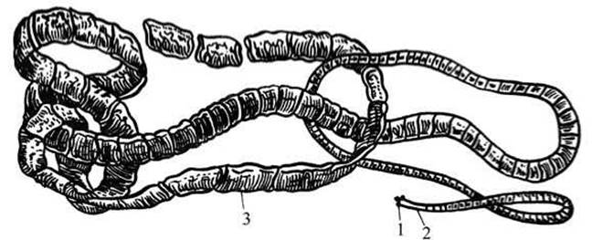
Fig. 37 Unarmed tapeworm (Taeniarhynchus saginatus):
1 — scolex (head); 2 — neck; 3 — middle (hermaphroditic) segments.
Study of the wet strobila mount. Examine the strobila of the unarmed tapeworm using a hand lens. Pay attention to the varying shapes of the segments at the anterior and posterior ends of the strobila. The ribbon-like body (strobila) reaches a length of 4–12 m and a width of 5–6 mm. Locate the head (scolex), the neck, and examine the segments (proglottids). Segment size increases with distance from the neck. The largest segments are found at the posterior end of the strobila (Fig. 37). Note that the entire body is covered by a tough, multi-layered cuticle. The worm's movements are powered by a Skin-muscular sac.
Two excretory canals are quite distinct. They originate in the neck region and run along the sides of the strobila, each terminating in an opening at the posterior segment of the worm. The canals in each segment are connected by transverse cross-links. Small branches (invisible in standard mounts) extend from the main canals and end in terminal Cells. In other words, the excretory organs operate on a protonephridial system principle.
Study of the microscopic scolex mount. Examine and sketch the prepared slide of the head.

Fig. 38 Head and Neck of the unarmed tapeworm (Taeniarhynchus saginatus)
The head should be examined under low magnification of the microscope.
It measures 1–2 mm in diameter and is equipped with four muscular suckers (Fig. 38) that the parasite uses to anchor itself to the host's intestinal mucosa. The parasite attaches to the host's mucosa so firmly that its removal can be difficult. During deworming, the parasite must be removed completely with its head. If the strobila is removed without the head, the worm's body will regenerate. The head lacks any structures for food ingestion: tapeworms completely lack a Digestive System (a regression associated with their parasitic lifestyle).
Nutrition occurs osmotically—by absorbing food digested by the host across the entire surface of the body.
The suckers can be easily spotted by slightly raising and lowering the microscope tube, adjusting the lighting, and using the fine adjustment knob.
Examine the neck located behind the scolex—this is the narrowest, unsegmented part of the strobila. The growth of the worm originates from the neck, which serves as a growth zone. From here, young proglottids bud off throughout the entire lifespan of the worm.
Make a drawing of the scolex with suckers and the neck.
Studying a slide of a hermaphroditic proglottid. Examine the stained slide of a proglottid from the middle part of the strobila using a hand magnifier and low microscope magnification. Locate the excretory canals running along the sides of the proglottid, which are connected by a transverse canal in the lower part of the proglottid (Fig. 39). Parallel to them, closer to the edge of the proglottid, run two nerve trunks originating from the cerebral ganglion located in the scolex (they are not always visible on standard slides).
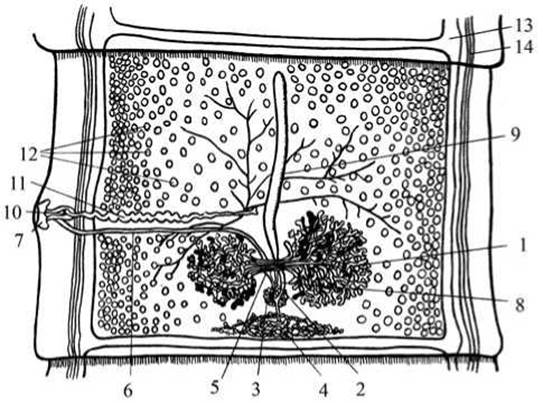
Fig. 39 Hermaphroditic proglottid of the unarmed tapeworm (Taeniarhynchus saginatus):
1 — bilobed Ovary; 2 — oviduct; 3 — Mehlis' gland; 4 — vitelline gland; 5 — seminal receptacle; 6 — Vagina; 7 — genital atrium; 8 — uterine pore; 9 — uterus; 10 — copulatory organ; 11 — vas deferens; 12 — Testes; 13 — excretory system canal; 14 — nerve cord.
The rest of the space within the proglottid parenchyma is almost entirely occupied by reproductive organs.
Examine the male and FEMALE REPRODUCTIVE ORGANS in each middle proglottid. Since each contains a full set of male and female reproductive organs, the middle proglottids are called hermaphroditic. The Structure of the reproductive apparatus resembles that of the Liver fluke.
The Male Reproductive System is represented by numerous testes scattered throughout the proglottid as small vesicles. Their efferent ducts (not visible on the slide) converge into the vas deferens, which appears as a thin, convoluted tube. The vas deferens empties into the ejaculatory duct, which penetrates the cirrus. The latter is located inside the cirrus pouch on the side of the proglottid. The male opening opens into the genital atrium.
The central part of the FEMALE REPRODUCTIVE SYSTEM is the ootype. It is connected by the oviduct to the bilobed ovary, and by the vitelline duct to the vitelline gland. The vagina extends from the ootype, running parallel to the vas deferens as a straight tube, and opens into the genital atrium adjacent to the cirrus.
Mehlis' gland also opens into the ootype. At the site of the genital atrium, the lateral wall of the proglottid protrudes outward, forming the genital papilla. The vitelline glands and Mehlis' gland supply the ootype with substances necessary for the complete formation of eggs.
Make a drawing of the hermaphroditic proglottid.
The Fertilization process involves either two individuals or different proglottids of the same tapeworm. Spermatozoa enter the vagina via the cirrus and then pass into the ootype, where they meet the egg cells.
Fertilized eggs (supplied with yolk and a shell) enter the uterus, which connects to the ootype and appears as a straight tube without an outlet pore (blindly ending).
Studying a slide of a mature proglottid. Sketch a mature proglottid under a magnifier, paying special attention to the shape of the uterus. The uterus is closed, meaning it has no connection to the external environment. When examining the uterus under low microscope magnification, one can see that it is packed with tapeworm eggs. Count the main lateral Branches of the uterus on one side using a magnifier.

Fig. 40 Mature proglottid of the unarmed tapeworm (Taeniarhynchus saginatus)
The shape of the uterus changes as it fills with eggs. Initially, it appears as an unbranched closed tube, after which numerous lateral branches develop. The uterus grows, gradually filling the entire body of the proglottid. All other reproductive organs, having fulfilled their purpose, gradually atrophy. A proglottid with an expanded uterus filled with eggs is called mature (Fig. 40). Mature proglottids are located in the posterior part of the strobila. They periodically detach from the worm one by one and enter the external environment along with the host's excrement. If the eggs are accidentally ingested by an intermediate host (cattle), a complex developmental cycle begins in its body, ending in The formation of larvae known as bladderworms (cysticercus). Humans become infected with the beef tapeworm by consuming undercooked or rare measles-infected beef.
Examination of measly beef and a cysticercus slide.
Examine fresh or preserved cattle meat infested with cysticerci using a hand magnifier. Examine and sketch a slide of the unarmed tapeworm cysticercus under low microscope magnification. Locate the scolex with suckers, the neck, and the bladder from which the head has everted.
Studying the developmental cycle of the unarmed tapeworm using the diagram (Fig. 41). The Development of the unarmed tapeworm proceeds with a change of hosts. The adult worm parasitizes the human small intestine. Mature proglottids detach from the strobila and pass out of the intestine with human feces. The mature eggs filling the uterus contain a fully formed spherical embryo armed with six hooks, known as an oncosphere.

Fig. 41 Diagram of the Development of the unarmed tapeworm
When grazing on grass or eating hay, the animal swallows mature proglottids and eggs. Under the action of gastric juice, the membrane of the oncosphere dissolves, and the embryo emerges into the animal's intestine. Here, using its hooks, it penetrates the intestinal mucosa and enters the bloodstream. Wandering through the bloodstream, the embryo is carried to the muscles, Heart, Lungs, liver, or other organs.
Here, the oncosphere loses its hooks and, as it develops, transforms into a distinctive larval stage—the cysticercus (bladderworm). The cysticercus is a pea-sized bladder containing fluid inside. The head is inverted into the cavity from one side of the bladder. Infection of the definitive host—the human—can occur only by consuming measly meat. In the human intestine, the cysticercus bladder is digested, the head everts, and it attaches to the intestinal wall using its suckers. The formation of proglottids begins from the neck—this is the growth of the strobila.
The pork tapeworm (Taenia solium).
The definitive host of the pork tapeworm is human, while intermediate hosts include pigs, wild boars, cats, dogs, and, rarely, humans. The disease caused by the presence of the pork tapeworm in the human intestine is known as taeniasis.
Materials and equipment. Since the primary host of the pork tapeworm, much like the beef tapeworm, is humans, the Methods and techniques for obtaining material for study are the same as those described for the beef tapeworm.
Required tools and materials include: a microscope; a dissecting magnifier; a hand lens; a wet mount of the pork tapeworm strobila; microscopic slides of the pork tapeworm scolex, a hermaphroditic proglottid, a mature proglottid, and a cysticercus (finna); fresh or preserved pork meat containing cysticerci; forceps; and a diagram of the life cycle.
Assignment. Examine and sketch the general appearance of the pork tapeworm. Examine and sketch the scolex, hermaphroditic and mature proglottids, and the cysticercus stage. Compare the Anatomy of the pork and beef tapeworms. Study the Life Cycle of the pork tapeworm and the preventive measures against taeniasis.
Examination of the wet mount of the pork tapeworm. Since the pork tapeworm is morphologically very similar to the previous specimen—the beef tapeworm—primary attention should be paid to the differences between them.
Examine the wet mount of the pork tapeworm strobila using a hand lens and write a Description of the parasite (Fig. 42). Note that the worm's body is tape-like and flattened dorsoventrally. The body color is white or yellowish, which is typical of internal parasites. The strobila is 2–3 m long and consists of about 900 proglottids. Examine the various shapes of the proglottids at the anterior and posterior ends of the strobila, and compare them with the strobila of the beef tapeworm.
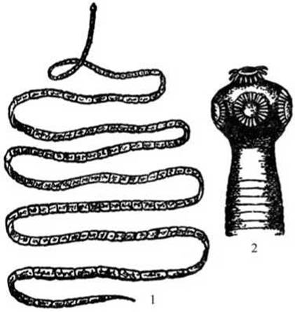
Fig. 42 General appearance of the pork tapeworm (Taenia solium):
1 — tapeworm body; 2 — scolex (head with suckers and a rostellum with hooks).
Examination of the scolex slide. By adjusting the illumination and focusing the microscope field of view, examine the stained slide of the pork tapeworm scolex under low magnification. Locate the four suckers and the prominent rostellum bearing a crown of cuticular hooks on the scolex (Fig. 42). The hooks are arranged in a double row. The rostellum with hooks distinguishes the scolex of the pork tapeworm from that of the beef (unarmed) tapeworm, hence the name "armed tapeworm." Locate the neck, which is the unsegmented region immediately following the scolex.
Draw the scolex.
Examination of the microscopic slide of a hermaphroditic (middle) proglottid. Examine the stained slide of a hermaphroditic proglottid using a dissecting stereomicroscope; low magnification of a compound microscope may be used to observe finer details. Observe the shape of the proglottid.

Fig. 43 Hermaphroditic proglottid of the pork tapeworm (Taenia solium):
1 — ovary; 2 — vitelline gland; 3 — Mehlis' gland; 4 — uterus (lacking an external opening); 5 — testes; 6 — vas deferens; 7 — vagina; 8 — genital atrium; 9 — excretory canal.
Sketch the outlines of the proglottid large in your sketchbook, and gradually add anatomical details as you study the slide. Under good illumination, the lateral excretory canals appear as white tubes running down the sides, connected at the posterior margin of each proglottid by a transverse canal.
Locate the vitelline gland just above the transverse excretory canal in the posterior part of the proglottid; it consists of numerous small, lobed follicular clusters (Fig. 43).
Positioned above the vitelline gland is the ovary, which consists of two main lobes and a smaller third (accessory) lobe. The presence of this third ovarian lobe distinguishes the armed tapeworm from the unarmed one. Locate Mehlis' gland and the uterus between the ovarian lobes. In some preparations, it can be observed that the uterus, filled with eggs, forms lateral blind branches. From the ootype, which is surrounded by a cluster of glandular cells (Mehlis' gland), a straight tube—the vagina—extends laterally and opens externally at the Base of the genital atrium. The genital atrium is clearly distinguishable as it is located on a prominent genital papilla on one of the lateral margins of the proglottid.
Examine the testes, which appear as small vesicles scattered in large numbers throughout the parenchyma of the proglottid, particularly in its anterior region. Very fine vas efferentia lead from each Testis, making them nearly invisible on the slide. All ducts merge into a single vas deferens, which is convoluted and thicker than the vagina, lying dorsal to it. Its terminal portion—the cirrus—opens into the genital atrium within the cirrus pouch.
Examination of the microscopic slide of a mature proglottid. Using a hand lens or dissecting magnifier, count the primary lateral branches of the uterus on one side. The mature proglottid of the pork tapeworm is typically characterized by having 7–12 lateral uterine branches on each side of the main uterine stem. Counting the uterine branches in a mature proglottid facilitates species identification of the parasite and Diagnosis of the infection. Note that in the mature proglottid, the uterine branches have crowded out almost all other structures of the reproductive system. The vagina, vas deferens, and genital atrium remain barely visible.
Examination of measly pork and a cysticercus slide.
Examine fresh or preserved measly pork. Note the numerous whitish structures visible between the muscle fibers—these are the cysticerci of the pork tapeworm.
Examine the microscopic slide of pork tapeworm cysticerci under low magnification. Locate the head with its suckers and crown of hooks, the beginning of the strobila, and the fluid-filled bladder.

Fig. 44 Life cycle of the pork tapeworm (Taenia solium):
1 — mature proglottid; 2 — egg containing a larva; 3 — six-hooked larva (oncosphere); 4 — bladder stage (cysticercus); 5 — cysticercus with everted scolex.
Study the developmental cycle of the armed tapeworm using the diagram (Fig. 44). The life cycle of the pork tapeworm proceeds generally similarly to that of the unarmed tapeworm.
The difference lies in the fact that the intermediate host of the armed tapeworm is typically the pig, though humans may serve as accidental intermediate hosts. In this case, A large number of cysticerci accumulate in human organs (eyes, Brain, muscles, heart, lungs, liver, etc.).
Just as in the life cycle of the beef tapeworm, mature proglottids are expelled with feces from the human intestine and ingested by pigs. In The Stomach of the intermediate host (the pig), the proglottids are digested, and oncospheres (small multicellular embryos equipped with six hooks) emerge from the released eggs. The oncospheres enter the intestine, use their hooks to burrow into the intestinal wall, penetrate Blood Vessels, and are carried by the blood or Lymph stream throughout the pig's body. In the muscles or other organs, the oncosphere develops into a cysticercus. Humans become infected by eating semi-cooked pork containing cysticerci. Inside the human intestine, the cysticercus develops into an adult armed tapeworm.
Prevention: veterinary inspection of pork and pork products intended for sale, Treatment of individuals suffering from taeniasis (as they pose a danger to themselves, other people, and animals), and zoohygienic measures to prevent pig infection (building latrines so that pigs have no access to them, maintaining yard cleanliness, etc.).
Echinococcus (Echinococcus granulosus).
The Significance of veterinary and sanitary inspection during animal slaughter becomes especially apparent to future veterinarians and livestock specialists when studying the echinococcus tapeworm.
Definite hosts of Echinococcus include dogs, wolves, jackals, and other canids. The larval stage takes place in the Internal Organs of intermediate hosts—cattle (cows), small ruminants (sheep), camels, pigs, and humans. The severe disease caused by the larval (cystic) stage of the echinococcus tapeworm, which most frequently localizes in the liver, lungs, and other internal organs, is called echinococcosis. Humans contract echinococcosis from dogs that have ingested discarded livers containing echinococcus cysticerci and other internal organs of cows or sheep.
Materials and equipment. Material is obtained by dissecting dogs. The adult worm is examined using a whole mount stained with alum carmine. Extreme caution is required when working with live adult specimens of Echinococcus, as their eggs can enter The Human Body and develop into the larval stage.
Required items: microscope; hand lens; whole mount of the tape stage of Echinococcus; wet mount of the echinococcus cysticercus; life cycle diagram.
Assignment. Examine and draw the bladder stage of Echinococcus. Study the STRUCTURE OF THE tape stage of Echinococcus and draw it. Review the life cycle of Echinococcus and preventive measures against echinococcosis.
Study of the whole mount of the tape stage of Echinococcus. Examine the whole microscopic mount of Echinococcus under low and high magnification of the microscope, darkening the field of view. Note that Echinococcus is a very small tapeworm (Fig. 45).
The length of its strobila is 3–4 mm, consisting of 3–4 proglottids. Locate 4 suckers and a rostellum on the head (scolex), bearing two rows of 26 to 50 hooks. The neck (growth zone) begins immediately behind the scolex. Note that in the first and youngest proglottid, the reproductive system is not yet developed. The penultimate proglottid contains a hermaphroditic reproductive system, which is not clearly visible on the mounts. The last and largest proglottid is mature. The reproductive system of Echinococcus closely resembles in structure that of the armed tapeworm.
Examine the mature proglottid of Echinococcus. It is filled with a sac-like uterus containing over 3,000 eggs.
Draw the echinococcus worm.
Study of the mount of the cystic (bladder) stage of Echinococcus. Examine the echinococcus cysticercus using a hand lens. It is a fluid-filled bladder. The outer wall of the bladder is Chitin-like and layered. The inner wall is parenchymatous. Locate smaller daughter bladders on the inner wall, inside which even smaller bladders with scolices develop. The number of scolices ranges from 10 to 30. New bladders constantly form on the inner wall; therefore, the size of the cysticercus continuously increases, and the bladder can grow to the size of an apple or even a child's head.
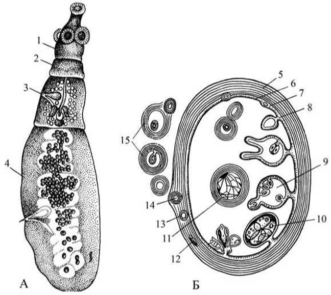
Fig. 45 Echinococcus (Echinococcus granulosus):
A — adult specimen; B — diagram of the structure of the echinococcal bladder;
1 — scolex with suckers and rostellum with hooks; 2 — immature proglottid; 3 — hermaphroditic proglottid; 4 — mature proglottid; 5 — bladder cuticle; 6 — germinative membrane; 7–11 — internal daughter bladders at various Selection/3.html">Stages of development; 12–15 — external daughter bladders at various stages of development.
Draw the echinococcus cysticercus.
Study of the echinococcus life cycle using the diagram.
Detaching from the strobila, the mature proglottid is expelled with the host's feces and acts as an egg dispenser. The eggs, along with grass, are ingested by livestock (Fig. 46).

Fig. 46 Life cycle of the echinococcus
Humans become infected by ingesting eggs that stick to their hands after touching the fur of a dog infected with the tapeworm form of Echinococcus. In the intestine of the intermediate host, an oncosphere (hexacanth embryo) hatches from the egg. Penetrating the intestinal wall, the oncosphere enters the bloodstream or Lymphatic system, is carried throughout the body into various organs—most commonly the liver, lungs, brain, and eyes—and there develops into a hydatid cyst (metacestode), which is the invasive larval stage of the tapeworm.
Definitive hosts—such as dogs, wolves, and jackals—become infected with the Echinococcus tapeworm when they consume animal organs infested with hydatid cysts. Inside the definitive host's intestine, the larval stage develops into a mature adult. A large number of parasites develop from a single ingested cyst, as each scolex gives rise to a tapeworm form.
Prevention: adherence to general hygiene rules—keeping hands clean, maintaining clean domestic dogs, deworming dogs, eliminating stray dogs, organizing veterinary supervision at slaughterhouses, destroying animal organs affected by cysts, and protecting premises, pastures, and livestock feed from contamination by dog feces.
Order Pseudophyllidea
This order comprises tapeworms whose scolex bears only two attachment grooves, known as bothria, and only occasionally solitary hooks. Their uterus features an opening, representing an open-type uterus. We will examine two representatives of this order: the broad fish tapeworm and the strap tapeworm.
Broad fish tapeworm (Diphyllobothrium latum).
The broad tapeworm leads a parasitic lifestyle. Its life cycle involves two intermediate hosts. The definitive hosts of the tapeworm can include humans, cats, dogs, arctic foxes, and other fish-eating mammals; the first intermediate hosts are lower crustaceans of the order Copepoda (such as Cyclops and Diaptomus), while the second intermediate hosts are fish (including pike, perch, ruff, and burbot). Parasitism of the broad tapeworm in the human intestine leads to a severe disease—diphyllobothriasis—which is frequently associated with pernicious anemia.
Materials and equipment. Diphyllobothriasis is widespread in regions where fishing constitutes a major source of freshwater harvesting. Material is most conveniently obtained from deworming clinics and hospitals. The expelled worm must be thoroughly washed in water and carefully unraveled. Subsequently, whole mounts should be prepared from the scolex and proglottids of various Regions of the strobila and stained.
Required equipment: microscope; dissecting loupe; hand lens; wet mount of the strobila; microscopic slides of the broad tapeworm's head, as well as its proglottids (hermaphroditic and gravid); and a diagram of the life cycle.
Task. Examine the External structure of the broad tapeworm. Study the structure of the scolex, as well as the middle and posterior proglottids. Compare the anatomy of the broad tapeworm with that of unarmed and armed tapeworms. Examine the life cycle of the broad tapeworm and preventive measures against diphyllobothriasis.
Examination of the wet mount. Examine the broad tapeworm. Note that it is the largest tapeworm: the length of its strobila ranges from 2 to 10–16 m, with 3,000–4,000 proglottids in the strobila. The body color is whitish-gray, and the scolex is elongated. Locate the unsegmented neck directly behind the scolex, and observe that the proglottids of the broad tapeworm differ from those of the pork and beef tapeworms in that the width of the proglottids exceeds their length.
Examination of the scolex slide. Under low magnification, examine the scolex of the broad tapeworm—it is elongated in shape, bearing specialized attachment grooves (bothria) on its narrow sides instead of circular suckers.
Draw the scolex of the broad tapeworm (Fig. 47).
Examination of the hermaphroditic proglottid slide. Study the structure of the reproductive system using a magnifying Glass and under low microscope magnification. The reproductive System of the broad tapeworm consists of the same components as those of the armed and unarmed tapeworms, but they are arranged differently. Examine the male reproductive system. It consists of small, rounded testes located along the lateral margins of the proglottid (Fig. 47), with thin, barely distinguishable sperm ducts extending from them and merging into a common ejaculatory duct that transitions into the cirrus. The cirrus is enclosed within a voluminous cirrus pouch, which opens externally in the middle of the upper third of the proglottid.
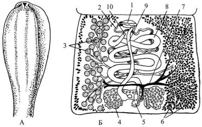
Fig. 47 Broad tapeworm (Diphyllobothrium latum):
A — scolex with bothria; B — hermaphroditic proglottid; 1 — copulatory organ; 2 — vas deferens; 3 — testes; 4 — ovary; 5 — ootype; 6 — vitelline gland; 7 — vitelline duct; 8 — uterus; 9 — vagina; 10 — uterine pore.
Examine the female reproductive system. It is represented by a bilobed ovary located in the center of the posterior third of the proglottid, and vitelline glands distributed along the lateral margins of the proglottid behind the testes (on some slides, distinguishing between testes and vitelline glands can be difficult). The vitelline ducts empty into the ootype. Extending from the ootype is a long, loop-forming tube—the uterus—which has a rosette-like shape (characteristic of the broad tapeworm). Note that the tubular uterus of the tapeworm, unlike that of cestodes of the family Taeniidae, features an opening along the midline of the proglottid (open-type uterus). The vaginal and uterine openings are difficult to visualize on the slide. Eggs of the broad tapeworm can continuously exit the uterus and enter the host's feces. The vagina, which opens next to the cirrus pouch, also originates from the ootype. Mehlis' gland also opens into the ootype.

Fig. 48 Gravid proglottid of the broad tapeworm (Diphyllobothrium latum)
Draw the hermaphroditic proglottid of the tapeworm.
Examination of the gravid proglottid slide. Using a magnifying glass and low microscope magnification, examine the structure of the gravid proglottid of the broad tapeworm (Fig. 48). In the gravid proglottids of the tapeworm, much like in other cestodes, only the uterus is retained. It possesses the rosette shape typical of this species. The uterus is packed with eggs. In some slides, testes and vitelline glands are still visible. The vitelline glands are located closer to the lateral margins and are smaller than the testes.
Draw the slide of the gravid tapeworm proglottid.
Study of the life cycle from the diagram. The life cycle of the broad tapeworm differs from that of taeniid tapeworms in that it proceeds with the involvement of two intermediate hosts (Fig. 49).
The eggs of the broad tapeworm are expelled into the environment along with the feces of the definitive host. Further development of the larval stages depends on the eggs reaching water and on a specific water Temperature (eggs die at temperatures below 6°C and above 30°C). Ciliated larvae, known as coracidia, hatch from the eggs in the water and use their cilia to swim. At a temperature of 10°C, the larva develops within 4–5 days. If a coracidium is ingested by a cyclops or diaptomus (freshwater lower crustaceans), it sheds its cilia in the crustacean's gut and penetrates its body cavity.
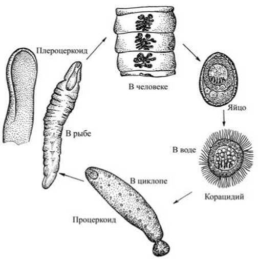
Fig. 49 Life cycle of the broad tapeworm (Diphyllobothrium latum)
The crustacean serves as the first intermediate host. Inside its body, the next larval stage of the worm develops—the procercoid, which retains 6 hooks on a spherical appendage at the posterior end of its body.
Fish (such as pike, perch, ruffe, and burbot) become the additional (second intermediate) hosts by preying on infected crustaceans. The crustaceans are digested in the fish's intestine, and the procercoids use their hooks to actively penetrate through the stomach wall into the fish's musculature (less commonly into The Liver and other organs). Here, the procercoids develop into the encysted stage—plerocercoids. Plerocercoids appear as white, unsegmented, motile worms up to 6 cm in length. Like adult tapeworms, the anterior end of a plerocercoid features two bothria (sucking grooves). In infected fish, plerocercoids can be seen through the skin as whitish, elongated nodules.

Fig. 50 Ligulid tapeworms:
A — swollen fish infected with ligulids; B — a cluster of ligulids emerging through a ruptured or dissected body wall; C — a plerocercoid extracted from the body cavity of a fish.
Humans and animals are infected by consuming insufficiently cooked, fried, raw, or undercooked fish containing plerocercoids. In the definitive host's intestine, the fish tissue is digested, and the living plerocercoids attach to the mucous membrane of the small intestine and develop into adult worms.
Prevention: consuming thoroughly cooked or fried fish, Processing fish with low temperature, pickling, or salting, and protecting water bodies from contamination with human and canine excrement.
Ligulid tapeworm (Ligula intestinalis).
Materials and equipment. The ligulid tapeworm (Fig. 50) is a parasite of birds and fish. Similar to the broad tapeworm, its life cycle involves two intermediate hosts In addition to the definitive host. The definitive hosts of the ligulid include herons, gulls, ducks, and others. The first intermediate host is lower crustaceans of the order Copepoda (cyclops, diaptomus), where the parasites occur at the procercoid stage, while the second intermediate host is fish, in which they live as plerocercoid larvae. The tapeworm predominantly infects bream, ruffe, roach, bleak, and pike. The disease caused by the presence of ligulid larvae in the body cavity of fish is called ligulosis. Birds become the definitive hosts by preying on fish infected with plerocercoids, and the tapeworms establish themselves in the intestines of these birds.
Required tools: hand lens, forceps, dissecting needles, live or preserved plerocercoids, wet mounts of fish, or live fish infected with ligulids (1 — fish with a distended abdomen, 2 — fish with ligulids protruding from its body cavity).
Task. Examine live or preserved ligulids at the plerocercoid larval stage, study and sketch their external morphology. Examine wet mounts of fish infected with plerocercoids.
Study of a live or preserved plerocercoid specimen. Examine a plerocercoid in a dissecting dish using a hand lens. Pay attention to its ribbon-like body shape, coloration, large size (up to 50–80 cm), and lack of body segmentation. Locate the bothria—two sucking grooves—at the anterior end of the body using the lens (Fig. 50).
Make a drawing of the ligulid at the plerocercoid stage, indicating the anterior end with the bothria. Examine a wet mount of a fish or a live fish afflicted with ligulosis. Note the distended abdomen. Inside the body cavity of such a fish, plerocercoids form a tangle of white ribbons. These fish typically remain near the water surface, often belly-up, unable to dive to the bottom.
Developing within the fish's body cavity, the ligulid compresses its internal organs; consequently, organ function is impaired, nutrition is compromised, and the fish's growth is stunted. In cases of heavy infection, the fish dies.
Last update: 13/08/2026
Editorial and Educational Adaptation: This material has been compiled based on the primary/original source text. The project team performed an editorial review, corrected technical inaccuracies, structured sections, and adapted the content for an educational format.
What was processed:
- elimination of formatting defects (OCR errors, structural breaks, corrupted characters);
- editorial organization of content;
- standardization of terminology in accordance with academic sources;
- verification of factual statements against the original source text.
All mentions of the author, publication year, and origin of the primary text have been preserved in accordance with the source.