Practical Guide to Zoology: Study Guide - T. A. Dauda 2014
Multicellular Animals
Flatworms
Class Digenea
The Class Trematoda consists entirely of parasites that inhabit the Internal Organs of invertebrate and vertebrate animals. The body shape of flukes is usually leaf-like. A characteristic feature is the presence of two suckers (oral and ventral), which gives the class its name. Their body size ranges from a few millimeters to 5-8 cm.
Any species of flukes can be used for laboratory practicals. Flukes parasitizing fish and frogs are the easiest to obtain. However, considering the specifics of agricultural universities, we will examine the Morphology, internal Structure, and life cycles of the lancet Liver fluke and the common liver fluke, which parasitize large and small ruminants as well as humans.
Lancet liver fluke (Dicrocoelium lanceatum)
parasitizes the liver of cattle and small ruminants (most commonly sheep), certain other mammals, and rarely humans. A characteristic life cycle and alternation of hosts are observed. The definitive hosts of the fluke are the animals listed above, in whose liver Bile ducts *Dicrocoelium* lives; the intermediate hosts are terrestrial gastropods of the genera *Helicella*, *Zebrina*, *Chondrula*, etc.; and the supplementary hosts are ants (*Formica fusca*, *F. rufibarbis*, etc.). The disease caused by the parasitism of *Dicrocoelium* in the bile ducts of the liver is called dicroceliasis.
Required equipment: Microscope; dissecting magnifier; whole-mount slide of the lancet liver fluke; life cycle diagram.
Task. Examine and draw the body shape and STRUCTURE OF THE lancet liver fluke: two suckers (oral and ventral), Digestive System (Mouth, Pharynx, Esophagus, two intestinal branches), and Reproductive System (cirrus pouch containing the cirrus, pair of Testes, Ovary, seminal receptacle, Uterus filled with eggs, and vitelline glands). Study The life cycle of the lancet liver fluke using the diagram.
Examination of the slide. Examine the whole-mount slide of the lancet liver fluke, stained with borax carmine, using a dissecting magnifier and low microscope magnification. Note that the body is flattened dorsoventrally and resembles a scalpel blade in shape. Body size is 0.5-1.2 cm. Locate the two suckers (Fig. 31): the oral sucker with the mouth opening at the anterior end of the body, and the ventral (attachment) sucker located in the first third of the body. Draw the body outline of *Dicrocoelium* and sketch the Organ Systems as you study them. Locate the mouth deep within the oral sucker, which leads into the pharynx, transitioning into a slender esophagus. Two straight intestinal branches extend from the esophagus and end blindly. The intestinal branches are often obscured by other organs and are not fully visible on the slide. There is no anus. Undigested food residues are eliminated through the mouth opening. The excretory system is of the protonephridial type (typical for Flatworms). It is not visible on a whole-mount slide as it requires special preparation. The Nervous system is of the ladder type and is also not visible on a standard slide.
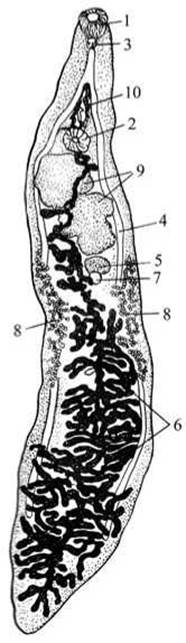
Fig. 31 Lancet liver fluke (Dicrocoelium lanceatum):
1 — oral sucker; 2 — ventral sucker; 3 — pharynx; 4 — intestinal branches;
5 — ovary; 6 — uterus; 7 — seminal receptacle; 8 — vitelline glands; 9 — testes; 10 — ejaculatory duct.
Examine the reproductive system—it is complex and occupies the largest portion of the worm's body. The lancet liver fluke is a hermaphrodite. The Male Reproductive System consists of two rounded testes located below the ventral sucker. The vasa deferentia extending from them (not visible on the slide) ascend and, above the ventral sucker, empty into the ejaculatory duct housed within the cirrus (copulatory organ). The cirrus is enclosed in a special sac, the cirrus pouch, situated between the oral and ventral suckers, beneath the intestinal bifurcation.
The FEMALE REPRODUCTIVE SYSTEM is represented by a single small, rounded ovary lying beneath the second Testis. A short oviduct connects to the seminal receptacle, which may be visible adjacent to the ovary. The uterus extends downwards as a long, looped tube occupying all the space between the intestinal branches. The walls of the uterus are thin and transparent, and the eggs filling the uterus are clearly visible on the slide. The uterus opens externally into the cirrus pouch near the cirrus. Botryoidal (grape-like) glands, the vitelline glands, are located along the sides of the body and produce nutritive material for the eggs and secretions for eggshell formation. The vitelline ducts, oviduct, Mehlis' gland, and the dorsal-directed Laurer's canal are not visible.
Study of the life cycle based on the diagram (Fig. 32). The adult *Dicrocoelium* lives in the liver of the definitive host.

Fig. 32 Life Cycle of Dicrocoelium lanceatum:
1 — lancet liver fluke; 2 — egg; 3 — miracidium; 4 — egg with hatching miracidium in the mollusk's esophagus; 5 — sporocysts; 6 — redia; 7 — cercaria; 8 — collective cysts; 9 — cysts on plant surfaces; 10 — cercaria emerging from a collective cyst; 11 — young fluke.
Mature eggs pass through the worm's genital pore into the host's hepatic bile ducts, then enter the intestine with the bile, and are excreted in the feces.
Eggs accidentally ingested by the intermediate host—a terrestrial gastropod crawling on pasture grass—already contain larvae at a specific stage of development. Within the mollusk's body, the larvae (miracidia) hatch from the eggs, penetrate its liver, and transform into sporocysts (sac-like larvae).
Within the sporocysts, new sporocysts (secondary sporocysts) or rediae develop from Germ Cells, and within them, cercariae (larvae possessing a tail and a piercing stylet) are formed. The cercariae emerge from the sporocysts, leave the liver, and actively penetrate the mollusk's Lungs, where they encyst and clump together to form mucous "collective cysts."
During the mollusk's Respiration, these cysts are expelled outwards and fall onto the grass. They can be ingested by ants. In the Abdominal cavity of the ants, the cercariae transform into encysted larvae known as metacercariae. Livestock become infected by ingesting them along with grass. Humans become infected through raw, unwashed vegetables.
Prevention: eradication of Mollusks, veterinary measures, and personal prophylaxis (consuming only thoroughly washed vegetables).
Common liver fluke (Fasciola hepatica).
The common liver fluke parasitizes the hepatic bile ducts of cattle, sheep, rabbits, and certain other herbivorous domestic and wild animals. Occasionally, the liver fluke is found in humans. It feeds on Blood and the breakdown products of liver cells. The Development of *Fasciola*, much like that of the lancet liver fluke, involves an alternation of hosts and generations. The definitive hosts of *Fasciola* are the animals listed above and humans, while the intermediate host is gastropod mollusks (such as the truncatula snail). The disease caused by the parasitism of *Fasciola* in the hepatic bile ducts is called fascioliasis.
Materials and equipment. For the practical session, one can use a fascioliasiscoveted liver obtained from a slaughterhouse and rejected by veterinary inspection. The sites of parasite infestation are noticeable even upon external inspection of the liver—the bile ducts are swollen, their walls thickened and calcified. By carefully slicing such ducts longitudinally, one can consistently find live parasites. Studies can be conducted both *in vivo* and on fixed, stained preparations.
Required equipment and materials: microscope; dissecting magnifier; hand lens; Glass slides, coverslips, and watch glasses; pipettes; forceps; dissecting needles; physiological saline; animal liver with live liver flukes or a wet mount of a sheep's liver with flukes; live or fixed flukes; stained whole mount; life cycle diagram.
Assignment. Examine and draw the external Morphology of the liver fluke. Study and illustrate the arrangement of its internal organs. Examine the life cycle of the liver fluke. Observe and sketch miracidia, rediae, and cercariae.
Observation of live flukes. Examine the animal liver (or liver wet mount) infected with live flukes. Note the localization of these parasites.
Examine a live liver fluke under a magnifying glass in a watch glass containing physiological saline (or a fixed fluke in a watch glass). Note that the body of the fluke is flat, leaf-shaped, grayish-white, and reaches 2–5 cm in length. The anterior end is distinctly set off, tapering into a cone that bears an oral sucker, which is clearly visible under a magnifying glass. Locate the somewhat larger ventral sucker situated slightly posterior to the oral one (Fig. 33). The genital opening lies between the suckers. At the posterior end of the body is the excretory pore, where the main channel of the excretory system terminates. Observe that the entire body of the worm is covered with a fairly thick cuticle.

Fig. 33 Liver fluke (Fasciola hepatica):
1 — oral sucker; 2 — ventral sucker; 3 — vitellaria; 4 — lumen of the excretory canal; 5 — genital pore.
Study of a stained whole mount. Examine the whole mount under a dissecting microscope and sketch it (inspect unclear details under low magnification of a compound microscope). Locate the mouth situated deep within the oral sucker; it leads into a muscular pharynx used to pump food into the gut (the fluke feeds primarily on the host's blood). The pharynx opens into a short esophagus, through which food enters the intestine, consisting of two main branches with numerous ramifications. The intestine ends blindly. There is no anus. Undigested food residues are eliminated through the mouth.
In standard preparations, the intestine is not always fully visible because it is partially obscured by the Organs of the reproductive system. It can be clearly observed in liver fluke preparations with the digestive tract injected with Berlin blue or alum carmine.
Outline the body contour and make drawings of the digestive and reproductive systems as you examine them on the slide (Fig. 34).
The reproductive system attains exceptional development and complexity. The liver fluke is a hermaphrodite. By moving the slide on the stage of the dissecting magnifier, locate the vitellaria (yolk glands), which are located along the sides of the body and appear as fine granularity, along with the longitudinal vitelline ducts. Transverse ducts extend from the longitudinal ones and empty into the vitelline reservoir (a small brown spot).

Fig. 34 Structure of the reproductive (A) and digestive (B) systems of the liver fluke:
1 — oral sucker; 2 — ventral sucker; 3 — branched intestine (left and right branches); 4 — copulatory organ; 5 — ovary; 6 — vitellaria; 7 — vitelline ducts; 8 — testes; 9 — vas deferens; 10 — uterus.
Beneath the transverse vitelline ducts in the posterior part of the body, locate the two highly branched testes. Trace the vasa deferentia emerging from the testes; above the ventral sucker, they merge and enter the cirrus pouch (they are not visible along their entire length). Below the cirrus pouch, posterior to the ventral sucker, lies the convoluted uterus, whose loops are packed with eggs at various stages of maturity (light, brown, black). The uterus opens via the genital pore near the cirrus pouch. Slightly above the vitelline reservoir, a prominent rounded structure is clearly visible — Mehlis' gland — which is a complex of numerous glands. To the right and behind the uterus, locate the branching unpaired ovary. The short oviduct extending from it and Laurer's canal are faintly discernible.
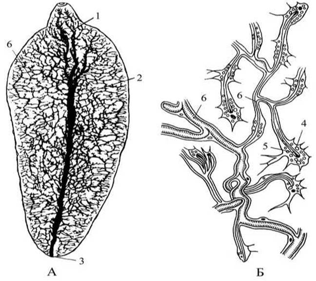
Fig. 35 Excretory System of the liver fluke:
A — arrangement of excretory canals in the body of the fluke; B — diagram of protonephridial structure: 1 — ventral sucker; 2 — main excretory canal; 3 — excretory pore of the excretory system; 4 — terminal Cell; 5 — flame bulb; 6 — collecting ducts.
The excretory organs are well developed (Fig. 35). In the preparation being examined, the main excretory canal shines through, most noticeable in the posterior half of the body and opening to the exterior via the excretory pore. In an injected preparation, one can observe that numerous lateral canals branch off from the main excretory trunk and break down into the finest branchlets penetrating all PARTS OF THE worm's body. Each branchlet begins with a large, star-shaped terminal cell that closes the lumen of the tubule.
Keep in mind that a bundle of cilia — a "flame bulb" — extends from The Cell into the lumen of the canal. The terminal cell collects harmful Metabolic waste products from the surrounding tissue that need to be removed from the Organism and excretes them through the tubule lumen. Due to the beating of the cilia bundle (the movement resembles the flickering of a candle flame), the fluid is propelled along the tubule. It collects from all the tubules into the main canal and is expelled from the fluke's body through the excretory pore. Excretory organs of this design are called protonephridia.
The nervous system is not visible in standard preparations. It consists of a paired ganglion located above the pharynx (supraesophageal ganglion), from which nerve cords extend. Note the two particularly well-developed lateral nerve cords. Sense Organs are absent, except for tactile nerve endings in the integument.
Study of the liver fluke's life cycle using the diagram (see Fig. 36). Mature fluke eggs leave the maternal organism via the uterine duct and enter the bile ducts of the parasite's host. Along with bile, the eggs pass into the host's intestine and are eliminated into the external environment with feces, where their development takes place. Proper Temperature, humidity, and light are essential for further development. The egg must inevitably reach Water. In the presence of light and at a temperature of 25–30 °C, a larva — the miracidium — develops inside the egg.

Fig. 36 Life cycle of the liver fluke
The developed miracidium actively hatches from the egg and enters the water. The larva possesses an eyespot and is covered with cilia, which it uses to swim. The next stage of the miracidium's development can proceed only within the body of the intermediate host — the pond snail (Limnaea truncatula).
The miracidium actively penetrates the liver or gonad of the marsh snail, sheds its cilia and eyespot, and transforms into the next larval stage—the sporocyst. Within the sac-like sporocyst, germ cells develop into the next larval generation known as rediae. They emerge from the sporocyst but remain within the mollusk's liver. Rediae possess an unbranched gut and germ cells. Inside the rediae, a third generation of larvae—cercariae—develops from the germ cells. Occasionally, daughter rediae develop first, which in turn produce cercariae.
Cercariae possess oral and ventral suckers, two intestinal branches, and a tail. Their reproductive system is underdeveloped. Cercariae leave the body of the intermediate host, swim freely in the water for a time using their tail, then attach to aquatic plants, shed their tail, and become enclosed in a tough protective cyst—a process called encystment. This spherical larval stage is known as an adolescent fluke or metacercaria (adolescaria). Further development can only occur within the body of the definitive host.
Infection of the host occurs when metacercariae are ingested with food or water. In the intestine of a human or animal, the protective cyst dissolves, and the young fluke migrates to the host's liver, where it develops into an adult worm. This mature adult stage of the fluke is called a marita.
Prevention involves stopping metacercariae from entering the body (as ingestion is the sole route of infection), clearing water bodies of snails, draining unnecessary stagnant waters, breeding waterfowl that feed on mollusks, and implementing targeted veterinary measures—most notably, the deworming of animals suffering from fascioliasis.
Last update: 13/08/2026
Editorial and Educational Adaptation: This material has been compiled based on the primary/original source text. The project team performed an editorial review, corrected technical inaccuracies, structured sections, and adapted the content for an educational format.
What was processed:
- elimination of formatting defects (OCR errors, structural breaks, corrupted characters);
- editorial organization of content;
- standardization of terminology in accordance with academic sources;
- verification of factual statements against the original source text.
All mentions of the author, publication year, and origin of the primary text have been preserved in accordance with the source.