Practical Course in Zoology: Textbook - T. A. Dauda 2014
Multicellular Animals
Flatworms
Class Turbellaria
Ciliates (Flatworms) are free-living marine, freshwater, or rarely terrestrial animals, with their entire body covered by a ciliated epithelium.
Class="center">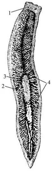
Fig. 30 White planarian flatworm (Dendrocoelum lacteum):
1 — eyes; 2 — Mouth; 3 — Pharynx; 4 — intestinal branches.
White planarian (Dendrocoelum lacteum).
Materials and equipment. The white planarian is a common species found in standing and running freshwaters. It lives under stones, on the underside of submerged fallen leaves, beneath the bark of snags, and in the leaf axils of aquatic plants. The white planarian adapts very well to aquarium conditions and feeds readily (for example, on Water lice—small crustaceans).
Studies are conducted using both live planarians and whole mounts.
Required items: Microscope; dissecting loupe; microscope slides and coverslips; depression slide; pipettes; forceps; dissecting needle; soft brush; live planarians in a watch Glass; whole mount of a white planarian.
Task. Examine and sketch the planarian. Pay attention to the eyes, mouth, pharynx, and intestine.
Observation of a live specimen. Examine a live planarian placed in water on a watch glass under a loupe. Observe its body shape, coloration, and movement. The body is flattened and elongated (Fig. 30). In live planarians, it is milk-white. The body length reaches 2.5 cm, and the width is 0.5 cm. Touch the planarian with a dissecting needle and observe its response to the stimulus. Using a brush, transfer the planarian into a drop of water on a depression slide, cover it with a coverslip, and examine it under low and then high microscope magnifications. Locate the black-pigmented eyes at the anterior end of the body, along with two small mobile lobes with rounded edges that function as tentacles. Move the slide to study The Structure of the Digestive System: the mouth opening is located on the ventral side, and the muscular pharynx and three-branched intestine are visible through the body wall.
Transfer the planarian onto a standard microscope slide, cover it with a coverslip, apply slight pressure to the glass, and examine it under low and then high microscope magnifications to observe the ciliary beating of the outer epithelial layer and rhabdites—specialized glistening rod-like structures within the epidermis of turbellarians.
Examination of the whole mount. Study the structural details of the planarian that could not be observed in the live specimen by using a prepared whole mount of the white planarian.
Draw the general appearance of the planarian and the details of its internal structure.
Last update: 13/08/2026
Editorial and Educational Adaptation: This material has been compiled based on the primary/original source text. The project team performed an editorial review, corrected technical inaccuracies, structured sections, and adapted the content for an educational format.
What was processed:
- elimination of formatting defects (OCR errors, structural breaks, corrupted characters);
- editorial organization of content;
- standardization of terminology in accordance with academic sources;
- verification of factual statements against the original source text.
All mentions of the author, publication year, and origin of the primary text have been preserved in accordance with the source.