BIOLOGY Volume 2 - A Guide to General Biology - 2004
13. TRANSPORT IN PLANTS
13.3. Transpiration and Water Movement Through Leaves
13.3.9. Stomata: Structure and Mechanism
Stomata are microscopic Pores in the epidermis that facilitate gas exchange. They are found predominantly on leaves, but also occur on stems. Each stoma is flanked by two guard Cells which, unlike ordinary epidermal cells, contain METABOLISM/14.html">Chloroplasts. Guard cells regulate the aperture of the stomatal pore by altering their turgor pressure. The Morphology of stomata and guard cells is clearly visible in scanning electron micrographs (Fig. 13.14).
Class="center">
Fig. 13.14. Scanning electron micrograph of stomata on the lower leaf surface.
In Section 6.1, we discussed the appearance of epidermal cells, guard cells, and stomata when viewed from above under a Light Microscope. Figure 13.15 shows a schematic cross-section of a stoma. As illustrated, the walls of the guard cells are unevenly thickened: The Cell wall adjacent to the stomatal pore, known as the ventral wall, is thicker than the opposite, dorsal wall. Furthermore, the Cellulose microfibrils within the wall are oriented in such a way that the ventral wall is less elastic than the dorsal wall. Certain microfibrils form hoop-like structures encircling the sausage-shaped guard cells (Fig. 13.15, B). These hoops are inelastic; as The Cell fills with Water and turgor increases, they prevent an increase in its diameter while allowing it to elongate only lengthwise. However, because the guard cells are joined at their ends and the thin dorsal walls stretch more easily than the thick ventral walls, the cells assume a crescent shape (Fig. 13.15). As a result, a gap opens between the two adjacent guard cells, known as the stomatal pore. A similar effect can be observed by inflating two elongated balloons joined at their ends, with adhesive tape applied along their contacting sides (simulating the inextensible ventral wall). To complete the analogy, one can loosely wrap them in a spiral with the same tape to simulate the cellulose hoops.
When guard cells lose water and turgor, the stomatal pore closes. The exact mechanism by which these changes in turgor pressure occur is still not fully understood.
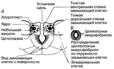
Fig. 13.15. A. Vertical cross-section through a stoma, also showing a portion of the lower leaf surface. B. Orientation of cellulose microfibrils in the guard cell walls.
According to the classical "sugar-starch" hypothesis, the concentration of water-soluble sugars in guard cells increases during daylight hours, thereby lowering their osmotic potential and driving the osmotic influx of water. However, no one has yet been able to demonstrate that guard cells accumulate a sufficient concentration of sugar to account for the Observed changes in osmotic potential.
It has recently been established that during the day, in the light, guard cells actively accumulate potassium cations and accompanying anions, which fulfill the role previously attributed to sugars. It remains unclear whether their electrical charges are fully balanced in the process. In some studied plants, an accumulation of large quantities of organic acid anions, particularly malate, has been observed in the light. At the same time, the starch grains that form in the chloroplasts of guard cells during the dark decrease in size. This occurs because starch is converted into malate in the light (requiring blue wavelengths of the spectrum), potentially via the following pathway:
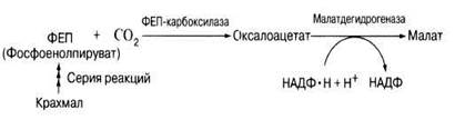
(Compare with the photosynthetic pathway in C4 plants; Section 7.9.)
In certain species, such as onions, starch is absent from guard cells. Consequently, when the stomata are open, malate does not accumulate, and cations appear to be taken up alongside inorganic ions such as chloride (Cl-).
In the dark, potassium (K+) effluxes from the guard cells into the surrounding epidermal cells. As a result, The water potential of the guard cells rises, causing water to move down the water potential gradient into the surrounding tissue. The turgor of the guard cells drops, altering their shape and causing the stomatal pore to close.
Several questions remain unanswered. For instance, why do potassium ions enter guard cells in the light? What is The Role of chloroplasts other than starch storage? It is possible that potassium uptake is driven by the activation of an ATPase localized in The Plasma Membrane. According to some data, this enzyme is activated by blue light. The ATPase may be required to pump protons (H+) out of the cell, while potassium cations move inward to maintain charge balance (a similar pump, discussed in Section 13.8.4, operates in the phloem). Indeed, consistent with this hypothesis, the pH inside guard cells drops in the light. In 1979, it was demonstrated that the chloroplasts of broad bean (Vicia faba) guard cells lack the Enzymes of The Calvin Cycle, and their thylakoid system is poorly developed, although chlorophyll is present. Consequently, conventional C3 Photosynthesis does not take place, and starch is not synthesized via this pathway. This likely explains why starch accumulates at night rather than during the day, as it does in ordinary photosynthetic cells.
Last update: 06/08/2026
Editorial and Educational Adaptation: This material has been compiled based on the primary/original source text. The project team performed an editorial review, corrected technical inaccuracies, structured sections, and adapted the content for an educational format.
What was processed:
- elimination of formatting defects (OCR errors, structural breaks, corrupted characters);
- editorial organization of content;
- standardization of terminology in accordance with academic sources;
- verification of factual statements against the original source text.
All mentions of the author, publication year, and origin of the primary text have been preserved in accordance with the source.