Biochemistry - The Chemical Reactions of Living Cells, Volume 2 - D. Metzler 1980
Coenzymes – specialized natural reagents of a distinct kind
Thiamine diphosphate
Observed changes in the optical properties of the coenzyme
Notably, many Coenzymes possess The ability to absorb light (see Chapter 13). This property manifests itself as an absorption spectrum and may also be accompanied by Circular Dichroism and fluorescence. The Optical Properties of vitamin B6 coenzyme forms are particularly sensitive to environmental changes and the ionization state of various groups within the molecule. For example, PMP in the neutral zwitterionic form, which predominates at pH 7, exhibits three strong Light absorption bands centered at 327, 253, and 217 nm [50].
Most of the various Ionic Forms of vitamin B6 derivatives feature three absorption bands located in approximately the same regions, yet the position and intensity of the bands vary for individual forms. For instance, the minor tautomer of PMP containing an uncharged ring exhibits a low-energy (long-wavelength) band at 283 nm [equation (8-27)]:
Class="center">
* Absorption band positions are given in both nanometers and wave numbers. The wave number  in cm-1 is equal to 107 divided by the wavelength in nanometers (see Chapter 13).
When both the ring nitrogen and the phenolic oxygen are protonated, the absorption band shifts to 294 nm (34.0∙103 cm-1), whereas if both groups are deprotonated, the resulting anion absorbs at 312 nm (21.1∙103 cm-1). Thus, studying the absorption spectrum of a coenzyme bound in an enzyme complex can answer the question of whether specific groups are protonated or deprotonated. For example, the peak of bound PMP at 330 nm in aspartate aminotransferase (Fig. 8-9) indicates the presence of the zwitterionic form [equation (8-27) 1).
Pyridoxal phosphate exists as an equilibrium mixture of aldehyde and hydrated aldehyde forms [as in equation (7-35)]. The aldehyde has a yellow color and absorbs at 390 nm (25.6∙103 cm-1, Fig. 8-9), whereas the aldehyde hydrate absorbs in approximately the same region as PMP. The absorption bands of PLP Schiff bases are shifted even further into the long-wavelength region, with N-protonated forms absorbing at 415–430 nm [(23–24)∙103 cm-1], while forms with a deprotonated C=N group absorb at shorter wavelengths.
1) However, a 5 nm shift in the absorption band compared to that of free PMP apparently indicates distinct Changes in the chromophore environment.
a. Imine groups in free Enzymes
When first investigating a highly purified preparation of aspartate aminotransferase (in 1957), Jenkins noted a striking fact: the bound coenzyme (at pH ~5) absorbed not at 390 nm like PLP, but at 430 nm, resembling a Schiff base (Fig. 8-9). Furthermore, as the pH was increased, the absorption band shifted to 363 nm. This result apparently indicates the dissociation of a proton (with pKa ≈ 6.3) accompanied by the Cleavage of a Hydrogen bond in a Schiff Base of the same type as shown in Fig. 8-5. It was soon demonstrated for this enzyme and several others that (as might be expected for a Schiff base) reduction with sodium borohydride converts the absorption spectrum into one resembling that of PMP and firmly attaches the coenzyme to the protein. Following complete Hydrochloric acid Hydrolysis of such borohydride-reduced Proteins, a fluorescent amino acid containing a reduced pyridoxyl residue was obtained; in all cases, it was identified as ε-pyridoxyllysine:


FIG. 8-9. Absorption spectra of Various Forms of aspartate aminotransferase and free pyridoxal phosphate. Absorption [see equation (13-5)] is plotted as a function of light quantum energy in units of wave numbers. The more widely used wavelength scale is also provided. In the spectra shown, wavelength more frequently increases from left to right; however, the format adopted here is becoming increasingly common. The transition of the enzyme from the form observed at low pH (pH < 5) to the form existing at high pH is controlled by a group with pKa ≈ 6.3. The addition of erythro-ß-hydroxyaspartate leads to a quinonoid form, whose spectrum is represented here at one-third of its true magnitude. THE SPECTRUM OF free PLP was obtained at pH 8.3. The apoenzyme spectrum (dashed line) is characterized by a small residual Absorption in the 300–400 nm region, the cause of which has not been elucidated. I — (+)erythro-ß-OH-Asp, 500 nm; II — 430 nm, at low pH; III — 390 nm, free PLP, at pH 8.3; IV — 363 nm, at high pH; V — 330 nm, PLP.
Thus, in the absence of substrates, PLP-containing enzymes harbor the coenzyme in the form of a Schiff base with the ε-amino group
of a protein Lysine residue1). A possible exception is 6-aminolevulinate synthase [equation (8-20)], in which an adduct between PLP and the active-center —SH group is postulated [51].
b. Transaldimination
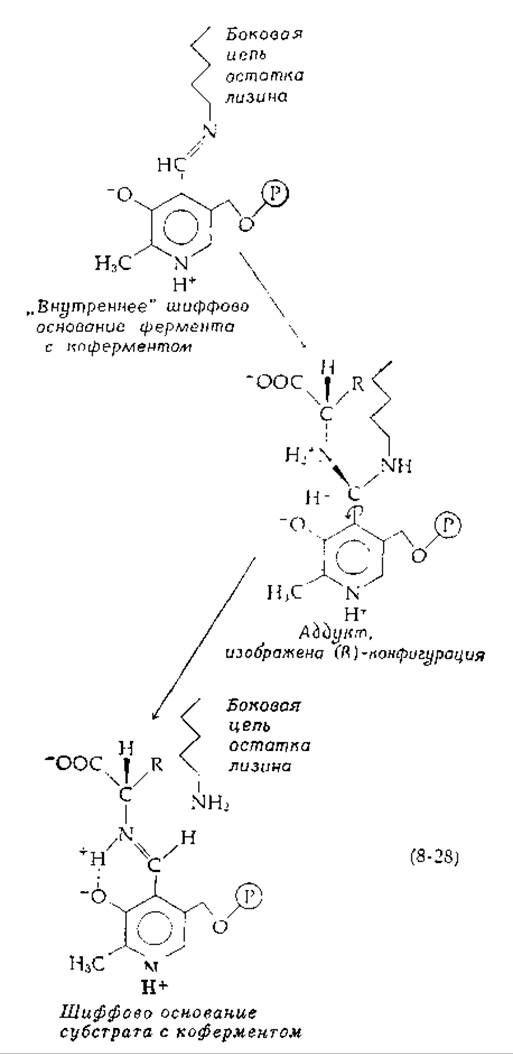
The formation of a PLP Schiff base with a substrate apparently occurs via the addition of the amino group not to the C=O group, but to the C=N group. This addition is followed by elimination, not of the —OH group, but of the ε-NH2 group [equation (8-28)]:
1) Reduction of Glycogen phosphorylase with sodium borohydride results in the attachment of PLP to a lysine side chain, yet the enzyme remains remarkably active.
This mechanism is presumably utilized by the enzyme because addition to —HC—NH+ proceeds faster [52] than addition to —HC=O. Transaldimination is confirmed by the temporary disappearance of positive circular dichroism (CD) (Chapter 13, Section B,5) in the coenzyme absorption band upon substrate addition. Following substrate conversion, circular dichroism reappears1).
c. Absorption bands at 500 nm
Most striking are the absorption spectra obtained under specific conditions for many PLP enzymes: they contain intense and unusually narrow bands in the 500 nm region (20∙103 cm-1). Such a band is observed in aspartate aminotransferase upon its action on erythro-ß-hydroxyaspartate (Fig. 8-9). This pseudosubstrate undergoes Transamination very slowly, and the accumulating form absorbing at 500 nm likely arises as an intermediate Structure in the normal catalytic cycle as well. A similar absorption spectrum is found upon the interaction of tryptophanase with the competitive inhibitor L-Alanine. Under the same conditions, the enzyme catalyzes the rapid exchange of the alanine α-hydrogen with 2H from 2H2O. Serine hydroxymethylase yields a band at 495–500 nm with both D-alanine and the normal reaction product, Glycine [53]. It is generally accepted that these bands originate from postulated quinonoid intermediate structures.
1) It remains unclear why the absorption bands of PLP in enzymes almost invariably exhibit positive CD. CD is apparently induced in a symmetrical chromophore (the light-absorbing group) by the electronically asymmetric environment of the protein. [An exception is the long-wavelength absorption bands of the quinonoid forms of substrate-coenzyme Imines described in the next section: they are typically associated with an intense negative CD extremum. — Transl. note]
Last update: 06/08/2026
Editorial and Educational Adaptation: This material has been compiled based on the primary/original source text. The project team performed an editorial review, corrected technical inaccuracies, structured sections, and adapted the content for an educational format.
What was processed:
- elimination of formatting defects (OCR errors, structural breaks, corrupted characters);
- editorial organization of content;
- standardization of terminology in accordance with academic sources;
- verification of factual statements against the original source text.
All mentions of the author, publication year, and origin of the primary text have been preserved in accordance with the source.