Chemistry and Biology of Proteins - F. Haurowitz 1953
Internal Structure of Globular Proteins
Optical Properties of Proteins
The Study of optical properties provides valuable data for elucidating The Structure of Organic compounds. In this regard, attempts have been made to investigate the Internal Structure of Proteins by determining refractive indices, as well as by studying optical rotation and absorption spectra.
When discussing issues related to Protein Electrophoresis, it was already noted that aqueous protein solutions have a higher refractive index than Water, and that this circumstance can be used to determine THE POSITION OF the moving boundary during electrophoresis. The refractive index of protein solutions increases linearly with an increase in protein concentration. The difference between the refractive index of a 1% protein solution and that of water is termed the specific refractive increment. This increment varies slightly depending on The Nature of the protein. For instance, it is 0.001901 for bovine serum albumin, 0.001887 for human serum albumin, 0.001876 for egg albumin, and 0.001875 for human y-globulins [106]. Temperature and the presence of salts in the solution do not significantly affect the specific refractive increment; therefore, the protein concentration in a solution can be determined quite rapidly by measuring the refractive index of the solution and subtracting the refractive index of the dialysate. It must be remembered, however, that the refractive increment of Lipoproteins, equal to 0.00171, is significantly lower than the corresponding value for lipid-free proteins [107].
The refraction caused by organic molecules represents the sum of the refractions of their constituent atoms. This holds true for proteins as well. Unfortunately, however, this circumstance cannot be utilized to study protein structures because the contents of carbon, nitrogen, hydrogen, and oxygen in various proteins are nearly identical. Furthermore, the number of individual atoms in a protein molecule is so large that THE CONTRIBUTION OF a single atom or atomic grouping to the overall molecular refraction is negligibly small. The same considerations apply to the ability of proteins to rotate a polarized beam of light [108, 109]. Since all Amino Acids, with the exception of Glycine, are optically active compounds, and the asymmetric carbon atoms of amino acids remain asymmetric within the peptide chain, Polypeptides and proteins are optically active compounds. The sign and magnitude of the specific rotation of a protein depend on the number of amino acids it contains and on their structure. Consequently, the total value resulting from the interaction of hundreds of amino acid molecules does not allow for any Conclusions regarding the arrangement of amino acids or other details concerning the internal STRUCTURE OF THE protein. The specific rotation of proteins reaches a minimum at their isoelectric point, fluctuating between —30 and —70° [109]. Considerably higher values, up to —313°, have been detected in gelatin gels [110].
Certain insights into Cell/13.html">Protein Structure have been obtained by investigating their absorption spectra in various spectral regions. Visible light is absorbed only by colored proteins, such as Hemoglobin, the yellow enzyme, visual purple, and other Chromoproteins. Determining absorption spectra in visible light has provided very valuable information regarding the structure of the colored prosthetic groups of these proteins; however, such determinations contribute nothing to the Assessment of the structure of their colorless protein carriers.
All proteins exhibit a distinct absorption spectrum in ultraviolet light, with an absorption maximum lying around 270 mμ [111, 112]. This maximum corresponds to the absorption maxima of Tryptophan, phenylalanine, and Tyrosine. Therefore, the intensity of the ultraviolet absorption spectrum can be used to gauge the content of these amino acids in various proteins. Hydrolysis of Proteins by Proteolytic Enzymes does not noticeably affect the nature of their absorption spectrum (Fig. 27) [113]. On this basis, it can be concluded that native proteins contain no specific light-absorbing structures that are absent in the hydrolysate. This also refutes the hypothesis of certain authors regarding the presence of a significant number of heterocyclic rings in native proteins.
Class="center">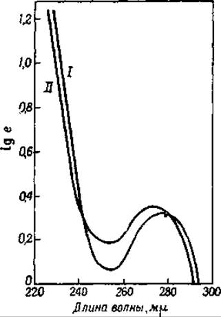
Fig. 27. Absorption spectra of serum globulin and the hydrolysate of this protein obtained by the action of Trypsin [113].
I — serum globulin; II — hydrolysate.
Absorption in infrared light is due to specific atomic groupings. A major difficulty in studying infrared absorption is that water also absorbs in this region of the spectrum. Consequently, studies must be conducted using dry protein films, which are never entirely homogeneous and, moreover, differ from one another in thickness and contain cracks. All these factors severely complicate the determination of infrared absorption intensity and significantly reduce the accuracy of measurements. Despite this, it has nevertheless been possible to obtain typical infrared absorption maxima
in the region for A number of proteins. Maxima at 3 μ and 3.22 μ are due to the NH groups of the peptide bond, while the maximum at 6 μ is due to CO groups. There is currently no evidence indicating that enolized peptide bonds —C(OH)=N— absorb in infrared light [114]. When determining the absorption intensity of silk fibers in the region of λ = 1.90 μ, it was found that the absorption band is more clearly expressed in the direction perpendicular to the long axis of the fiber. On this basis, it was hypothesized that the CO groups in silk fiber are arranged perpendicularly to this axis [116, 117]. Conversely, the Analysis of proteins in polarized infrared light led to the Conclusion that the NH groups are arranged parallel to the long axis of the fiber [115].
Last update: 06/08/2026
Editorial and Educational Adaptation: This material has been compiled based on the primary/original source text. The project team performed an editorial review, corrected technical inaccuracies, structured sections, and adapted the content for an educational format.
What was processed:
- elimination of formatting defects (OCR errors, structural breaks, corrupted characters);
- editorial organization of content;
- standardization of terminology in accordance with academic sources;
- verification of factual statements against the original source text.
All mentions of the author, publication year, and origin of the primary text have been preserved in accordance with the source.