Chemistry and Biology of Proteins - F. Haurowitz 1953
Internal Structure of Globular Proteins
Dielectric Constant of Protein Solutions
Determining the dielectric constant can provide insight into the polar Properties of the compounds under investigation. This is done by measuring the electrostatic forces between two charged capacitor plates, first in a vacuum and then in a solution of the test substance. If the electrostatic force in a vacuum is denoted as v0, and in the solution of the test substance as vp, it turns out that vp is always less than v0. The ratio v0/vp is called the dielectric constant.
The magnitude of the dielectric constant appears to be determined by whether the molecules possess a polar or nonpolar Structure, since high dielectric constants have been found for solutions of polar compounds, and low ones for solutions of nonpolar compounds. Zwitterions are highly polar compounds, so one would expect them to have high dielectric constants. Unfortunately, there are no direct Methods for determining the dielectric constants of Amino Acids and Proteins, as neither amino acids nor Proteins can be converted into a liquid state without destruction. The melting point of amino acids and proteins is so high that their molecules decompose before melting occurs. Valuable results have nevertheless been obtained by determining the dielectric constants of aqueous protein solutions. Water itself has a high dielectric constant, reaching up to 80 at 20 °C. By contrast, the dielectric constant of alcohol is 24, of ethyl ether 4.3, and of paraffin less than 2. The high dielectric constant of water is due to its polarity (see Chapter VI). When organic molecules are dissolved in water, the dielectric constant of water generally decreases. However, when amino acids or proteins are dissolved in water, the dielectric constant of the water increases. This atypical Effect of Amino acids and proteins is due to the fact that they are highly polar compounds. The increase in the dielectric constant, which varies for individual amino acids, Peptides, and proteins, is proportional to the concentration of the compound in the solution. If the dielectric constant of water is denoted as Dw, the dielectric constant of the test solution as D, and the molar concentration of the solute as C, then the molar increment of the dielectric constant will be equal to lm = (Dp — Dw)/C. The increase caused by dissolving amino acids in water ranges between 22 and 28, whereas peptides cause a much more significant increase. For peptides containing 2, 3, 4, 5, and 6 amino acids, the following values were obtained respectively: 70, 113, 159, 215, 239 [122]. It must be remembered, however, that molar solutions of larger peptides contain more substance per unit volume. If the above values are divided by the number of Glycine residues contained in the specified peptides, the following values are obtained for a single glycine molecule: 35, 38, 40, 43, and 39. These data show that the increase in the dielectric constant of water is identical when equal weight amounts of triglycine and hexaglycine are dissolved in water.
The increase in the dielectric constant of water caused by dissolving proteins in it varies over a very wide range. Since protein concentration in solution is not conventionally expressed in moles, the molar increment of the constant im must be replaced by i1 — the increment per 1 g of protein. In this case, the equation given above takes the following form: i1 = (Dp — Dw)/c, where c is the concentration in grams per liter. The values im and i1 found for various amino acids and proteins are listed in Table 10.
When calculating the dielectric constant increment per 1 g of amino acid, values ranging between 0.18 and 0.36 were found—i.e., values of the same order of magnitude as those for protein solutions. It is still rather difficult to explain the fact that the dielectric constant of water increases by the same amount upon the dissolution of 1 g of Amino Acid and 1 g of protein. This is possibly due to two mutually compensating effects. When Amino acids are dissolved in water, we are dealing with A large number of small dipoles, whereas when proteins are dissolved, we deal with a small number of large dipoles. The higher the dipole moment, the greater the increment in the dielectric constant should be.
Class="center">Table 10 Increase in the dielectric constant of water upon the dissolution of proteins
|
Dielectric constant increment |
Substance |
Dielectric constant increment i1 |
||
|
Substance |
im |
i1 |
||
|
Glycine |
22.6 |
0,30 |
Egg albumin |
0,1 |
|
23,2 |
0,26 |
Gliadin |
0,1 |
|
|
Valine |
25 |
0,21 |
Serum albumin |
0,17 |
|
Leucine |
25 |
0,19 |
0,3 |
|
|
Diglycine |
70,6 |
0,54 |
Carboxyhemoglobin |
0,33 |
|
Triglycine |
1131 |
0,60 |
Zein |
0,4 |
|
Tetraglycine |
159 |
0,65 |
Edestin |
0,7 |
|
Pentaglycine |
215 |
0,71 |
Serum pseudoglobulin.................. |
1,1 |
|
Hexaglycine |
239 |
0,65 |
Lactoglobulin |
1,5 |
The dipole moment of α-amino acids can be determined by measuring the distance between the positive and negative groups. This distance is approximately 3 Å (3∙10-8 cm), and the electron charge is 4.8∙10-10 electrostatic units. From these data, it can be calculated that the dipole moment of α-amino acids is approximately 15∙10-18 electrostatic units, or 15 debyes. Since the distance between the positively and negatively charged groups is the same in all α-amino acids, their dipole moment will also be the same; and because the increase in the dielectric constant is proportional to the dipole moment, it is not surprising that all Amino acids have a dielectric constant of the same order of magnitude.
In protein molecules, only a small number of amino acids contain free positively or negatively charged groups. As a first approximation, it can be assumed that out of every 10 amino acids making up a protein, only one has a positively charged group and one has a negatively charged group. It follows that the number of charges in 1 g of protein is approximately 0.1 times the number of charges in 1 g of the corresponding amino acid mixture. Since the increase in the dielectric constant of water caused by dissolving proteins does not noticeably differ from the increase caused by dissolving amino acids, it must be assumed that the distances between charged groups in proteins are significantly greater than in amino acids. Only under this condition will proteins possess a dipole moment as high as that of amino acids. However, it should not be assumed that all positively charged groups are located on one side of the protein molecule and all negative ones on the opposite side. If this were the case, the increase in the dielectric constant would be much greater than what is actually observed. It can be concluded that the values found for proteins indicate that the electrical charges in their molecules are arranged with a high degree of Symmetry [124].
The dielectric constant of protein solutions varies depending on the pH of the solution, reaching a maximum at the isoelectric point—that is, when the electrical charges in the protein also reach their maximum (see Chapter V). The dielectric constant also depends on the frequency of the field. By using fields of varying frequency to determine the dielectric constant, one can gain insight into the axial ratio of the protein molecule.
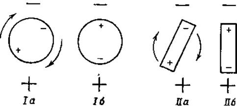
Fig. 28. Orientation of dipoles in an electric field.
The principles of this method are briefly outlined below.
When a polar molecule (dipole) is placed in an electric field, it orients itself in such a way that its positive pole points toward the external negative pole (Fig. 28), and its negative pole toward the external positive pole. Furthermore, if the polar molecule is spherical in shape and the distances of the poles from the center of the sphere are equal, the molecule will rotate around its axis, as shown in Fig. 28 (positions Ia and Ib). If, however, the polar molecule is not spherical, the entire molecule will move under the Influence of the electrostatic forces acting on both of its poles and will assume the orientation shown in Fig. 28 (positions IIa and IIb). The time required for such movement to take place can be determined by varying the frequency of the external alternating electric field. When this frequency exceeds a certain value, the protein molecule is no longer able to rapidly change its position and therefore will no longer contribute to the increase in the dielectric constant of the solution.
The time required for molecular orientation, known as the relaxation time, is relatively short for small molecules and long for large molecules. For small water molecules it is approximately 10-11 s, for amino acid molecules 10-10 s, and for large protein molecules from 10-8 to 10-6 s. At an electric field frequency of 103 Hz (wavelength 3∙107 cm), amino acids and proteins cause the aforementioned increase in the dielectric constant. At a frequency of 107 Hz (wavelength 3∙103 cm) and higher, however, proteins do not cause an increase in the dielectric constant of water. In fact, a slight decrease in this constant is even observed, because a certain fraction of the water bound by the protein loses its ability to follow the oscillations of the electric field [119].
By varying the field frequency from high to low, one can establish the critical frequency that corresponds to the relaxation time. If this critical frequency is equal to n, the relaxation time will be t = 1/2πn, and the molecular volume will be Vm = RTt/3η, where η is the viscosity of the solution. If the particle is not spherical but forms an ellipsoid, two values rather than one are found for the relaxation time: one value corresponding to rotation around the long axis, and the other around the short axis. Determining these two relaxation time values makes it possible to calculate the axial ratio (a/b) of protein molecules in aqueous solution. The a/b values found by this method for various proteins are listed below (protein Hydration was not taken into account in the calculation) [121, 123, 125, 126]:
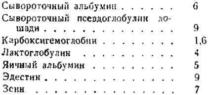
In calculating the above values, a simplification was made by assuming that the molecular shape is strictly ellipsoidal. However, this does not always correspond to reality. For example, the Hemoglobin molecule has the shape of a cylinder with slightly convex bases [2].
The dipoles depicted in Fig. 28 are permanent dipoles. It should be noted, however, that molecules of any compounds, including typical nonpolar compounds, become dipoles if placed in an electric field. This is due to the displacement of their electrons toward the external positive pole and a corresponding shift of the nuclei toward the negative pole. Such dipoles arising in an external electric field are called induced dipoles. The total dipole moment of protein molecules, like the dipole moment of molecules of Other Compounds, also includes the induced dipole moment.
Up to this point, we have considered proteins as rigid structures with fixed dipole moments. It is necessary, however, to account for the possibility that certain peptide chains within the globular protein molecule possess a certain degree of freedom and can undergo vibrational or rotational movements. In this case, the relaxation time of their polar elements will be shorter than the relaxation time of the macromolecule as a whole [127]. It is possible that some of these groups may cause an increase in the dielectric constant increment observed at certain intermediate frequencies. At present, we are not yet able to precisely determine the distribution of electrical charges within the globular protein molecule. Although low dielectric constant increments indicate that the charges are evenly distributed within the molecule, a difference still exists between the charge of the globule surface and that of its interior. Differences of up to 0.78 have been found between pHi (surface pH) and pH (interior pH of the molecule) [128].
By investigating the reactions occurring between proteins and other oppositely charged macromolecules, some data can be obtained regarding the distribution of ionic groups between the surface and the interior of the protein molecule. For example, it has been found that globulins form precipitates upon reacting with strongly alkaline protamines dissolved in a neutral medium. Albumins do not form such precipitates under the same conditions [129]. Based on these data, it was concluded that in globulins, negatively charged groups are located On the surface of their molecules (exogroups), whereas in albumins, these groups reside inside the corpuscular particles (endogroups). Both exogroups and endogroups can react with small oppositely charged ions (e.g., hydrogen ions), but only exogroups can react with large oppositely charged ions. This method thus provides a way to differentiate between exo- and endogroups.
Last update: 06/08/2026
Editorial and Educational Adaptation: This material has been compiled based on the primary/original source text. The project team performed an editorial review, corrected technical inaccuracies, structured sections, and adapted the content for an educational format.
What was processed:
- elimination of formatting defects (OCR errors, structural breaks, corrupted characters);
- editorial organization of content;
- standardization of terminology in accordance with academic sources;
- verification of factual statements against the original source text.
All mentions of the author, publication year, and origin of the primary text have been preserved in accordance with the source.