Molecular Biology of the Cell - Volume 3 - Alberts B., Bray D., Lewis J., Raff M., Roberts K., Watson J. 1994
From Cells to Multicellular Organisms
Features of Plant Cells
Transport of Substances Between Cells
In the previous section, we discussed how a rigid Cell wall poses very specific challenges for the GROWTH AND DEVELOPMENT of plant Cells. Moreover, it severely limits the opportunities for interaction between cells walled up within Tissues, both with each other and with their surrounding environment. However, plant cells have devised ingenious ways to overcome these limitations. Direct cell-to-cell communication is just as crucial for multicellular plants as it is for Multicellular animals; consequently, specialized channels have evolved that connect the Cytoplasm of a plant cell to that of its neighbors, while ensuring the controlled passage of ions and small molecules. In addition, in vascular plants, long strands of cylindrical cells are joined end-to-end via perforations, forming extended tubes that facilitate the flow of Water and nutrients.
Class="center">
Fig. 20-19. Specialized layers of small cells at the Base of the petiole, which participate in leaf abscission. Two or three layers of small cells secrete Enzymes that trigger cell wall degradation. The petiole detaches from the stem at the point where The Cell walls are thinnest. Several lignified cells cross the entire abscission layer. The cells remaining in the Separation zone deposit suberin, forming a protective layer over the wound.

Fig. 20-20. A. Cytoplasmic channels called plasmodesmata pierce the cell walls and interconnect all plant cells into a single continuous living system. B. A plasmodesma is lined by The Plasma Membrane, which is continuous with the membranes of both adjacent cells. Typically, the lumen of the plasmodesma contains a thin, cylindrical Structure known as the desmotubule, which is derived from The Endoplasmic reticulum.
20.2.1. Plant cells are interconnected by specialized cytoplasmic bridges known as plasmodesmata [9]
With the exception of very few specialized cell types, all living cells in a vascular plant are connected to their neighbors by fine cytoplasmic channels called plasmodesmata, which traverse the intervening cell walls. As shown in Fig. 20-20, within each plasmodesma, the plasma membrane of one cell is continuous with that of the adjacent cell. The plasmodesma itself is a membrane-lined cylindrical channel with a diameter of 20 to 40 nm. Running axially through the center of the channel from one cell to the other is a narrower cylindrical structure—the desmotubule—whose lumen, as revealed by Electron Microscopy, communicates with the cavities of the endoplasmic reticulum in both adjacent cells (Fig. 20-21). The space between the outer surface of the desmotubule and the plasma membrane lining the plasmodesma is filled with Cytosol (Fig. 20-20). Very often, this annular layer of cytoplasm constricts at both ends of the channel. These constrictions likely play a vital regulatory role, as it is precisely here that each cell can potentially control the passage of molecules into or out of neighboring cells.
Typically, plasmodesmata form around elements of the endoplasmic reticulum that become trapped within the newly forming cell wall during cytokinesis, separating the mother cell (see Fig. 13-71). However, plasmodesmata are also found in the walls of non-sister cells. Furthermore, the number of plasmodesmata can increase during cell growth, indicating that they can also form de novo.

Fig. 20-21. Cytology/cytology/93.html">ELECTRON MICROGRAPHS OF plasmodesmata. A. Longitudinal section of a plasmodesma in an aquatic fern. The plasma membrane lines the pore and is continuous from one cell to the next. The Endoplasmic reticulum and the associated central desmotubule are visible. B. The same plasmodesma in cross-section. (Courtesy of R. Overall.)
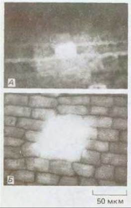
Fig. 20-22. Determining the functional size limit of plasmodesmatal channels using fluorescent Peptides of different sizes. A. A fluorescent peptide with a molecular mass greater than 850 daltons. B. A fluorescent peptide with a molecular mass of less than 850 daltons. In both cases, the peptide was injected into a single epidermal cell of *Potamogeton pusillus*. Note that the large peptide has barely moved, whereas the smaller peptide has diffused into many neighboring cells. (Courtesy of R. W. Goodwin.)
20.2.2. Plasmodesmata allow molecules to pass directly from one cell to another [9, 10]
What evidence, besides their characteristic structure, Supports the proposed role of plasmodesmata in Intercellular Communication? Indirect evidence comes from the fact that plasmodesmata are particularly abundant in the walls of cells grouped in zones of intense secretion, such as floral nectaries. In such cells, there may be 15 or more plasmodesmata per 1 µm3 of cell wall surface area, whereas in other cell types this number is often less than one.
The most direct evidence for intercellular transport via plasmodesmata has been obtained from dye-injection and electrical-coupling experiments. For example, fluorescent Dyes cross the plasma membrane only with great difficulty; however, when injected via a microcapillary into a single *Elodea* leaf cell, they quite rapidly appear in adjacent cells. Similarly, when electrical pulses are applied inside one cell, these pulses can be detected (albeit with some attenuation) by electrodes in neighboring cells. The degree of electrical signal attenuation depends on the density of plasmodesmata and the number of cells between the electrodes. Moreover, an electrode applied to the outer surface of the plasma membrane fails to detect signals injected inside the cell, indicating that they propagate via intracellular pathways.
Thus, it appears likely that plasmodesmata mediate The transport of solutes between adjacent plant cells, much like Gap Junctions mediate intercellular transport in animals (see Section 14.1.5). Data obtained from microinjections of fluorescent dyes coupled to peptides of various sizes indicate that molecules with a mass of up to roughly 800 daltons can move through plasmodesmata (Fig. 20-22), which closely matches the size exclusion limit of gap junctions. A body of evidence suggests that transport through plasmodesmata is regulated in a complex manner. For instance, dye-injection experiments have shown that, despite the presence of seemingly normal plasmodesmata, the movement of even low-molecular-weight substances is restricted between certain tissue systems. For example, dye migration is not detected between ROOT cap cells and root tip cells, or between epidermal and cortical cells in both roots and shoots. The mechanisms restricting cell-to-cell communication in these cases remain unknown, although there is some evidence pointing to the possible involvement of Ca2+ ions and protein phosphorylation. On the other hand, certain plant Viruses have been shown to induce the dilation of plasmodesmata, thereby facilitating their own cell-to-cell movement (Fig. 20-23). For example, transport of tobacco mosaic virus through plasmodesmata is known to depend on a single viral protein (P30) with a molecular mass of 30,000: a defective virus that is normally unable to move from Cell to Cell in a wild-type plant does so successfully in transgenic tobacco plants expressing the corresponding Gene.

Fig. 20-23. This electron micrograph shows small, spherical Viral Particles passing through the cytoplasm from one plant cell to another. These plant viruses have a diameter of 25 nm, which significantly exceeds the size exclusion limit of these channels. (Courtesy of K. Plaskitt.)

Scheme 20-2. KEY FEATURES OF early angiosperm development.

Fig. 20-24. Highly schematic representation of a group of plant cells interconnected by plasmodesmata. The plasma membrane lining the plasmodesmata divides the entire plant volume into two compartments: extracellular and intracellular (the symplast). For clarity, cellular Organelles are omitted from the diagram.
20.2.3. In higher plants, biological fluids are partitioned into two major compartments: intracellular and extracellular
Plasmodesmata connect the plasma membrane and cytoplasm of adjacent cells, integrating them into a complex community of living protoplasts. Consequently, the entire plant body can be viewed as a two-compartment system: 1) the intracellular compartment—known as the symplast—consisting of the interconnected network of protoplasts (including the protoplasts of phloem sieve tubes) and bounded by the continuous plasma membrane of all living cells; and 2) the extracellular compartment, or apoplast, which encompasses all cell walls and dead, empty xylem conducting elements, as well as the water contained within both (Fig. 20-24). Both compartments possess their own transport systems, although they can communicate at specific points and undergo local modifications to regulate Metabolic exchange between them.
20.2.4. Photosynthesizing and absorbing cells are interconnected by Vascular Tissues comprising xylem and phloem [11]
Multicellular Organization in plants, as in animals, enables a division of labor in which different cell types Complement each other through the specialization acquired during differentiation. Two crucial plant-specific Functions are performed by photosynthetic cells, which contain METABOLISM/14.html">Chloroplasts and serve as a source of organic molecules—particularly sucrose—for the entire Organism, and absorbing cells, which take up water and dissolved minerals from the environment. In most higher plants, these two functions cannot be carried out by the same cells, since the former requires light, whereas the latter takes place deep within the dark soil. Each of these processes also requires A number of other conditions. Photosynthesis, for example, must occur within a specialized microenvironment where relative humidity and carbon dioxide levels are strictly regulated. This is achieved by means of Stomata—specialized Pores in the cuticle-covered leaf epidermis that open and close depending on the turgor pressure of the guard cells (see Fig. 20-11). Conversely, efficient uptake of soil nutrients requires a massive absorptive surface area provided by roots, as well as membrane transport systems, often supplemented by the transport systems of symbiotic microorganisms. Thus, photosynthetic and absorbing cells nourish one another and, together, supply all other PARTS OF THE plant with the organic and inorganic substances required for biosynthetic processes. To ensure long-distance transport of these substances, photosynthetic and absorbing tissues are connected to the xylem and phloem, which form a branching network of conducting elements. Each of these consists of chains of cylindrical cells joined end-to-end to form microscopic pipelines (see Panel 20-1 and Fig. 20-16).
20.2.5. Water and Dissolved Solutes Move Through the Xylem [12]
The xylem is a complex component of the vascular tissue system. Mature xylem vessel elements are dead cells devoid of cytoplasm. Their lateral walls are heavily lignified and feature secondary thickenings on their inner surfaces. These pipelines transport water and dissolved inorganic ions from the roots to the rest of the plant (Fig. 20-25). The xylem also provides structural support, particularly in woody plants. Fluid flow in the xylem is unidirectional—toward sites of water evaporation. Water is pulled upward into these capillary-like tubes through Transpiration. Lignin is deposited around xylem vessels in a manner that creates highly crush-resistant structures, which is critical for tubes carrying fluid under negative pressure. Without such reinforcement, these long pipelines would simply collapse like thin cocktail straws.
Xylem vessels transport primarily mineral salts and nitrogenous compounds, which are presumably delivered into the system via active secretion by root parenchyma cells.

Fig. 20-25. The two main conducting systems, xylem and phloem, transport water and dissolved substances throughout the plant. This is a highly simplified diagram; in particular, it omits the intensive water exchange that occurs between the xylem and phloem across their lateral walls.

Fig. 20-26. A. The vascular network of a primitive plant, the aquatic fern Azolla (schematic transverse section of the root axis). The arrangement of vascular structures in this plant is remarkably simple: there are four xylem vessels and four phloem sieve tube elements. In most higher plants, the distribution of vascular cells is far more complex. Note that the xylem and phloem are surrounded by endodermal cells, and that the Casparian strips, strategically positioned within the endodermis, prevent water from leaking out of The Vascular System via the apoplast. B. The Casparian strip between two root endodermal cells, analogous to those shown in panel A. Unlike a conventional primary cell wall, the Casparian strip has a smooth texture due to its high content of suberin; the hydrophobic properties of suberin render these Regions of the cell wall impermeable to water. (B, after W. Gunning and M. Steer, Ultrastructure and Biology of Plant Cells, London: Arnold, 1975.)
The leakage of solutes in the reverse direction via the apoplast at the entry points of the xylem is blocked by so-called Casparian strips, which are functionally analogous to the tight junctions between adjacent animal epithelial cells (Fig. 20-26). At the exit points of the xylem pathways, specialized parenchyma cells equipped with specific membrane-localized transport Proteins pump the dissolved solutes into the photosynthetic tissues. Most of the solvent (water) flowing through the xylem vessels eventually evaporates, primarily from The surface of the leaf photosynthetic tissues.
Because the loading of solutes into the root xylem occurs continuously, it can generate an osmotic pressure gradient that drives the delivery of solutes to leaves and shoots even when weather conditions inhibit transpiration. The droplets of water often seen on the tips of lawn grasses in the early morning are secreted precisely As a result of this phenomenon of root pressure driven by osmosis. (For a description of some of the cell types involved in this process, see Panel 20-1.)
20.2.6. Sugars Are Transported by Pressure Flow in the Phloem [13]
The phloem is a complex tissue system that transports dissolved organic nutrients, primarily sucrose, from the photosynthetic leaf cells to the rest of the plant (see Fig. 20-25). The main conducting component of the phloem is the sieve tube—a long Column of living cylindrical cells that communicate with one another through pores in their end walls (sieve plates). Sucrose enters the cells at the upper end of the sieve tube and moves downward as a concentrated solution (typically 10-25%), passing from one cell to the next. Thick-walled cells form a tube well-suited for the transport of fluids under high pressure (up to 30 atm). Although mature sieve elements are living cells with a functional plasma membrane (and thus constitute part of the symplast), they have lost their nuclei and some of their cytoplasm. Their survival is maintained by associated companion cells, which transfer nutrients and other molecules in both directions—into and out of the sieve tubes—via clusters of plasmodesmata located in their shared lateral walls (see Panel 20-1).
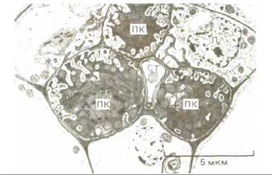
Fig. 20-27. Specialized transfer cells in a minor leaf vein. A phloem sieve tube element (ST) is surrounded by three transfer cells (TC). Infoldings of The cell wall, lined by the plasma membrane, increase the surface area of these cells twentyfold. Transfer cells are found in regions of the plant where The rate of solute Transport Across the plasma membrane is exceptionally high—for example, where inorganic ions are pumped from the xylem into tissues or, as in this case, where sucrose is loaded into the phloem.
Phloem transport is a considerably more complex process than xylem transport because it is not restricted to a single direction: dissolved organic solutes, mainly sucrose, are translocated from their sites of synthesis to sites of utilization and storage, regardless of where these locations lie. Sucrose is actively transported into and out of the sieve tubes by specialized transfer cells located in the source and sink regions, respectively (Fig. 20-27). The accumulation of sugars at the source increases the osmotic influx of water into the phloem at these sites, generating the hydrostatic pressure required to drive a rapid mass flow of fluid through the sieve tubes toward metabolic sinks. At the sinks, the sugar is largely unloaded, and the water is removed osmotically (primarily into the xylem). Fluid moves through the phloem at a velocity of about 1 m/h, which is vastly greater than can be achieved by diffusion.
It is worth noting that fluid transport in plants has at least two distinctive features that set it apart from the analogous process in animals. First, animals possess a single transport system—the Circulatory system—whereas plants have two distinct systems: the phloem and the xylem. Second, in plants, fluids do not circulate in a closed loop like Blood in animals; instead, a continuous, unidirectional stream of water is maintained from the roots to the leaves via two "open pipelines" (see Fig. 20-25).
The presence of a rigid, relatively impermeable cell wall largely dictates the specific ways in which plant cells interact with one another and with their environment. All living plant cells are interconnected by plasmodesmata—minute cytoplasmic channels lined by the plasma membrane that traverse the cell walls and allow many dissolved substances to pass directly from cell to cell. Thus, all living protoplasts of a plant organism constitute a single continuous system known as the symplast. The remaining space, occupied by cell walls and dead, "empty" cells through which most of the plant's water is transported, is called the apoplast. Photosynthetic plant cells produce sugars that are distributed to all other plant Organs and tissues via living phloem cells, which form part of the symplast. Root cells absorb water and dissolved minerals from the soil, which are then transported to the leaves through xylem vessels formed by dead cells that belong to the apoplast. Once again, physical forces—in this case, transpiration—drive the continuous ascent of the water column from roots to leaves.
Last update: 12/08/2026
Editorial and Educational Adaptation: This material has been compiled based on the primary/original source text. The project team performed an editorial review, corrected technical inaccuracies, structured sections, and adapted the content for an educational format.
What was processed:
- elimination of formatting defects (OCR errors, structural breaks, corrupted characters);
- editorial organization of content;
- standardization of terminology in accordance with academic sources;
- verification of factual statements against the original source text.
All mentions of the author, publication year, and origin of the primary text have been preserved in accordance with the source.