BIOCHEMISTRY - L. Stryer - 1984
VOLUME 2
PART II GENERATION AND STORAGE OF METABOLIC ENERGY
CHAPTER 13. THE CITRIC ACID CYCLE
13.14. Stereospecific Hydrogen Transfer by NAD+-Linked Dehydrogenases
In the 1950s, Brigit Vennesland, Frank Westheimer, and their coworkers performed an elegant experiment investigating the stereospecificity of hydrogen transfer by NAD+-dehydrogenases. The substrate for the reaction catalyzed by Alcohol dehydrogenase was ethanol labeled with two deuterium atoms at C-1. It was established that the reduced coenzyme contained one deuterium atom per molecule, while the second deuterium atom was incorporated into acetaldehyde. Thus, no deuterium was lost or transferred to the solvent; rather, it was transferred directly from the substrate to NAD+.
Class="center">
The deuterated reduced coenzyme formed in this reaction was subsequently used to reduce acetaldehyde. The result was striking: the deuterium was transferred entirely from the coenzyme to the substrate and could no longer be detected within NAD+.
These reactions demonstrate the stereospecificity of the transfer catalyzed by alcohol dehydrogenase. The positions occupied by the two hydrogen atoms at C-4 in NADH are nonequivalent. One of them (HA) is positioned in front of, and the second (HB) behind, the plane of the nicotinamide ring. In other words, C-4 represents a prochiral center. Alcohol dehydrogenase, being a chiral reagent, distinguishes between the A and B positions at C-4. Deuterium is transferred from deuterated ethanol exclusively to the A position. In the reverse reaction, the deuterium atom is removed from the A position and transferred directly to acetaldehyde.

Some dehydrogenases, such as glyceraldehyde-3-phosphate dehydrogenase, transfer hydrogen to the B position. Thus, There are two classes of NAD+ (and NADP+)-linked dehydrogenases: A-stereospecific and B-stereospecific. A comparison of the three-dimensional structures of NAD+-dependent dehydrogenases reveals that the difference in stereospecificity between the A- and B-type Enzymes arises from a 180° Rotation of the nicotinamide ring relative to the adjacent ribose moiety. As a result of this 180° shift, the opposite face of the nicotinamide ring becomes exposed and, consequently, reactive in A- and B-dehydrogenases. As might be expected, all known dehydrogenases are stereospecific. When a dehydrogenase reacts with a series of substrates, the stereospecificity of hydrogen transfer remains identical for all of them. The evolutionary conservation of NAD+-binding sites is underscored by the fact that the stereospecificity of a given dehydrogenase is independent of the Organism's species (for instance, Yeast and horse Liver Alcohol dehydrogenases share the same stereospecificity). Furthermore, the stereospecificity of a specific reaction catalyzed by enzymes utilizing both NAD+ and NADP+ proves to be identical for both Coenzymes.
Fig. 13.13. Model and formula of the nicotinamide component of NADH, showing that HA and HB lie on opposite sides of the ring. According to the RS nomenclature, HA is pro-R, and HB is pro-S.

13.15. Lethal Synthesis: The Conversion of Fluoroacetate to Fluorocitrate
The Citric Acid Cycle is blocked in animals that consume the leaves of Dichapetalum cymosum, a poisonous South African plant. Within an hour, the citrate content in most Organs increases more than ten-fold. Poisoned animals experience convulsions and typically die shortly thereafter. The toxic agent in these leaves is fluoroacetate, which is also used as a rat poison. However, fluoroacetate has no effect on purified enzymes of The Citric Acid cycle. Why, then, is this cycle blocked upon the ingestion of fluoroacetate in vivo? The explanation is that, within the organism, fluoroacetate undergoes enzymatic conversion into fluorocitrate, a potent inhibitor of aconitase. Fluoroacetate is activated to fluoroacetyl-CoA, which then condenses with oxaloacetate to form fluorocitrate.
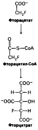
The Active Site of aconitase contains an Fe2+ ion, which normally forms chelates with the oxygen atom of the hydroxyl group and two oxygen atoms of the carboxyl groups of citrate. In contrast, fluorocitrate binds in such a way that its fluorine atom forms a chelate with the iron ion (Fig. 13.14). The high electronegativity of fluorine promotes strong interaction with Fe2+, leading to Enzyme Inhibition. It is important to note that The conversion of fluoroacetate to fluorocitrate is not an isolated instance of lethal synthesis. Many chemical substances, relatively harmless in themselves, can be converted by enzymatic action into highly deleterious compounds. For example, certain polycyclic aromatic Hydrocarbons un-
dergo enzymatic modification in vivo into potent mutagens and carcinogens (Sec. 20.21).
Fig. 13.14. Proposed binding model of citrate and fluorocitrate to the active site of aconitase: A — binding of citrate, leading to catalysis; B — binding of fluorocitrate, leading to inhibition. The fluorine atom of fluorocitrate forms a chelate with the iron ion in the active site.

13.16. The Citric Acid Cycle as a Source of Precursors for Biosynthetic Processes
Up to this point, our Structure/133.html">Discussion of the citric acid cycle has focused on it as a major degradative pathway responsible for ATP generation. However, the citric acid cycle also serves another role: it supplies intermediates for biosynthetic pathways (Fig. 13.15). For instance, most of the carbon atoms in Porphyrins are derived from succinyl-CoA. Many Amino Acids are derived from α-oxoglutarate and oxaloacetate. We will examine The Biosynthesis of these compounds in subsequent chapters. Here, it is essential to emphasize the crucial point that the withdrawal of citric acid cycle intermediates for biosynthetic purposes must necessarily be balanced by their replenishment. Suppose, for example, that oxaloacetate is converted into amino acids used for METABOLISM/35.html">Protein Biosynthesis. If de novo synthesis of oxaloacetate does not occur concurrently, the operation of the citric acid cycle will halt, because acetyl-CoA can enter the cycle only upon Condensation with oxaloacetate. How is oxaloacetate replenished? Mammals lack the enzymatic machinery required to convert acetyl-CoA into oxaloacetate or any other citric acid cycle intermediate. In these organisms, oxaloacetate is produced via the carboxylation of Pyruvate, catalyzed by pyruvate carboxylase (Sec. 15.15).
Pyruvate + CO2 + ATP + H2O ⇄ Oxaloacetate + ADP + Pi + 2H+.
The carboxylation of pyruvate is an example of an anaplerotic reaction (from the Greek meaning 'to fill up').
Fig. 13.15. The Role of the citric acid cycle in biosynthetic processes. Intermediates withdrawn from the cycle for biosynthetic needs (indicated by red arrows) are replenished by The formation of oxaloacetate from pyruvate.

Last update: 06/08/2026
Editorial and Educational Adaptation: This material has been compiled based on the primary/original source text. The project team performed an editorial review, corrected technical inaccuracies, structured sections, and adapted the content for an educational format.
What was processed:
- elimination of formatting defects (OCR errors, structural breaks, corrupted characters);
- editorial organization of content;
- standardization of terminology in accordance with academic sources;
- verification of factual statements against the original source text.
All mentions of the author, publication year, and origin of the primary text have been preserved in accordance with the source.