BIOCHEMISTRY - L. Stryer - 1984
VOLUME 2
PART II GENERATION AND STORAGE OF METABOLIC ENERGY
CHAPTER 13. THE CITRIC ACID CYCLE
13.11. Variations on a Multienzyme Theme: The Alpha-Ketoglutarate Dehydrogenase Complex
The oxidative decarboxylation of α-ketoglutarate has much in common with the corresponding process for Pyruvate:
α-Ketoglutarate + CoA + NAD+ → Succinyl-CoA + СO2 + NADH,
Pyruvate + CoA + NAD+ → Acetyl-CoA + СO2 + NADH.
Both reactions involve the same Cofactors: TPP, lipoamide, CoA, FAD, and NAD+. Essentially, the Oxidative Decarboxylation of α-ketoglutarate is catalyzed by an enzyme complex structurally similar to the pyruvate dehydrogenase complex. The α-ketoglutarate dehydrogenase complex comprises Three types of Enzymes: the α-ketoglutarate dehydrogenase (A'), transsuccinylase (B'), and dihydrolipoyl dehydrogenase (C') components. Furthermore, A' binds to B' and B' binds to C', but A' does not bind directly to C'. Thus, the core of the complex consists of transsuccinylase (analogously to transacetylase).
The α-ketoglutarate dehydrogenase component (A') and transsuccinylase (B') differ from the corresponding enzymes (A and B) of the pyruvate dehydrogenase complex. At the same time, the dihydrolipoyl dehydrogenase parts (C and C') of both complexes are identical. Reconstitution experiments with these systems have shown that the complex formed by A, B, and C' is as active in the oxidative decarboxylation of pyruvate as the complex composed of A, B, and C. Similarly, C and C' are interchangeable in forming the reconstituted complex that carries out the oxidative decarboxylation of α-ketoglutarate.
As noted earlier, Chymotrypsin, Trypsin, Thrombin, and Elastase are homologous enzymes. Here we see that the pyruvate and α-ketoglutarate dehydrogenase complexes represent homologous enzyme associations. The structural and mechanistic features that ensure coordinated catalysis at the entrance to The Citric Acid Cycle are employed once again later in the operation of this cycle.
13.12. Beriberi Is Caused by Thiamine Deficiency
"A very painful affliction that strikes people is called beriberi (meaning 'sheep') by the locals. I have seen those stricken with this disease. Their knees tremble, they lift their legs high, and they walk like sheep. This is a type of paralysis or rather tremor. Patients exhibit abnormalities in movement patterns, impaired sensation in the hands and feet, and sometimes the entire body..." Thus beriberi was described by the Danish physician Jacobus Bonitus in 1630 while working on the island of Java.
Beriberi is caused by a dietary deficiency of thiamine (also known as vitamin B1). The disease was and remains a serious problem for the inhabitants of the Far East because rice, their staple food, is poor in thiamine. The problem is further aggravated by the consumption of polished rice, since only the outer layer of the rice grain contains appreciable amounts of thiamine. Beriberi is occasionally encountered in cases of severe malnutrition in alcoholics. The disease is characterized by neurological symptoms and Heart Failure. Damage to the Peripheral Nervous system manifests as pain in the extremities, Muscle weakness, and impaired cutaneous sensitivity. The Heart may be enlarged and Cardiac Output reduced.
How does thiamine deficiency cause these symptoms? Thiamine pyrophosphate serves as the prosthetic group for three important enzymes: pyruvate dehydrogenase, α-ketoglutarate dehydrogenase, and transketolase. Transketolase transfers two-carbon units from one sugar to another; its role in the Pentose Phosphate Pathway will be discussed later. A common feature of enzymatic reactions utilizing TPP is The transfer of an activated aldehyde moiety. In beriberi, Blood levels of pyruvate and α-ketoglutarate exceed normal values. The increase in pyruvate levels is especially pronounced after glucose administration. Consistently, the enzymatic activities of the pyruvate and α-ketoglutarate dehydrogenase complexes in vivo are reduced compared to normal. In addition, erythrocyte transketolase activity is decreased in beriberi, and the determination of this enzyme's activity can be used for diagnosing the disease.
13.13. Symmetrical Molecules Can React Asymmetrically
Let us trace The Fate of a specific carbon atom in The Citric Acid cycle. Suppose that in oxaloacetate, the carboxyl carbon most distant from the oxo group is labeled with the 14C isotope. Analysis of the resulting α-ketoglutarate will show that no loss of the radioactive label occurs. Decarboxylation of α-ketoglutarate will then yield succinate devoid of radioactivity, with all the label residing in the released СO2. The fact that all the label is found within СO2 came as a complete surprise. The citrate molecule is symmetrical. Therefore, it was believed that its two —СН2СОО - groups should react identically. Consequently, it was further assumed that for every citrate molecule converted via pathway 1, there must be another citrate molecule converted via pathway 2. If this were so, only half of the label should appear in the СO2.
Class="center">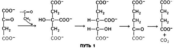

These experiments, conducted in 1941, were interpreted to mean that because the transformation of the label is asymmetric, citrate (or any other symmetrical compound) cannot be an intermediate in The formation of α-ketoglutarate. This interpretation seemed indisputable until 1948, when Alexander Ogston astutely pointed out the fallacy in the assertion that two identical groups in a symmetrical molecule cannot be differentiated by an enzyme: "On the contrary, it is possible that an asymmetric enzyme acting on a symmetrical compound may distinguish between its identical groups... the asymmetric distribution of an isotope in a reaction product cannot be taken as an argument against its formation from a symmetrical precursor."
Let us analyze Ogston's proposition. For simplicity, let us consider a molecule in which two hydrogen atoms, group X, and another group Y are bonded to a tetrahedral carbon atom. Let us designate one hydrogen as HA and the second as HB. Now suppose that the enzyme binds three groups of this substrate: X, Y, and H. Can it differentiate HA from HB? Figure 13.12 illustrates the binding of X, Y, and HA to three sites on the enzyme. Note that X, Y, and HB cannot be bound to this Active Site; or rather, two groups can be bound, but not all three. Thus, the fates of HA and HB must be different.
Fig. 13.12. HA and HB are sterically nonequivalent when the substrate CXYH2 is bound to the enzyme at three sites
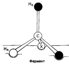
Table 13.3. Commonly used radioactive isotopes

It should be noted that HA and HB are sterically non-equivalent, even though the CXUH2 molecule lacks optical activity. Similarly, the —CH2COO groups of citrate are sterically non-equivalent, despite the absence of optical activity in citrate. The Symmetry rules that determine whether a compound contains indistinguishable substituents differ from the rules determining whether it is optically inactive: 1) a molecule is optically inactive if it is superimposable on its mirror image; 2) a molecule possesses indistinguishable substituents only if these groups coincide upon rotation while the rest of the Structure remains unchanged.
Sterically non-equivalent groups, such as HA and HB, almost always behave differently in enzymatic reactions. The basis for the differentiation of these groups is that the enzyme holds the substrate in a specific orientation. Three-point attachment, as shown in Fig. 13.12, is a clear but not the only way to achieve this specific substrate orientation.
Chirality — "I call any geometrical figure, or group of points, chiral, and say that it has chirality, if its image in a plane mirror, ideally realized, cannot be brought into coincidence with itself."
Kelvin (1893)
Derived from the Greek cheir – hand.
The terms "chiral" and "prochiral" are now widely used in describing the stereochemistry of molecules. A chiral molecule possesses Asymmetry and, consequently, is optically active. A prochiral molecule, such as citrate or CXUH2, lacks asymmetry and is accordingly optically inactive. However, it can become chiral As a result of just a single step. A prochiral molecule (such as CXUHAHB) is converted into a chiral one (CXYZHB) upon replacement of one of its identical atoms or groups (in this example, HA). The prefixes R and S are used to specifically designate chiral and Prochiral Centers, as described in the Appendix to this chapter (p. 69).
Last update: 06/08/2026
Editorial and Educational Adaptation: This material has been compiled based on the primary/original source text. The project team performed an editorial review, corrected technical inaccuracies, structured sections, and adapted the content for an educational format.
What was processed:
- elimination of formatting defects (OCR errors, structural breaks, corrupted characters);
- editorial organization of content;
- standardization of terminology in accordance with academic sources;
- verification of factual statements against the original source text.
All mentions of the author, publication year, and origin of the primary text have been preserved in accordance with the source.