MODERN BOTANY - P. RAVEN - 1990
SECTION I. THE PLANT CELL
CHAPTER 4. MOVEMENT OF SUBSTANCES INTO AND OUT OF CELLS
Endocytosis and Exocytosis
During endocytosis, substances enter The Cell through the invagination of The Plasma Membrane. The resulting small pouch-like structures pinch off from the plasma membrane and are carried into the Cytoplasm along with the substances enclosed within them.
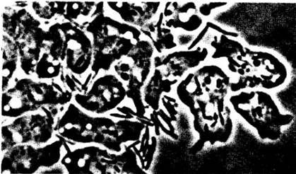
The uptake of solid particles, such as Bacteria, is called phagocytosis, from the Greek word phagein meaning "to eat"; many single-celled organisms, such as amoebas, feed in precisely this manner. Among the organisms discussed in this book, phagocytosis is characteristic of plasmodial and cellular slime Molds (Fig. 4-11).
The uptake of dissolved substances, as opposed to solid particles, is sometimes designated by the specific term pinocytosis (from the Greek pinein meaning "to drink"), although in principle it is the same process as phagocytosis. Pinocytosis occurs not only in unicellular organisms, but also in multicellular plants and animals.
Class="center">Fig. 4-11. The amoeboid feeding stage of the cellular slime mold Dictyostelium aureum. The amoebas were feeding via phagocytosis. Note the bacterial Cells Escherichia coli inside and outside the amoebas
Phagocytosis and pinocytosis can also occur in reverse. Many substances are exported from cells in vesicles or specialized vacuoles. A prime example is the involvement of dictyosome vesicles in Cell wall formation, as described in Chapter 2. These vesicles, loaded with cell wall components, move toward the cell surface. When they reach the plasma membrane, the membrane surrounding them fuses with it, and the Contents of the vesicles are incorporated into the forming cell wall. Reverse endocytosis is termed exocytosis.
Although phagocytosis and pinocytosis at first glance appear different from membrane transport systems involving carrier molecules, they share a common fundamental basis. All three mechanisms depend on the ability of the membrane to "recognize" specific molecules.
Transport Through Plasmodesmata
As described in Chapter 2, plant cells are connected by fine strands of cytoplasm known as plasmodesmata, through which substances move from Cell to Cell. The term symplast is used to refer to this interconnected protoplast system together with its plasmodesmata. Movement of substances via the plasmodesmata is called symplastic transport. In contrast, the movement of substances through the continuous system of cell walls, or apoplast, surrounding the symplast is referred to as apoplastic transport.
Plasmodesmata can provide a more efficient exchange of metabolites between adjacent cells than the less direct alternative pathway through a plasma membrane, a cell wall, and a second plasma membrane. It is believed that Cells and Tissues distant from direct nutrient sources can be supplied by simple diffusion or via plasmodesmata. Possibly, certain substances move through plasmodesmata to and from the xylem and phloem (the Vascular Tissues of plants). Substances may pass through desmotubules connected to The Endoplasmic reticulum of neighboring cells and/or through the channels surrounding the desmotubules, provided that these desmotubules and/or channels are not constricted or blocked (Fig. 4-12).
Fig. 4-12. Diagram of possible structural variations in plasmodesmata. A. The desmotubule and the annular cytoplasmic sleeve located between the desmotubule and the plasma membrane lining the plasmodesmal canal are open. B. The annular sleeve is open, and the desmotubule is closed. C. The desmotubule is open, and the annular sleeve is constricted at both ends

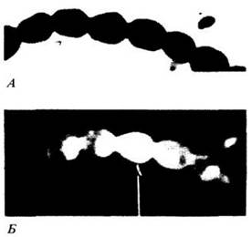
Evidence for intercellular transport via plasmodesmata has been obtained from experiments using fluorescent Dyes and the recording of electrical impulses. The former demonstrated how a dye, which barely penetrates the plasma membrane, moves from the cell into which it was microinjected into adjacent and neighboring cells (Fig. 4-13). Electrical impulses applied inside a cell are recorded by electrodes inserted into neighboring cells. The magnitude of the measured electrical signal varies depending on the density of plasmodesmata, as well as the number and size of the cells between the two electrodes. Whether plasmodesmata can actively regulate the movement of substances from cell to cell remains unresolved, although some researchers believe that certain plasmodesmata may possess "Valves."
Fig. 4-13. Staminal hairs of Setcreasea purpurea before (A) and after (B) the microinjection of a fluorescent dye (fluorescein disodium salt) into one of the cells. Following the Introduction of the dye into the Cell Cytoplasm (indicated by the arrow), it has moved into the cytoplasm of neighboring cells. B. Two minutes after injection. Because the plasma membrane is impermeable to the dye, it moves from cell to cell through the plasmodesmata traversing the walls of adjacent cells
Last update: 07/08/2026
Editorial and Educational Adaptation: This material has been compiled based on the primary/original source text. The project team performed an editorial review, corrected technical inaccuracies, structured sections, and adapted the content for an educational format.
What was processed:
- elimination of formatting defects (OCR errors, structural breaks, corrupted characters);
- editorial organization of content;
- standardization of terminology in accordance with academic sources;
- verification of factual statements against the original source text.
All mentions of the author, publication year, and origin of the primary text have been preserved in accordance with the source.