Biochemistry - The Chemical Reactions of Living Cells, Volume 2 - D. Metzler 1980
Biosynthesis: how new molecules are formed
Polysaccharide synthesis
Lipid carriers
A cluster of carbohydrate residues with a specific Structure forms the repeating structural unit of the Salmonella O-antigen (Fig. 5-11). Recall that this O-antigen is linked to a complex lipopolysaccharide. Both this lipopolysaccharide and the O-antigen are presumably synthesized within the bacterial Cell by Enzymes associated with the cytoplasmic membrane [13, 14]. The fully assembled lipopolysaccharide structure is then transported from the inner bacterial membrane to the outer membrane. While research on the Synthesis of the lipopolysaccharide "core" is still in its early stages, The Biosynthesis of the O-antigen has been studied in relatively great detail; we present an outline of this process in Fig. 12-3. A distinctive feature of this biosynthetic pathway is its dependence on a specialized polyprenol alcohol, undecaprenol (bactoprenol). This C55 compound, containing two trans double bonds and 9 cis double bonds, belongs to the family of isoprenoid Lipids found in All living organisms, where they act as carriers in biosynthetic processes occurring on or within cell membranes.
Class="center">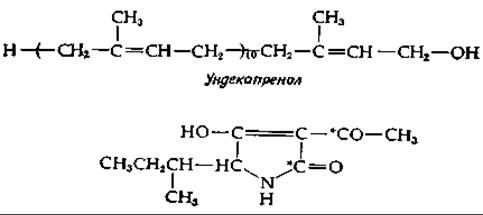
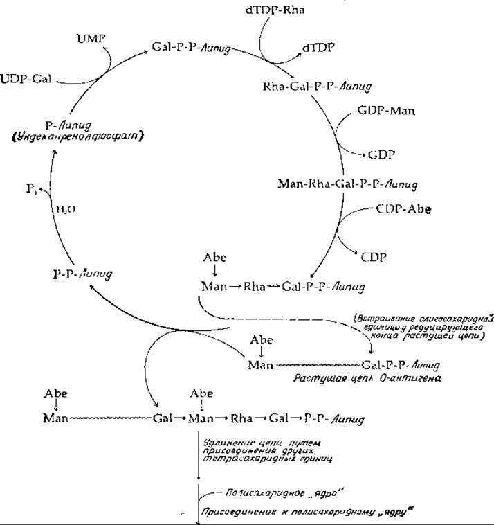
FIG. 12-3. Biosynthesis of the O-antigen polysaccharide in Salmonella typhimurium.
The Selection/23.html">Homologous Series of related compounds known as dolichols contains 16–20 prenyl residues, of which the terminal residue bearing the OH group is fully saturated [15]. Like undecaprenol [15a], these compounds form phosphate esters. The phosphate group of the resulting ester apparently projects into the Cytoplasm, whereas the lipid chain is firmly anchored in the membrane. Naturally, this picture is incomplete. The specific geometry of polyprenyl chains indicates that they perform some more complex structural and mechanical function that remains to be discovered.
During O-antigen synthesis, undecaprenyl phosphate (abbreviated as P-lipid in Fig. 12-3) reacts with UDP-galactose, transferring a phosphogalactosyl residue to the lipid carrier. Subsequently, through a sequential series of transfer Reactions Catalyzed by three additional transferases, an oligosaccharide residue is formed—representing the repeating unit of the O-antigen structure. In the case of the antigen shown in Fig. 12-3, one enzyme transfers a rhamnose residue, a second transfers mannose, and a third transfers abequose from their respective nucleotide sugars. Next, the entire growing O-antigen chain, attached to a second undecaprenyl diphosphate molecule, is joined to the tail of the already assembled oligosaccharide residue on another undecaprenol molecule, as illustrated in Fig. 12-3. Thus, the oligosaccharide is incorporated into the growing chain at its reducing end. Chain elongation proceeds via The transfer of the entire chain to the next tetrasaccharide unit. With each Addition of an oligosaccharide unit, undecaprenyl diphosphate is released; its terminal phosphoryl group is then cleaved off by the action of a phosphatase to regenerate the initial carrier, undecaprenyl (mono)phosphate. Finally, once the synthesized O-antigen reaches sufficient length, it is attached to the lipopolysaccharide core structure.
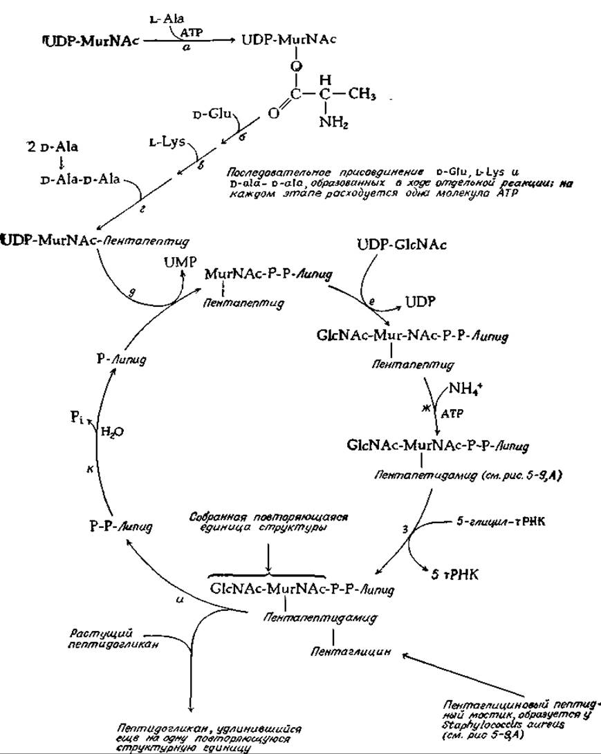
FIG. 12-4. Biosynthesis of peptidoglycan in Staphylococcus aureus. The structure of peptidoglycan is shown in Fig. 5-9, L.
Undecaprenyl phosphate is also essential for another biosynthetic cycle (Fig. 12-4)—namely, The formation of Introduction/37.html">Bacterial Cell wall peptidoglycans (Fig. 5-9). The first step of this process is the synthesis of UDP-N-acetylmuramic acid according to equation (12-5). Next, L-Alanine is added to the OH group of the muramic acid lactyl unit via a typical ATP-dependent reaction (Fig. 12-4, reaction a). This is followed by the sequential ATP-dependent additions of D-glutamic acid, L-Lysine, and D-alanyl-D-alanine. The complete structure assembled in this manner is then transferred to undecaprenyl phosphate (reaction β). Subsequently, N-acetylglucosamine is added through the action of a specific transferase (reaction e), and in some cases, the free α-carboxyl group of the glutamic acid residue binds an ammonium ion, a process driven by the energy of the ATP phosphate bond (reaction ж). Next (reaction з), five glycyl units are added sequentially, with glycyl-tRNA serving as the donor in each instance. The assembled repeating structure, together with the attached peptide chain required for cross-linking (Supplement 7-G), is then transferred to the growing chain (reaction и). Here again, as in the synthesis of dextrans and the Salmonella O-antigen, chain growth proceeds from the reducing end (via an insertion mechanism). This releases polyprenyl diphosphate, and the catalytic cycle is completed by the action of a phosphatase (reaction к).
The polyprenol carrier plays a somewhat different role in the synthesis of Yeast mannan, a polysaccharide. In the main chain of mannan, carbohydrate residues are linked by α-1,6 bonds, but There are also short side chains (consisting of 1–3 mannose residues) attached via α-1,3 and α-1,2 bonds. During mannan synthesis, yeast Cells apparently transfer mannosyl units from GDP-mannose to the phosphate esters of dolichols containing 14–18 prenyl residues [16]. The lipid carrier then transfers the mannose residues to the growing mannan chain. Substantial evidence points to the existence of a similar mechanism for the transfer of mannosyl and N-acetylglucosaminyl units to Glycoproteins in animal Tissues [17–19]. It has been suggested [19] that this yields intermediates with the structure (α-Man)4-(1—>4)-ß-Man-(1—>4)-GlcNac-(1—>4)-GlcNac-P-P-dolichol. Although the participation of polyprenol alcohols in proteoglycan synthesis has not been definitively proven (Section B 1), it is highly probable that they function as carriers during the assembly of the oligosaccharide units of glycoproteins (as shown in Fig. 12-3) [19a]. The synthesized Oligosaccharides are attached to amino acid residues in protein side chains to form glycoproteins, which are subsequently either secreted by the cells or incorporated into The Plasma Membrane.
Last update: 06/08/2026
Editorial and Educational Adaptation: This material has been compiled based on the primary/original source text. The project team performed an editorial review, corrected technical inaccuracies, structured sections, and adapted the content for an educational format.
What was processed:
- elimination of formatting defects (OCR errors, structural breaks, corrupted characters);
- editorial organization of content;
- standardization of terminology in accordance with academic sources;
- verification of factual statements against the original source text.
All mentions of the author, publication year, and origin of the primary text have been preserved in accordance with the source.