Lecture Notes in Human Anatomy - Hryhorieva O.A., Svitlytskyi A.O. 2020
Anatomy of the Musculoskeletal System
Introduction to Myology
Structure and Classification of Muscles
Lecture Outline:
1. Introduction to Myology.
3. General structural plan of a Muscle.
4. Classification of Muscles.
5. Muscle Groups of The Human Body: Deep, Superficial, and Internal Muscles.
Myology (Myologia) is The Study of muscles. Skeletal muscles represent the active component of the human Locomotor Apparatus. They enable diverse movements between skeletal parts, body displacement in space, and the fixation of the body and its segments in specific positions (such as maintaining an upright posture). Muscles facilitate the mechanisms of breathing, chewing, swallowing, and speech. They influence the position and function of Internal Organs, promote Blood AND Lymph Circulation, and participate in METABOLISM and thermoregulation. Muscles also serve as important analyzers that perceive body position in space and the relative arrangement of its parts. The human body contains approximately 400 muscles. In men, muscles account for 42% of body weight, in women 35%, and in athletes 45–55%. More than 50% of total muscle weight is located in the lower limbs, 25–30% in the upper limbs, and 20–25% in the trunk and HEAD regions. In a living Organism, muscles are constantly in a state of slight tension, or tone, maintained by impulses originating from the Central Nervous system (CNS). In Response to nerve impulses, muscles contract and exert a formative influence on bones, joints, and internal organs.
Muscle Structure. A muscle is an organ representing an integral entity with its own distinct structure, function, and Location within the organism. As an organ, a muscle is composed of Striated Muscle tissue forming its basis, Cytology/practical/45.html">Dense Connective Tissue, Blood Vessels, and nerves. Muscles contain nerve endings—receptors—and are innervated by nerve fibers. Each muscle features a central, contractile part
known as the belly, and tendinous extremities (tendons) for attachment. The muscle belly consists of muscle bundles. A muscle bundle is a group of muscle fibers enclosed in connective tissue (perimysium). A muscle fiber is a single muscle Cell of striated muscle tissue. Externally, the muscle belly is covered by a tough connective tissue sheath called the fascia. Tendons are formed under the Influence of the magnitude and direction of muscle force—the greater this force, the more robustly the tendon develops. Consequently, each muscle possesses characteristic tendon size and shape. Tendons are white and shiny, whereas the muscle itself is red. Tendons are exceptionally strong; for instance, the Achilles (calcaneal) tendon can withstand loads of up to 400 kg, and the tendon of the quadriceps muscle up to 600 kg. Tendons attach to the periosteum of bones, to the Skin, or to organs. The Accessory apparatus of muscles comprises anatomical structures that assist muscles in performing their functions. These include fascias, synovial bursae, tendon sheaths, intermuscular septa, and sesamoid bones. Fascia is a connective tissue membrane composed of Collagen and elastic fibers, located On the surface of a muscle or deep beneath it.
Class="center">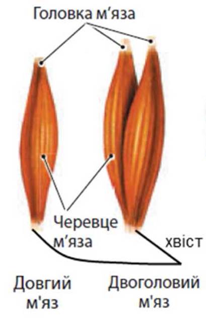
Synovial bursae are thin-walled connective tissue sacs filled with fluid, located beneath muscles, between a muscle and a bone, or between a muscle and a tendon, where they serve to reduce friction. Sesamoid bones develop within the thickness of tendons; they alter the angle at which a muscle approaches a bone and increase the muscle's leverage. The Patella is the largest sesamoid bone. The accessory apparatus of muscles provides additional support—a soft Skeleton—determines the direction of muscle pull, facilitates isolated contraction, prevents displacement during contraction, increases muscle strength, and promotes blood and lymph return.
Muscle Classification. All muscles are classified according to their shape, the direction of their muscle fibers, the number of bellies (heads) or tendons, their location, their relationship to joints, and their functions. Based on shape, muscles are divided into long, short, and broad. In long muscles, the longitudinal dimension exceeds the transverse one. These muscles always contract fully, have a small attachment area on the bone, are located primarily in the extremities, and provide a wide range of limb movement. In short muscles, the longitudinal dimension only slightly exceeds the transverse one. These muscles are found in body regions where the range of motion is limited (between individual vertebrae, between the Occipital bone and the atlas, etc.). Broad muscles are located mainly in the trunk and limb girdles. These muscles contain bundles of muscle fibers running in various directions, capable of contracting either as a whole or in separate parts, and they possess a large bone attachment area. Unlike other muscles, broad muscles perform not only a locomotor function but also postural and protective functions. For example, Abdominal muscles not only participate in trunk movements and breathing acts but also reinforce the abdominal wall, thereby supporting internal organs.
According to the direction of muscle fibers, a distinction is made between muscles with parallel fibers running along the belly, and those with transverse, oblique, and circular fibers. Muscles with circular fibers surround orifices and constrict them upon contraction; such muscles are called sphincters. Simple muscles feature a single belly and two tendons. Muscles with two, three, or four bellies and multiple tendons are classified as complex. Based on their anatomical location in the human body, muscles are subdivided into superficial, deep, external, internal, medial, and lateral. Regarding their relationship to joints, muscles are classified as monoarticular, biarticular, or multiarticular, depending on whether they cross one, two, or multiple joints. Functionally, muscles are grouped as follows: for limb movements—flexors, extensors, pronators, supinators, abductors, and adductors; for trunk movements—flexors, extensors, lateral flexors (right/left), and rotators (right/left); regarding specific body parts—elevators, depressors, protractors, and retractors; and regarding orifices—sphincters and dilators.
Muscles belonging to the same functional group perform the same motor function and are called synergists. Muscles performing opposing actions are termed antagonists—for instance, flexor muscles act as antagonists to extensor muscles.
Muscle activity manifests either in the fixation of the body and its parts or in movement. The former is termed static work, and the latter dynamic work. Static work is also known as holding work, whereas dynamic work involves the Displacement of the body or its parts.
Age-Related Characteristics of Muscles. Up to the age of 7, muscles grow primarily in length, while transverse dimensions change very little. During Puberty, muscle length increases more rapidly than thickness, resulting in only a minor gain in muscle strength during this period. Functionally more loaded muscles grow faster than those used less frequently. The ratio between flexors and extensors changes with age. In young children during their first years of life, flexors and extensors are equally developed, with the exception of FOOT muscles; gradually, extensor muscles begin to predominate in the lower limbs, and flexors in the upper limbs. Muscle strength also changes. The peak increase in grip strength in boys is observed at 15–16 years of age, and in girls at 12; the greatest gain in back strength (strength of the spinal extensors) occurs at 16–18 years in boys and 14–16 years in girls; respiratory muscle strength increases up to age 17 in boys and up to 12–13 years in girls.
Last update: 08/08/2026
Editorial and Educational Adaptation: This material has been compiled based on the primary/original source text. The project team performed an editorial review, corrected technical inaccuracies, structured sections, and adapted the content for an educational format.
What was processed:
- elimination of formatting defects (OCR errors, structural breaks, corrupted characters);
- editorial organization of content;
- standardization of terminology in accordance with academic sources;
- verification of factual statements against the original source text.
All mentions of the author, publication year, and origin of the primary text have been preserved in accordance with the source.