General and Sports Anatomy - L.V. Kapilevich, K.V. Davletyarova 2008
Anatomy of the Musculoskeletal System
Myology
Myology is The Study of Muscles.
The total Muscle mass accounts for 35–40% of body weight.
Skeletal muscles consist of Striated Muscle tissue.
Classification of Muscles:
1. by shape and Structure (Figure 9): fusiform, biceps, triceps, digastric, ribbon-like, unipennate, bipennate, deltoid, rhomboid, quadratus, trapezius;
2. by function: flexor, extensor, abductor, pronator, supinator;
3. by attachment sites: brachioradialis, sternocleidomastoid;
4. by fiber direction: straight, oblique, transverse;
5. by association: corrugator supercilii, sartorius;
6. by joints involved: monoarticular, biarticular, multiarticular.
A Muscle consists of bundles of fibers surrounded by loose Connective Tissue (endomysium/internal perimysium), and the entire muscle is enclosed externally by the epimysium (outer perimysium). Blood Vessels and nerves branch within the connective tissue. A muscle has a belly (body) that transitions into a tendon and attaches to bones. Tendons are formed from Collagen fibers. Broad and flat tendons are known as aponeuroses. Muscles are externally covered by fascia (fibrous membranes).
Over the tendons, the fascia thickens to form synovial sheaths. Two layers of fascia fuse to form a tendinous mesentery filled with synovial fluid.
Synovial bursae may form around muscles, also filled with a mucous substance that facilitates gliding. The fascia forms the neuromuscular hilum (muscle portal) through which nerves and vessels enter.
Class="center">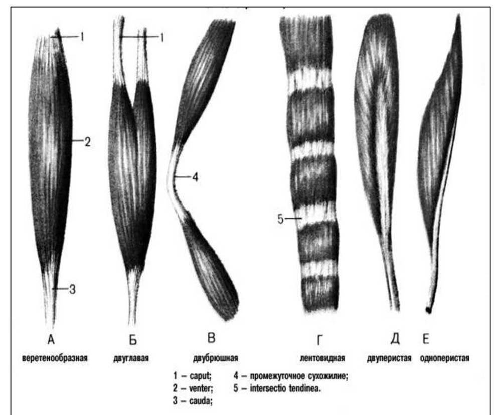
Figure 9. Muscle shapes.
Muscles that participate in the same movement are synergists. As a rule, they work together.
Antagonists perform movements in opposite directions. For example, the muscle group that flexes the FOOT is antagonistic to the muscles that extend the foot.
Muscle Function and Work
Depending on The ratio of the force generated by the muscle to the resistance of the object interacting with it, Three types of muscular work are distinguished (Table 1).
Table 1 Types of muscular work
|
Type of muscle work |
Muscle torque / force moment |
|
concentric (overcoming) |
force is greater than resistance |
|
eccentric (yielding) |
force is less than resistance |
|
isometric (holding) |
force is equal to resistance |
Levers
A first-class lever (lever of equilibrium) has forces applied on opposite sides of the fulcrum and acting in the same direction (Figure 10).
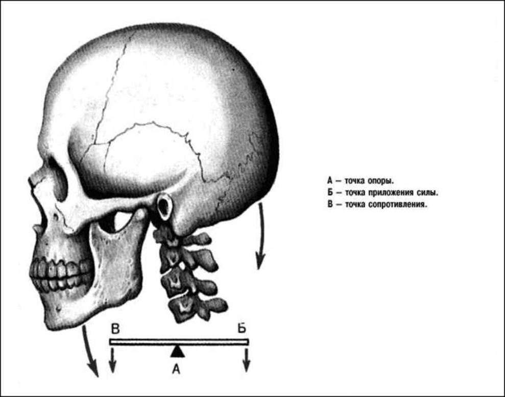
Figure 10. Balance lever.
Class 2 lever – forces act on the same side of the fulcrum. There are two types of this lever:
1. Class 2a power lever: the force arm of the muscle is longer than the gravity force arm (e.g., the foot when rising onto the forefeet). Lever movements are limited; There is a mechanical advantage in force, but a disadvantage in amplitude and speed (Figure 11);
2. Class 2b speed lever: the muscle arm is shorter than the gravity force arm (Figure 12). The arm ratio is 1:10, meaning the load is 10 times greater. When lifting a 16 kg kettlebell, the load on the muscle reaches 160 kg. There is a mechanical disadvantage in force, but an advantage in movement amplitude and speed.
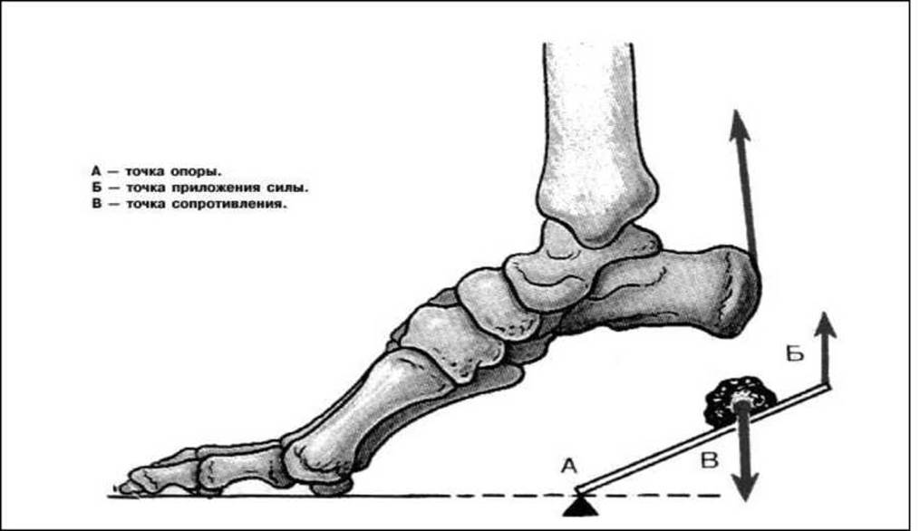
Figure 11. Power lever.
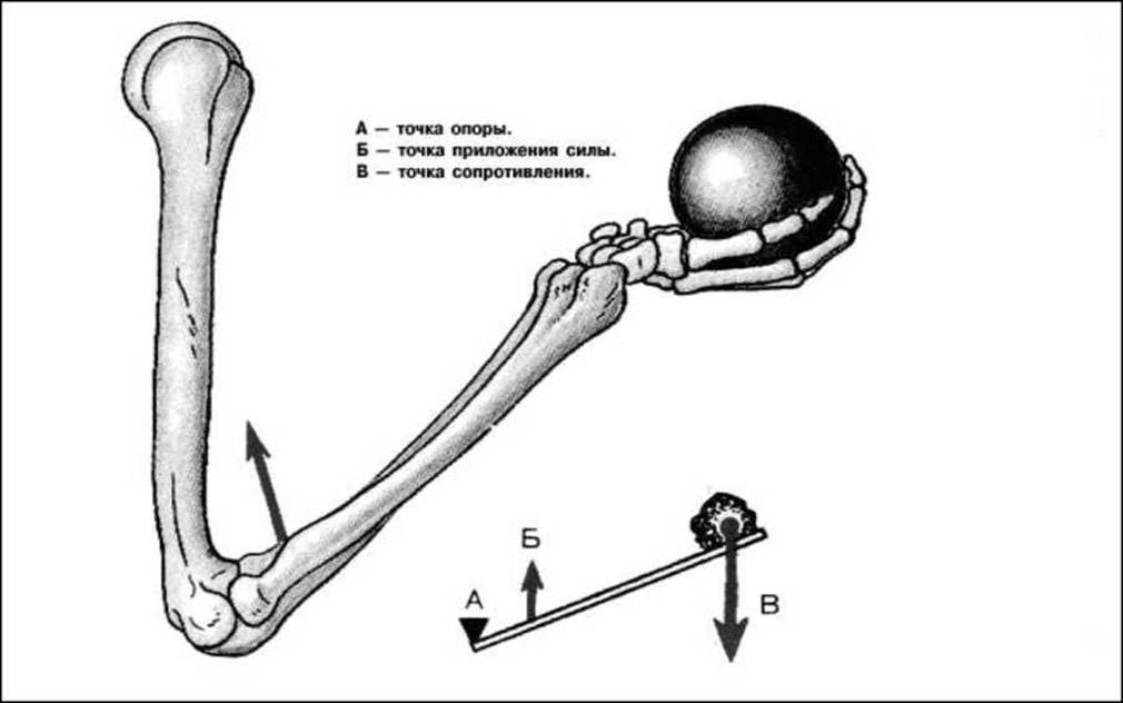
Figure 12. Speed lever.
Muscles of the Upper Limb
Movements of the Pectoral Girdle
Forward movement:
1. Pectoralis major (Figure 13). Attachment: crest of the greater tubercle of the humerus – medial part of the clavicle, anterior surface of the Sternum, cartilaginous parts of Ribs 1–5, and the anterior wall of the rectus abdominis sheath. Main function: movement of the arm.
2. Pectoralis minor. Located beneath the pectoralis major. Attachment: ribs 2, 3, 4, and 5 – coracoid process of the scapula.
3. Serratus anterior (Figure 14). Located on the lateral surface of the chest. Attachment: ribs 1–9 – medial border of the scapula. Main function: moves the scapula forward and upward.
Backward movement:
1. Latissimus dorsi (Figures 13, 15). Attachment: spinous processes of the lower 5–6 thoracic vertebrae, all lumbar and upper sacral vertebrae, posterior part of the iliac crest, lower 4 ribs – crest of the lesser tubercle of the humerus. Main function: adduction of the arm; well-developed in skiers and rowers.
2. Rhomboid muscles – Major and minor. Attachment: spinous processes of the lower two cervical and upper four thoracic vertebrae – medial border of the scapula. Function: adduction of the scapula, but can also extend and bend the spine.
3. Trapezius (Figures 13, 15). Triangular in shape. Attachment: Occipital bone, nuchal ligament, spinous processes of the thoracic vertebrae – pectoral girdle, clavicle, acromion of the scapula, spine of the scapula. Functions: moves the scapula backward; extends the spine and HEAD backward (both muscles) or tilts them to the side (one muscle).
Upward movement:
1. Sternocleidomastoid muscle (Figure 13). Has two heads. Attachment: manubrium of the sternum, clavicle – mastoid process of the Temporal bone. When the neck and head are fixed, it elevates the clavicle and, consequently, the scapula. Tilts the head to its own side and turns it to the opposite side. Acting together, both muscles pull the head backward.
2. Levator scapulae. Attachment: transverse processes of cervical vertebrae 1–4 – medial border of the scapula. Lies beneath the trapezius and sternocleidomastoid muscles.
3. Upper fibers of the trapezius muscle (see above).
4. Rhomboid muscles (see above).
Downward movement.
1. Subclavius. Attachment: 1st rib — Inferior surface of the acromial end of the clavicle. Pulls the clavicle downward and forward.
2. Pectoralis minor (see above).
3. Lower fibers of the trapezius (see above).
4. Lower digitations of the serratus anterior (see above).

Figure 13. Human muscles (anterior view).

Figure 14. Human muscles (anterior view).

Figure 15. Human muscles (posterior view).
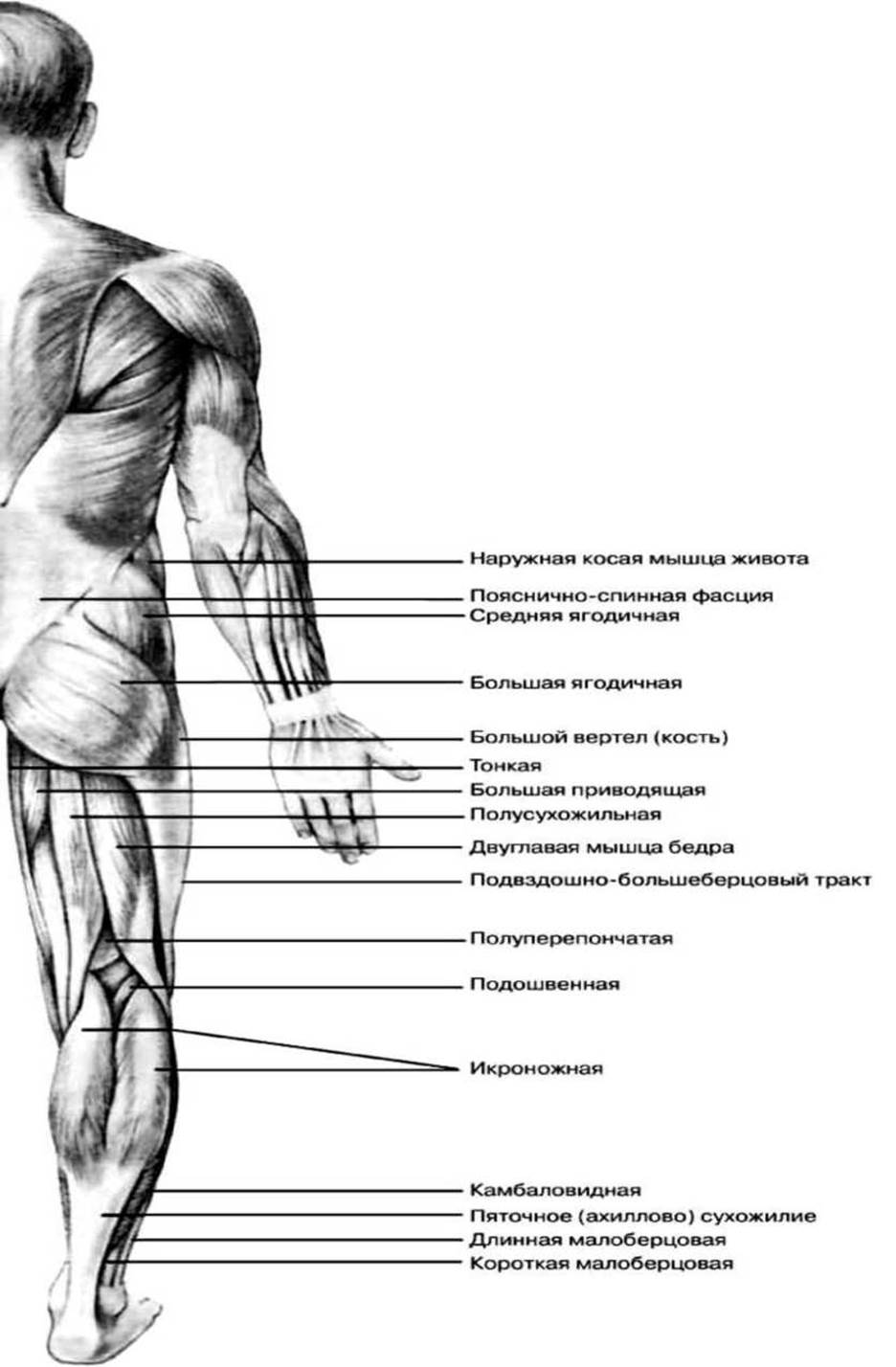
Figure 16. Human muscles (posterior view).
Rotation of the scapula.
Inward, toward the spine: pectoralis minor, lower part of the rhomboideus major.
Outward, away from the spine: serratus anterior, teres major.
Movements at the shoulder joint
Abduction of the arm: deltoid, supraspinatus.
1. Supraspinatus muscle. Located beneath the deltoid. Attachment: supraspinous fossa of the scapula — greater tubercle of the humerus.
2. Deltoid (Figures 13, 15, 17). Attachment: clavicle, acromion, and spine of the scapula — deltoid tuberosity of the humerus. Contraction of the anterior or posterior part alone produces forward and backward movement, respectively. Contraction of the whole muscle causes abduction.
Adduction of the arm. There are no dedicated muscles crossing the sagittal axis of the joint; therefore, According to the parallelogram rule, adduction is performed by the pectoralis major located anteriorly, and the latissimus dorsi and teres major located posteriorly, along with several accessory muscles:
1. Teres minor (Figure 15) — located inferior to the infraspinatus, shares the same attachment points.
2. Infraspinatus (Figure 15). Attachment: infraspinous fossa of the scapula — greater tubercle of the humerus.
3. Subscapularis. Attachment: subscapular fossa — lesser tubercle of the humerus.
4. Teres major (Figure 15). Shares a common tendon with the latissimus dorsi. Attachment: inferior angle of the scapula — crest of the lesser tubercle of the humerus.
Flexion of the arm. The muscles cross the frontal axis of the joint and are located anterior to it.
1. Coracobrachialis muscle. Attachment: coracoid process of the scapula, fusing with the short head of the biceps brachii to insert into the humerus.
2. Other muscles involved: deltoid, pectoralis major, and biceps brachii.
Shoulder extension. Crossing the frontal axis and located posteriorly: deltoid, latissimus dorsi, infraspinatus, teres minor, teres major, and the long head of the triceps brachii.
Shoulder pronation (medial rotation). Crossing the vertical axis of the joint from the anterior side: subscapularis, pectoralis major, anterior part of the deltoid, latissimus dorsi, teres major, and coracobrachialis.
Shoulder supination (lateral rotation). Crossing the vertical axis from the posterior side: infraspinatus, teres minor, and posterior part of the deltoid.
Circumduction of the shoulder is performed sequentially by all surrounding muscles. Although there are no muscles on the inferomedial side of the joint, the axillary fossa allows for full shoulder adduction.
Movements in the elbow joint
Forearm movements: flexion and extension, pronation and supination.
Forearm flexion.
1. Brachialis muscle (Figures 13, 15). A single-joint muscle. Attachment: lower half of the anterior surface of the humerus to the ulnar tuberosity.
2. Biceps brachii (Figures 13, 17). Located on the anterior aspect of the arm and easily palpable. The long head originates from the supraglenoid tubercle of the scapula, and the short head from the coracoid process. Distally, it inserts into the radial tuberosity. Functions: flexes the shoulder; the long head also abducts, while the short head adducts. Together, they stabilize the shoulder in the vertical plane. At the forearm: flexion and partial supination (though an aponeurosis extending from the tendon toward the ulna diminishes the supinating effect).
3. Pronator teres (Figure 13) — runs obliquely downward and laterally. Functions: flexion and pronation. Attachment: medial epicondyle of the humerus to the mid-shaft of the radius (lateral side).
4. Brachioradialis (Figures 13, 15). Attachment: lateral epicondyle of the humerus to the radius (just proximal to the styloid process). Performs flexion, as well as pronation when fully supinated, or supination when fully pronated.
Forearm extension.
1. Triceps brachii (Figures 13, 15, 17). Consists of three heads: the long head (attached to the infraglenoid tubercle of the scapula), and the medial and lateral heads (attached to the posterior surface of the humerus). Distally, it inserts into the olecranon of the ulna. Extends the elbow joint, while the long head also assists in shoulder extension.
2. Anconeus muscle (Figure 15). Attachment: lateral epicondyle of the humerus to the posterior surface of the proximal ulna. Extends the forearm.
Forearm pronation.
1. Pronator teres (see above).
2. Pronator quadratus. Located across the distal BONES OF THE forearm, running from the ulna to the radius.
3. Brachioradialis (see above).
Forearm supination.
1. Supinator muscle. Lies deep across the forearm bones. Attachment: lateral epicondyle of the humerus to the proximal third of the radius. Function: rotates the radius laterally at the proximal and distal radioulnar joints.
2. Brachioradialis muscle (see above).
3. Biceps brachii (see above).
Movements in the wrist joint and hand
Wrist flexion involves six muscles of the anterior forearm and hand.
1. Flexor carpi radialis (Figures 13, 17). Attachment: medial epicondyle of the humerus – Base of the 2nd metacarpal bone. Flexion at the radiocarpal joint; pronation and flexion at the elbow joint.
2. Palmaris longus (Figure 13). Attachment: medial epicondyle of the humerus – palmar aponeurosis.
3. Flexor carpi ulnaris (Figures 13, 15, 17). Attachment: medial epicondyle of the humerus – pisiform bone, with ligaments extending from it to the hook of the hamate and the 5th metacarpal bone.
4. Flexor digitorum superficialis (Figure 13). Attachment: medial epicondyle of the humerus, radius, and ulna – Phalanges of digits 2–5 (has 4 tendons). Flexes the middle phalanges and the entire hand, and makes a fist. When the hand is extended, this muscle stretches, making it harder to bend the fingers. If you want to pull an object out of someone's grasp, you extend their wrist (backward).
5. Flexor digitorum profundus. Attachment: upper part of the palmar surface of the ulna – distal phalanges of digits 2–5. Flexes and adducts the fingers; because the tendons fan out, it is difficult to bend spread fingers.
6. Flexor pollicis longus. Attachment: palmar surface of the ulna – distal phalanx of the thumb.
Wrist extension involves seven muscles of the posterior forearm.
1. Extensor carpi radialis longus (Figures 13, 15). Attachment: humerus, lateral border, and lateral epicondyle – base of the 2nd metacarpal bone.
2. Extensor carpi radialis brevis (Figures 13, 15). Attachment: lateral epicondyle of the humerus – base of the 3rd metacarpal bone. Extension and abduction of the hand.
3. Extensor carpi ulnaris (Figure 15). Attachment: lateral epicondyle of the humerus, forearm ligaments – base of the 5th metacarpal bone. Extension and adduction.
4. Extensor digitorum (Figure 15). Attachment: lateral epicondyle of the humerus, forearm ligaments – phalanges of digits 2–5 (4 tendons), reinforced by intertendinous connections. Each splits into 3 slips: the middle slip attaches to the middle phalanx, and the lateral slips attach to the distal phalanges. During forceful wrist flexion, the fingers extend due to passive tension of this muscle.
5. Extensor indicis. Attachment: dorsal surface of the ulna, interosseous membrane – 2nd digit, distal and middle phalanges.
6. Extensor digiti minimi (Figure 15). Attachment: lateral epicondyle of the humerus, forearm ligaments – dorsal aponeurosis of the 5th digit.
Bone adduction – there are no muscles on the medial side of the forearm. Adduction occurs via the parallelogram rule through the action of the flexor carpi ulnaris and extensor carpi ulnaris.
Hand abduction:
1. Flexor carpi radialis (see above).
2. Extensor carpi radialis longus (see above).
3. Extensor carpi radialis brevis (see above).
4. Abductor pollicis longus (Figure 15). Fusiform in shape. Attachment: radius, ulna, and interosseous membrane – base of the 1st metacarpal bone. Abducts the thumb, and when antagonists contract, the entire hand.
5. Extensor pollicis longus. Attachment: posterior surface of the radius and ulna, interosseous membrane – distal phalanx of the thumb.
6. Extensor pollicis brevis (Figure 15). Attachment: posterior surface of the radius and ulna, interosseous membrane – phalanges of the thumb (the long extensor attaches to the distal phalanx, the short extensor to the proximal).
Intrinsic Muscles of the hand
Middle Group of hand muscles:
1. Lumbricals. Extend to all digits except the 1st. Attachment: tendons of the flexor digitorum profundus – proximal phalanges. Flex the proximal phalanges of digits 2–5.
2. Palmar interossei (three in number). Attachment: bases of 2nd–5th metacarpals to the joint capsules of the metacarpophalangeal joints. Function: flexion and adduction toward the middle finger.
3. Dorsal interossei (four). Attachment: adjacent surfaces of the Metacarpal bones to the extensor expansion of the digits. The middle finger has two; the 2nd has one (radial); the 4th has one (ulnar). While flexing the proximal phalanges of the 2nd–5th digits, they simultaneously extend the distal phalanges and abduct the 2nd and 4th digits away from the 3rd.
Thenar muscle group:
They form the thenar eminence (ball of the thumb).
1. Abductor pollicis brevis. Attachment: flexor retinaculum and scaphoid bone to the proximal phalanx. Abduction of the thumb.
2. Flexor pollicis brevis. Attachment: flexor retinaculum and trapezium to the sesamoid bone. Flexion of the 1st phalanx and extension of the 2nd phalanx; opposition of the thumb.
3. Opponens pollicis. Attachment: flexor retinaculum and trapezium to the 1st metacarpal bone. Opposition of the thumb.
4. Adductor pollicis. It has two heads: transverse (attached to the shaft of the 3rd metacarpal) and oblique (attached to the bases of the 2nd and 3rd metacarpals). Its other end attaches to the sesamoid bone and the capsule of the metacarpophalangeal joint. Adduction and opposition.
Hypothenar muscle group:
1. Palmaris brevis. One of the few cutaneous muscles. Attachment: palmar aponeurosis to the Skin. Protects Blood Vessels and nerves when clenching the fist and during impacts to the hand.
2. Abductor digiti minimi. Attachment: pisiform bone to the base of the proximal phalanx. Abduction and flexion of the proximal phalanx, and extension of the middle and distal phalanges.
3. Flexor digiti minimi brevis. Attachment: flexor retinaculum and hamate bone to the ulnar margin of the base of the proximal phalanx. Flexion and adduction.
4. Opponens digiti minimi. Attachment: flexor retinaculum and hamate bone to the shaft and head of the 5th metacarpal bone. Adduction of the little finger.
Connective tissue structures of the upper limb
The superficial Fascia of the entire upper limb lies directly beneath the skin. In addition, each upper limb muscle has its own proper (deep) fascia.
The deep Fasciae of the arm and forearm form the flexor and extensor compartments (sheaths).
In the hand: on the palmar side are the synovial sheaths of the flexor tendons, covered by the palmar aponeurosis; on the dorsal side are the extensor sheaths; and around the wrist is the transverse carpal ligament, which forms the carpal tunnel.
Movements at the hip joint
Hip flexion:
1. Iliopsoas. Consists of three parts.
✵ Psoas major. Attached by one end to the bodies and transverse processes of the lumbar vertebrae and the body of the XII thoracic vertebra. Inferiorly, it merges with the iliacus muscle.
✵ Iliacus (Figure 14) is attached by one end to the iliac fossa and by the other to the lesser trochanter.
✵ Psoas minor — attached by one end to the bodies of the XII thoracic and I lumbar vertebrae, and by the second to the pelvic fascia. These muscles pass from the trunk to the thigh beneath the inguinal ligament through the muscular lacuna. It lies adjacent to the hip joint. Function: flexion and lateral rotation (supination) of the hip; if the thigh is fixed, it flexes THE Vertebral Column.
2. Sartorius muscle (Figures 14, 18) — the longest muscle in The Human Body. Attachment: anterior superior iliac spine, running obliquely across the anterior and Medial surface of the thigh to the tibial tuberosity. Flexion of the hip and knee, lateral rotation (supination) of the thigh, and medial rotation (pronation) of the leg.
3. Pectineus muscle (Figure 14). Attachment: superior ramus of the pubis to the linea aspera of the Femur. Flexion, adduction, and lateral rotation (supination) of the hip.
4. Tensor fasciae latae (Figure 14). Attachment: anterior superior iliac spine to the iliotibial tract (iliotibial band), which in turn attaches to the lateral condyle of the Tibia. Function: flexion, medial rotation (pronation), and abduction of the hip.
5. Rectus femoris (Figures 14, 18). One of the heads of the quadriceps femoris muscle (see below).
Hip Extension:
1. Gluteus maximus (Figures 16, 18). Located on the posterior aspect of the hip joint. Attachment: sacrum and posterior surface of the ilium to the gluteal tuberosity of the femur and the fascia lata. Stabilizes the body in an upright position; extends and laterally rotates the hip; extends the trunk from a flexed position.
2. Biceps femoris (Figures 16, 18). Attachment: ischial tuberosity (long head), linea aspera of the femur (short head) to the head of the Fibula. Extends the hip; flexes and extends the knee (leg).
3. Semimembranosus (Figures 16, 18). Attachment: ischial tuberosity to the medial condyle of the tibia. Hip extension, knee flexion.
4. Semitendinosus (Figures 16, 18). Attachment: ischial tuberosity to the tibial tuberosity. Hip extension, knee flexion, and medial rotation of the leg.
5. Adductor magnus — see below.
Hip Abduction:
These muscles are located on the lateral side of the hip joint.
1. Gluteus medius (Figures 16, 18). Located deep to the gluteus maximus. Attachment: external surface of the ilium to the greater trochanter.
2. Gluteus minimus. Attachment similar to the preceding muscle, lying deep to it.
3. Piriformis. Passes through the greater sciatic foramen. Attachment: anterior surface of the sacrum to the greater trochanter.
4. Obturator internus. Located within the lesser pelvis. Attachment: obturator membrane to the trochanteric fossa.
5. Superior and inferior gemelli. Attachment: ischial spine (superior), ischial tuberosity (inferior) to the tendon of the obturator internus. Abduction and lateral rotation of the hip.
6. Tensor fasciae latae (see above).
Hip Adduction:
These muscles are located medially to the hip joint.
1. Pectineus (see above).
2. Gracilis (Figure 14). Attachment: inferior ramus of the pubis to the tibial tuberosity; together with the sartorius and semitendinosus, it forms the superficial pes anserinus. Adduction of the hip and flexion of the knee.
3. Adductor longus (Figure 14). Attachment: superior ramus of the pubis to the linea aspera of the femur. Adduction of the hip.
4. Adductor brevis. Attachment: inferior ramus of the pubis to the linea aspera of the femur.
5. Adductor magnus (Figures 14, 16). Attachment: ischial tuberosity to the linea aspera of the femur. Assists in hip extension, but only from a flexed position.
Lateral Rotation (Supination) of the Hip:
They cross the vertical axis of the joint obliquely.
1. Iliopsoas (see above)
2. Quadratus femoris. Attachment: ischial tuberosity - greater trochanter.
3. Gluteal muscles (see above).
4. Sartorius (see above).
5. Obturator internus (see above).
6. Piriformis (see above).
7. Gemelli (see above).
8. Obturator externus. Triangular in shape. Attachment: obturator membrane - trochanteric fossa and pubic bone.
Hip pronation is performed by the tensor fasciae latae, anterior fibers of the gluteus medius, anterior fibers of the gluteus minimus, semitendinosus, and semimembranosus.
Movements in the knee joint
Leg flexion:
They cross the transverse axis of the joint and are located posteriorly.
1. Biceps femoris (see above)
2. Semitendinosus (see above).
3. Semimembranosus (see above).
4. Sartorius (see above).
5. Gracilis (see above).
6. Popliteus. Short muscle. Attachment: lateral femoral condyle - tibia.
7. Gastrocnemius (see below).
8. Plantaris (see below).
Leg extension:
1. Quadriceps femoris. It consists of four parts.
✵ Rectus femoris (Figures 14, 18). Attachment: anterior inferior iliac spine; it is a biarticular muscle.
✵ Vastus lateralis (Figure 14). Attachment: lateral side of the femur - Patella, from which the patellar ligament extends to the tibial tuberosity.
✵ Vastus medialis (Figures 14, 18). Attachment: lateral aspect of the femur – patella.
✵ Vastus intermedius. Attachment: between the other two superior heads – patella.
Pronation of the lower leg:
There are no dedicated muscles; pronation is only possible as flexion occurs and is performed by the medial flexors: semitendinosus, semimembranosus, sartorius, gracilis, medial head of the gastrocnemius, and popliteus.
Supination of the lower leg:
lateral flexor muscles; possible as flexion occurs at the knee joint.
1. Biceps femoris (see above).
2. Lateral head of the gastrocnemius.
These muscles are weaker than the pronators.
Foot Movements
Plantarflexion (flexion) of the foot:
1. Triceps surae. It has three heads.
✵ Gastrocnemius (Figures 14, 16, 18). It has two heads: lateral and medial. Attachment: femoral condyles – via the Achilles tendon to the calcaneus. It participates in knee joint movement.
✵ Soleus (Figure 16). Single-joint muscle. Attachment: posterior surface of the tibia – via the Achilles tendon to the calcaneus. It plays a major role in maintaining an upright stance; the tendon has a safety margin exceeding 500 kg.
2. Plantaris (Figure 16). Attachment: lateral femoral condyle – via the Achilles tendon to the calcaneus. Rudimentary, absent in 12% of people.
3. Tibialis posterior. Attachment: posterior surface of the interosseous membrane – navicular bone. Performs plantarflexion, adduction, and supination of the foot.
4. Flexor hallucis longus. Attachment: posterior surface of the fibula – distal phalanx of the great toe on the plantar side. Flexes the great toe and the entire foot.
5. Flexor digitorum longus (Figure 14). Attachment: posterior surface of the tibia – four tendons (distal phalanges of digits 2–5). Plantarflexion and supination of the foot, flexion of the toes.
6. Peroneus longus (see below).
7. Peroneus brevis (see below).
Dorsiflexion (extension) of the foot:
1. Tibialis anterior (Figures 14, 18). Attachment: anterior surface of the tibia – 1st metatarsal bone.
2. Extensor digitorum longus (Figures 14, 18). Attachment: upper margin of the tibia and crural fascia – five tendons (distal phalanges of digits 2–5 and 5th metatarsal bone). Extends the toes and the foot.
3. Extensor hallucis longus. Attachment: medial surface of the fibula and interosseous membrane – base of the distal phalanx of the great toe.
Adduction of the foot:
There are no dedicated muscles; they operate according to the parallelogram rule:
1. Tibialis anterior (see above).
2. Tibialis posterior (see above).
Abduction of the foot:
There are no dedicated muscles; they operate according to the parallelogram rule:
1. Peroneus brevis (see below).
2. Peroneus longus (see below).
Pronation of the foot:
1. Peroneus longus muscle (Figures 14, 16, 18) has a pennate structure. Attachment: head of the fibula, lateral condyle of the tibia, and crural fascia—the tendon wraps around the lateral malleolus and attaches to the base of the 1st and 2nd Metatarsal Bones and the cuneiform bone. Flexion, pronation, and abduction of the foot.
2. Peroneus brevis muscle (Figures 14, 16, 18). Attachment: lateral surface of the fibula—5th metatarsal bone.
3. Third fibular muscle (Peroneus tertius). Not present in everyone. It is essentially an additional tendon of the extensor digitorum longus extending to the base of the 5th metatarsal bone.
Supination of the foot.
1. Tibialis anterior (see above).
2. Extensor hallucis longus (see above).
Movements of the toes
I. Plantar surface.
1. Medial group. Attachment: bones and ligaments of the sole—1st digit. Abductor hallucis muscle. Flexor hallucis brevis. Adductor hallucis muscle.
2. Lateral group. Attachment: calcaneus and plantar aponeurosis—proximal phalanx of the 5th toe. Abductor digiti minimi muscle. Flexor digiti minimi brevis.
3. Intermediate group.
✵ Quadratus plantae muscle. Attachment: calcaneus—tendon of the flexor digitorum longus. By pulling on its tendon, it alters the line of pull.
✵ Lumbricals—four in number. Attachment: tendons of the flexor digitorum longus—dorsal aponeurosis. Flex the proximal phalanges, extend the distal ones.
✵ Flexor digitorum brevis. Attachment: calcaneus and plantar aponeurosis—4 tendons to the middle phalanges of the 2nd–5th toes.
✵ Interosseous muscles of the foot.
✵ Dorsal—four in number. Attachment: Tarsal Bones (each from two adjacent ones)—proximal phalanges of the middle toes (except the 1st and 5th).
✵ Plantar—three in number. Attachment: tarsal bones—proximal phalanges of the corresponding toes. Flex the proximal phalanges and extend the distal ones. Additionally, they shift the toes laterally.
II. Dorsal aspect.
1. Extensor digitorum brevis. Attachment: calcaneus to the tendons of the 2nd–4th toes.
2. Extensor hallucis brevis. Attachment: calcaneus to the proximal phalanx of the hallux.
3. Third fibular (peroneus tertius) muscle (see above).
Connective tissue structures of the lower limb muscles
Main fascias: superficial fascia and deep fascia — the fascia lata of the thigh. Other notable fascias include the popliteal fascia, crural fascia, and pedal fascia, which forms the plantar aponeurosis. In the ankle region, the dorsal aspect features anterior, lateral, and medial groups of synovial tendon sheaths.
Muscles of the Trunk and neck
Main functions: maintaining upright posture, spinal movements, supporting abdominal Organs, and respiratory movements.
Movements of the vertebral column
Extension of the vertebral column:
1. Trapezius muscle (see above).
2. Serratus posterior muscles, superior and inferior. Superior attachment: spinous processes of the 2 lower cervical and 2 upper thoracic vertebrae to the posterior surface of the 2nd–5th ribs. Inferior attachment: spinous processes of the last 2 thoracic and upper 2 lumbar vertebrae to the lower 4 ribs.
3. Erector spinae muscle. Running along the entire vertebral column from the sacrum to the Skull, it lies between the transverse and spinous processes. Attachment: arising from the sacrum, it is divided into three columns: the iliocostalis (lateral), the longissimus (intermediate), and the spinalis (medial), extending to the mastoid process and cranial base.
4. Splenius capitis and cervicis muscles. Attachment: nuchal ligament, spinous processes of the 3rd–7th cervical and 1st–6th thoracic vertebrae to the mastoid process of the temporal bone and transverse processes of the 2nd–3rd cervical vertebrae.
5. Transversospinales muscle group. Located beneath the longissimus and spinalis muscles, extending from the sacrum to the occipital bone and consisting of individual bundles passing from the transverse processes of some vertebrae to the spinous processes of others. It forms three layers: superficial — semispinalis, intermediate — multifidus, deep — rotators.
6. Levatores costarum muscles. From the transverse processes of the thoracic vertebrae to the ribs.
7. Suboccipital muscles:
✵ Rectus capitis posterior major and minor. From the occipital bone downward to the posterior tubercle of the atlas and the spinous process of the axis.
✵ Obliquus capitis superior. From the occipital bone to the transverse process of the atlas.
✵ Obliquus capitis inferior. From the transverse process of the atlas to the spinous process of the axis.
Main functions — extension, rotation, and lateral flexion of the head.
Flexion of the vertebral column:
1. Sternocleidomastoid (see above).
2. Scalene muscles (see below).
3. Longus colli and longus capitis muscles (see below).
4. Rectus abdominis (see below).
5. Oblique Abdominal muscles (see below).
6. Iliopsoas (see above).
Lateral flexion:
1. Levator scapulae (see above).
2. Quadratus lumborum (see below).
3. Intercostal muscles (see below).
4. Intertransversarii muscles.
Rotation / Twisting.
1. Sternocleidomastoid (see above).
2. Stylohyoid (see above).
3. Trapezius (see above).
4. Scalenes (see below).
5. External oblique (see below).
6. Rotatores muscles (see above).
7. Iliopsoas (see above).
Superficial:
1. Platysma. It attaches not to bones, but to the Cervical fascia — extending from the angle of the Mouth to the clavicle. Its primary function is to tighten the skin of the neck, enhance venous blood return, and depress the angle of the mouth.
2. Sternocleidomastoid (see above).
Muscles attaching to the Hyoid bone (HB).
They participate in flexion of the Vertebral Column and depression of the Mandible.
1. Omohyoid. From the superior border of the scapula to the HB.
2. Sternohyoid (Figure 13). From the manubrium of the sternum and the clavicle to the HB.
3. Sternothyroid muscle (Figure 13). Origin and insertion: from the manubrium of the sternum to the thyroid Cartilage of the Larynx — the Adam's apple.
4. Thyrohyoid muscle: from the thyroid cartilage to the hyoid bone.
1. Digastric muscle. Attachment: mastoid process to the mandible; an intermediate tendon lies between the two bellies and attaches to the hyoid bone.
2. Stylohyoid muscle. Attachment: styloid process of the temporal bone to the hyoid bone.
3. Mylohyoid muscle. Forms the floor of the Oral Cavity. Attachment: body of the mandible to the hyoid bone.
4. Geniohyoid muscle. Located superior to the third muscle. Attachment: mental spine to the hyoid bone.
Deep muscles:
Lateral group
1. Scalene muscles:
✵ Anterior scalene. Attachment: transverse processes of the 3rd–6th cervical vertebrae to the 1st rib.
✵ Middle scalene. Attachment: transverse processes of all cervical vertebrae to the 1st rib.
✵ Posterior scalene. Attachment: transverse processes of the 5th–6th cervical vertebrae to the 2nd rib.
They tilt and flex the spine, rotate it, and elevate the ribs during inspiration.
Medial group
1. Longus colli muscle. Attachment: bodies of the 2nd–3rd thoracic and 5th–7th cervical vertebrae to the 2nd–4th cervical vertebrae and transverse processes of the lower 3 cervical vertebrae.
2. Longus capitis muscle. Attachment: transverse processes of the 3rd–6th cervical vertebrae to the external base of the skull.
3. Anterior and lateral rectus capitis muscles. Attachment: transverse processes of the atlas to the basilar part of the occipital bone.
Connective tissue structures of the neck
The superficial layer is very poorly developed.
The pretracheal layer encloses all the muscles of the neck and the organs located anterior to the deep neck muscles and the vertebral column.
The prevertebral layer covers the deep neck muscles and the vertebral column anteriorly.
Abdomen: extends from the inferior margin of the sternum to the inguinal ligament, Pubic Symphysis, and iliac crests.
1. Rectus abdominis muscle (Figure 14). Located anteriorly. Attachment: xiphoid process and 5th–7th costal cartilages to the superior margin of the pubic bone. It features three to four tendinous intersections. Performs various functions: trunk flexion, and forms the abdominal press.
2. Pyramidalis muscle. Triangular in shape. Attachment: pubic bone - linea alba. Tenses the linea alba. May be absent.
3. External oblique muscle of the abdomen (Figures 14, 16). Flexes and rotates the spine. Has eight digitations. Attachment: from the lower eight ribs, runs anteroinferiorly, and continues into the aponeurosis and linea alba inferiorly, forming the inguinal ligament.
4. Internal oblique muscle of the abdomen. Located deep to the external oblique, originates from the thoracolumbar fascia, and attaches to the linea alba and the lower three ribs. Flexes and rotates the spine.
5. Transversus abdominis muscle. Located deep to the obliques. Attachment: cartilages of the lower six ribs - linea alba and pubic bone. Strengthens the abdominal wall and pulls the ribs downward.
Posterior wall:
6. Quadratus lumborum muscle. Attachment: iliac crest and transverse processes of the lower lumbar vertebrae - 12th rib and upper lumbar vertebrae. Flexes the spine laterally and depresses the rib.
Connective tissue structures of the abdomen
The subcutaneous fascia is well-developed, covering the abdominal press externally. The deep fascia of the abdomen consists of several layers corresponding to the muscles of the abdominal wall.
Linea alba - extends from the xiphoid process to the pubic symphysis, formed by the interlacing of the aponeurotic fibers of all abdominal muscles.
Umbilical ring - located along the linea alba.
Inguinal Canal - a slit-like passage between the abdominal muscles; in men, it transmits the Spermatic Cord, while in women, it transmits the round ligament of the Uterus.
The linea alba, umbilical ring, and inguinal canal are weak points of the abdomen (areas of least resistance of the abdominal wall) and serve as potential sites for hernia formation.
The Thorax expands in three directions: anteroposterior and transverse, due to the lateral, upward, and forward movement of the ribs. The vertical dimension is managed by the Diaphragm.
Primary:
1. Diaphragm. A dome-shaped structure facing upward; upon contraction, it pulls on three parts: sternal, costal, and lumbar. It features aortic and esophageal openings, as well as the opening for the INFERIOR VENA CAVA. Its excursion is 3–4 cm, which may reach 6–8 cm in athletes.
2. External and internal intercostal muscles. Fill the intercostal spaces. The external fibers run inferoanteriorly, while the internal fibers run superoanteriorly. Both participate in inspiration and expiration, with the external predominating during inspiration and the internal during expiration.
3. Serratus posterior superior muscle (see above).
4. Serratus posterior inferior muscle (see above).
5. Quadratus lumborum muscle (see above).
6. Iliocostalis muscle (see above).
Accessory muscles of inspiration: scalene muscles, sternocleidomastoid, pectoralis minor, subclavius, pectoralis major, serratus anterior, anterior neck muscles, erector spinae.
Indirectly involved in inspiration: trapezius, rhomboids, levator scapulae.
None; passive exhalation is a quiet process.
Accessory muscles:
1. Abdominal muscles—antagonists of the diaphragm.
2. Internal and external intercostal muscles (see above).
3. Subcostal muscles. Thin, triangular muscles located on the inner surface of the ribs (attaching to the ribs).
4. Transversus thoracis muscle. Originates from the xiphoid process, runs along the posterior surface of the costal cartilages, and attaches to ribs 2–6 near their junction with the cartilages.
5. Serratus posterior inferior (see above).
6. Quadratus lumborum (see above).
7. Iliocostalis (see above).
Elevation of the mandible:
1. Temporalis (Figure 19). Attachment: temporal fossa to the coronoid process of the mandible.
2. Masseter (Figure 19). Attachment: zygomatic arch to the ramus of the mandible.
3. Medial pterygoid. Attachment: pterygoid process of the Sphenoid bone to the angle of the mandible.
Depression of the mandible:
1. Digastric (see above).
2. Platysma (see above).
3. Infrahyoid muscles (see above).
Protraction (forward movement):
1. Lateral pterygoid. Attachment: sphenoid bone to the neck of the mandible.
Retraction (backward movement):
1. Posterior part of the temporalis.
2. Digastric.
3. Certain suprahyoid muscles.
Lateral movements:
1. Lateral pterygoid. Moves the jaw to the opposite side upon isolated contraction.
Facial Muscles
Some originate from bones and insert into the skin, while others lie entirely within soft Tissues. Their primary function is to dilate and constrict bodily openings.
1. Epicranial muscle (Figure 19). It consists of occipital and frontal bellies connected by the epicranial aponeurosis (a fibrous sheet covering the entire cranial vault). The frontal belly extends from the aponeurosis to the skin of the eyebrow region. The occipital belly extends from the mastoid process to the aponeurosis. Function: raises the eyebrows.
2. Corrugator supercilii muscle (Figure 19). Forms vertical wrinkles between the eyebrows.
3. Orbicularis oculi muscle (Figure 19). Closes the palpebral fissure.
4. Procerus muscle (Figure 19). Located over the dorsum of the Nose, it forms transverse skin folds across the bridge of the nose.
5. Levator palpebrae superioris muscle. Opens the eye.
6. Nasalis muscle. Narrows the nostrils.
Muscles of the Oral Region
Sphincters (Closers):
1. Orbicularis oris muscle (Figure 19). Forms the upper and lower Lips, attaches to the skin at the corners of the mouth, and closes the oral cavity.
Elevators of the upper lip and the angle of the mouth:
1. Zygomaticus major muscle (Figure 19). Extends from the Zygomatic bone to the skin at the angle of the mouth.
2. Levator labii superioris alaeque nasi muscle (Figure 19). Extends from the frontal process of the Maxilla to the skin of the upper lip and the alar cartilage of the nose.
3. Risorius muscle. Lies within soft tissues; upon contraction, it forms dimples in the skin.
Depressors of the lower lip and the angle of the mouth
1. Depressor labii inferioris muscle (Figure 19).
2. Depressor anguli oris muscle.
Buccinator muscle: quadrangular in shape, it forms the muscular foundation of the cheek. It attaches to the mandible and the alveolar process of the maxilla, terminating in the skin and mucosa of the Cheeks and lips. It pulls the angle of the mouth laterally, pressing the lips and cheeks firmly against the Gums.
Muscles moving the auricle. Located in the scalp near the auricle and within the auricle itself; they are virtually rudimentary in humans.
Connective Tissue Structures of the Head
The superficial layer is underdeveloped.
The deep layer is well-developed only in specific areas (associated with facial and masticatory muscles).
The temporal fascia covers the temporal muscle.
The masseteric fascia covers the masseter muscle.
The buccopharyngeal fascia covers the buccinator muscle and the Pharynx.
1. Name the muscles involved in flexion of the knee joint.
2. What is the function of the connective tissue structures of the limbs?
3. Which muscles are involved in the act of inspiration?

Figure 17. Muscles of the upper limb.

Figure 18. Muscles of the lower limb.
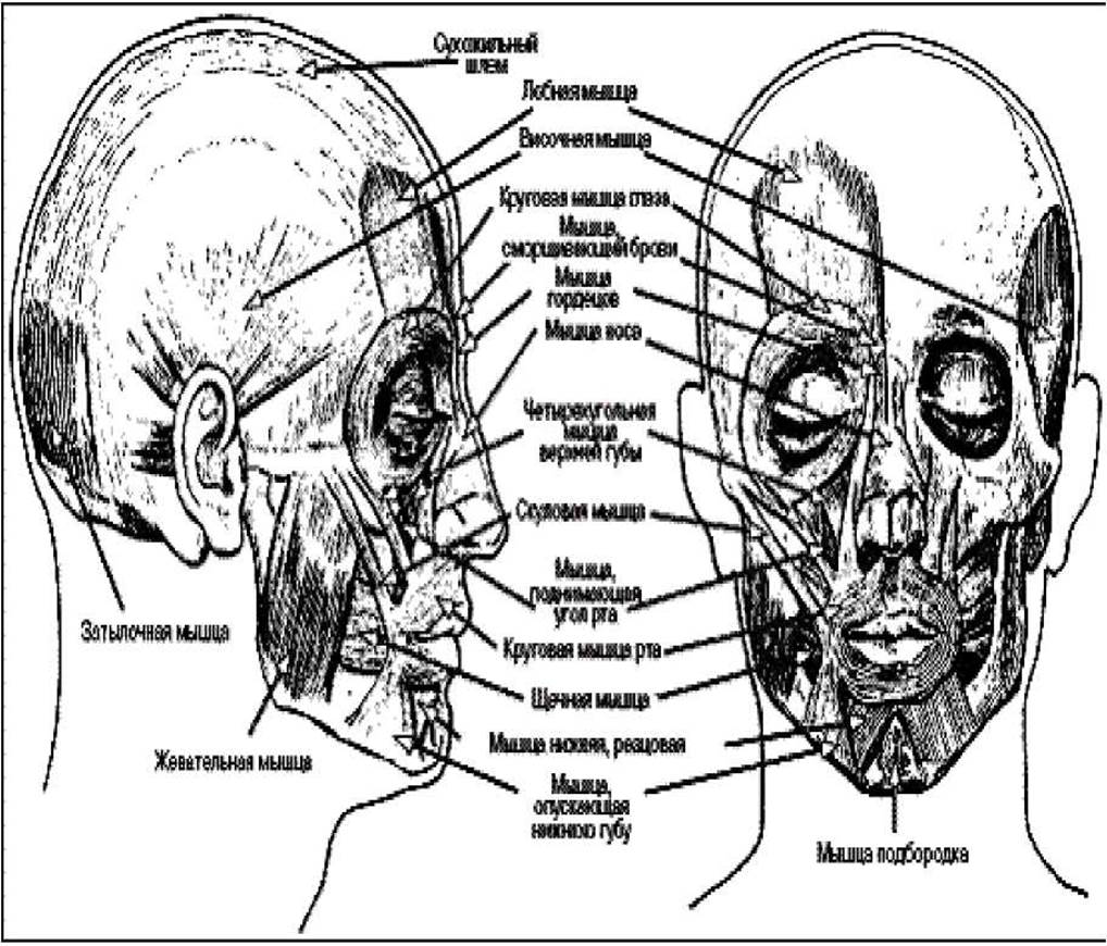
Figure 19. Muscles of the head.
Last update: 08/08/2026
Editorial and Educational Adaptation: This material has been compiled based on the primary/original source text. The project team performed an editorial review, corrected technical inaccuracies, structured sections, and adapted the content for an educational format.
What was processed:
- elimination of formatting defects (OCR errors, structural breaks, corrupted characters);
- editorial organization of content;
- standardization of terminology in accordance with academic sources;
- verification of factual statements against the original source text.
All mentions of the author, publication year, and origin of the primary text have been preserved in accordance with the source.