HUMAN ANATOMY ATLAS - G.L. Bilich - 2014
Muscular System
STRUCTURE AND CLASSIFICATION OF MUSCLES
A Muscle as an organ consists of bundles of striated muscle fibers. Striated (skeletal) Muscle tissue (textus muscularis striatus, s. skeletalis) is formed by cylindrical muscle fibers ranging from 1 to 40 mm in length and up to 0,1 mm in thickness. Each fiber is a complex consisting of a myosymplast and myosatellitocytes covered by a common sheath — the sarcolemma (from Greek sarcos — flesh), which is reinforced with fine Connective Tissue fibers and appears as a thin dark band under light Cell/15.html">Microscopy (Fig. 216). Beneath the sarcolemma of the muscle fiber lie numerous ellipsoid-shaped nuclei containing one or two nucleoli and A large number of granular Endoplasmic reticulum elements. Centrioles are absent. Approximately 2/3 of the dry mass of the myosymplast consists of cylindrical myofibrils running through the Cytoplasm (sarcoplasm).
Muscle fibers exhibit transverse striation: dark anisotropic bands (A-bands) alternate with light isotropic bands (I-bands). The A-band is divided by a lighter zone (H-band), in the center of which runs the mesophragma (M-line). The I-band is divided by a dark Z-line (telophragma — Amici disks (syn.: Krause transverse lines, Dobie lines, Z-lines, telophragms, telophragmae) (Amici, Giovanni Battista, 1786–1863 — Italian botanist, optician, and microscopy scientist, inventor of the Microscope immersion objective; Dobie, William Murray, 1828–1915 — English physician and anatomist)). Muscle fibers contain contractile elements — myofibrils, among which a distinction is made between thick (Myosin) filaments, 10–15 nm in diameter and 1,5 µm in length, occupying the A-band, and thin (Actin) filaments, 5–8 nm in diameter and 1 µm in length, located in the I-band and attached to the telophragms. The section of a myofibril located between two telophragms represents a sarcomere — the contractile unit, approximately 2,5 µm in length (Fig. 217).
Each myofibril is surrounded by an agranular sarcoplasmic reticulum consisting of reticular and tubular elements. The former surround the central part of the sarcomere as an openwork mesh, while the latter encompass most of the sarcomere as parallel tubules and are located on both sides of the reticular elements. The tubular elements of The endoplasmic reticulum transition on both sides of the A-band into terminal cisternae. At the boundary between the A- and I-bands, the sarcolemma invaginates to form T-tubules (transverse tubules), which branch within the fiber and anastomose exclusively in a horizontal direction.
Class="center">
Fig. 216. Striated muscle tissue
Openings of the T-tubules are visible On the surface of the sarcolemma. Two terminal cisternae and a transverse tubule contact one another, forming triads. The networks surrounding the sarcomeres communicate with each other.
Small polygonal groups of myofibril bundles, separated by myofibril-free sarcoplasm and visible on cross-sections of skeletal and cardiac muscle fibers under a Light Microscope, are termed Cohnheim's fields (syn. myofibrillar fields) (Cohnheim, Julius Friederich, 1839–1884 — German pathologist).
Each striated fiber is covered externally to the sarcolemma by a delicate connective tissue sheath — the endomysium (endomysium). Bundles of fibers of various sizes are surrounded and separated from each other by thin layers of connective tissue that form the internal perimysium (perimysium internum). The entire muscle is covered by the external perimysium, or epimysium (perimysium externum), which, together with the connective tissue structures of the endomysium and internal perimysium, transitions into a tendon (tendo) that attaches to the bone (Fig. 218).

Fig. 217. Striated muscle fibers (A — two myofibrils of a muscle fiber [diagram], B — myofibrils, scanning electron micrograph, C — myofibrils, transmission electron micrograph)
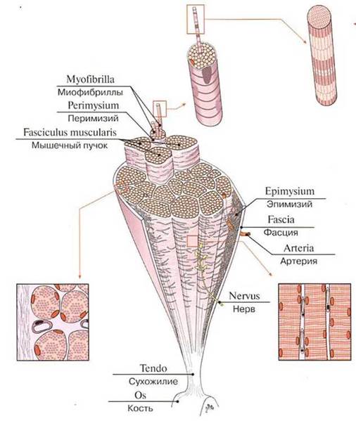
Fig. 218. Structure of a Skeletal Muscle
Tendons are extremely strong and withstand tremendous loads. The tensile strength of a tendon reaches 6–10 kg/mm2. For example, the tendon of the quadriceps femoris can withstand a stretching force of 600 kg, and the calcaneal tendon (Achilles tendon) can withstand 400 kg. This is ensured by dense regular Fibrous connective tissue forming the tendons, which consist of parallel bundles of Collagen fibers interspersed with tendon Cells — tendinocytes and fibroblasts. Tendinocytes (cellulae tendineae) feature an elongated Nucleus and a small amount of cytoplasm. These cells are equipped with processes that penetrate between the tendon fibers (fibrae tendineae). First-order collagen fiber bundles are enveloped by loose fibrous irregular connective tissue (endotendineum). The connective tissue internal peritendineum (peritendineum) surrounds several first-order connective tissue bundles (fasciculi tendineae), forming second-order bundles. The tendon is covered externally by the external peritendineum — a sheath made of Dense Fibrous Connective tissue. Blood Vessels (capillaries) and nerve fibers pass through the connective tissue layers between the tendon fibers.
Muscle bundles form the muscle belly (venter), which transitions into its tendon (tendo). The proximally located muscle HEAD (caput) originates on one bone, while the distal end — the tendon, or tail (cauda) — attaches to another bone. Here, the connective tissue fibers of the muscle or tendon firmly fuse with the periosteum (or perichondrium) and even penetrate into the bone (Sharpey's fibers of the periosteum) (Fig. 219).

Fig. 219. Origin and insertion of a muscle (diagram)
It is generally accepted that THE ORIGIN OF a muscle is located more proximally (or closer to the median axis of the body) than its insertion point, which is located more distally (or farther from the median axis of the body). The origin of a contracting muscle remains stationary; this is its fixed point (punctum fixum). On the other bone to which the muscle attaches is the movable point (punctum mobile). When the muscle contracts, it changes its position. During certain movements, the fixed and movable points exchange roles.
The tendons of various Muscles differ in structure. For instance, limb muscles typically transition into narrow and long tendons. A broad and flat tendon — a tendinous expansion, or aponeurosis (aponeurosis) — is characteristic of muscles involved in forming the walls of Body Cavities. Some muscles have two bellies and are termed digastric muscles. When a muscle features several intermediate tendons along its length, they are called tendinous intersections (intersectiones tendineae) (e.g., the rectus abdominis muscle).
Classification of Muscles. Muscles are categorized based on their Location, shape, direction of muscle fibers, relationship to joints, and function (Table 35).
Table 35. Classification of muscles
Muscle categorization |
Muscle categorization |
By shape: |
By function: |
fusiform |
flexor |
quadrate |
extensor |
triangular |
rotator-levator |
ribbon-like |
constrictor (sphincter) |
circular |
abductor |
By number of heads: |
adductor |
biceps |
By location: |
triceps |
superficial |
quadriceps |
deep |
By direction of muscle fascicles: |
medial |
unipennate |
lateral |
bipennate |
By number of bellies: |
multipennate |
digastric |
Muscles are subdivided into superficial (located subcutaneously) and deep, medial and lateral, external and internal (situated on the corresponding sides of the limbs or within body cavity walls).
Muscles vary significantly in shape and structure (Fig. 220). Fusiform and ribbon-like (strap-like) muscles are the most common. Fusiform muscles typically occur in the limbs, where they attach to bones that function as long and short levers (e.g., the biceps brachii, the abductor pollicis longus, etc.). Ribbon-like muscles appear as wide, thin muscular sheets (for instance, the latissimus dorsi and the rectus abdominis). These muscles primarily contribute to forming the walls of the trunk, abdominal, and thoracic cavities. The muscle bundles of both fusiform and ribbon-like muscles run parallel to their long axis.
Skeletal muscle architecture is diverse. If muscle bundles lie on one side of a tendon to which they attach at an angle, the muscle is termed unipennate, or semipennate (musculi unipennati, s. musculi semipennati), such as the vastus medialis. When muscle bundles attach to a tendon from both sides, the muscle is bipennate or pennate (musculi bipennati, s. musculi pennati), as seen in the rectus femoris. In multipennate muscles (musculi multipennati), muscle bundles intertwine and attach to the tendon from multiple directions (for example, the deltoid muscle).
Certain muscles consist of multiple parts, featuring two, three, or four heads, or several tendons ("tails"). Muscles with two or more heads originate from adjacent bones or from different points on a single bone. These heads subsequently converge to form a common muscle belly and a common tendon. Such muscles are named accordingly based on their structure (biceps, triceps, quadriceps). Conversely, a single common belly may give rise to several tendons that attach to different bones (e.g., the flexor digitorum longus). In some muscles, the constituent muscle bundles are arranged in a circular pattern. These circular muscles typically surround natural body orifices (such as the Mouth and anus) and function as sphincters (musculus sphincter).

Fig. 220. Muscle shapes:
I — fusiform muscle; II — unipennate muscle; III — bipennate muscle; IV — biceps muscle; V — multipennate muscle; VI — triangular muscle; VII — circular muscle; VIII — digastric muscle; IX — broad muscle with an aponeurosis; X — serratus muscle; XI — quadrilateral muscle; XII — muscle with tendinous intersections; 1 — tendon; 2 — muscle belly; 3 — head; 4 — aponeurosis; 5 — tendinous intersection
The spatial relationship between muscles and joints varies, dictated by their structure, topography, and function. Some muscles attach to adjacent bones and act across a single joint only; these are uniarticular muscles. Other muscles span two or more joints, functioning as biarticular and multiarticular muscles. Multiarticular muscles are generally longer than uniarticular ones and lie more superficially—typically originating on the BONES OF THE forearm or leg and inserting onto the bones of the hand or FOOT, or the Phalanges. Additionally, certain muscles originate and insert on bones that do not articulate via joints (e.g., the stylohyoid and mylohyoid muscles). Facial muscles and the Muscles of the Perineum also fall into this category.
Accessory apparatus of muscles. Muscles possess numerous accessory structures, including fasciae, fibrous and synovial tendon sheaths, synovial bursae, and muscular pulleys. Fascia (from Latin fascia meaning "band") is a connective tissue sheath that envelops a muscle (Fig. 221). Formed of dense regular connective tissue, fasciae feature bundles of collagen fibers arranged into a network that maximally resists tension. Fasciae compartmentalize individual muscles and muscle groups, perform mechanical Functions, and facilitate Muscle Action by providing support to the muscle belly during contraction while reducing mutual friction. Fascial sheaths also prevent the spread of purulent-inflammatory processes beyond a single fascial compartment (Fig. 222). Typically, muscles connect to fasciae via loose irregular connective tissue; however, some muscles originate directly from fascia and are firmly fused with them (as seen in the leg and forearm).
Fasciae are categorized into superficial, deep, and proper fasciae (Fig. 223). The superficial fascia runs continuously beneath the subcutaneous tissue, separating it from the underlying muscles. The thin proper fascia completely encloses its designated muscle. When muscles are arranged in multiple layers, a deep fascia (layer) lies between adjacent muscles. In many regions, fasciae attach to bones, extending intermuscular septa that partition groups of muscles with distinct functions.
Fascial nodes are localized thickenings formed at the junctions where fasciae intersect. They reinforce the fascial sheaths of Vessels and nerves, protecting them from compression. By blending firmly with bones, fasciae Complement the skeletal framework, forming a soft scaffolding or soft Skeleton.

Fig. 221. Muscle fascia:
1 — Biceps brachii; 2 — Medial intermuscular septum of arm; 3 — Triceps brachii; 4 — Humerus; 5 — Lateral intermuscular septum of arm; 6 — Brachialis; 7 — Fascia of individual muscle; Muscle sheath; 8 — Brachial fascia; 9 — Skin
The structure of fasciae depends on Muscle Function and the mechanical loads experienced during contraction. In areas of well-developed musculature, fasciae are denser and resemble broad tendinous sheets (e.g., the fascia lata of the thigh, the crural fascia). Conversely, muscles subject to lighter loads are surrounded by looser fascia. Where fasciae pass over neurovascular bundles, they thicken to form tendinous arches (arcus tendineus). In the ankle and wrist regions, thickened fasciae attach to bony prominences to form retinacula (retinaculum) that secure tendons and muscles. The underlying spaces—composed of osteofibrous and fibrous sheaths or canals (Vagina fibrosa tendinis)—transmit the tendons. In some cases, a fibrous sheath encloses multiple tendons, whereas in others, each tendon possesses its independent sheath. Retinacula prevent tendons from lateral displacement During Muscle contraction.

Fig. 222. Osteofascial and fascial sheaths of muscles in the lower third of the right thigh:
1 — Medial femoral intermuscular septum; 2 — Femur; Thigh bone; 3 — Fascia lata; 4 — Lateral femoral intermuscular septum; 5 — Sciatic nerve; 6 — Femoral artery and vein

Fig. 223. Superficial and deep Fascia of the arm, anterior view:
1 — Lateral intermuscular septum of arm; 2 — Humerus; 3 — Brachial fascia; 4 — Brachial fascia, superficial part; 5 — Subcutaneous tissue; 6 — Skin; 7 — Biceps brachii; 8 — Brachialis; 9 — Medial intermuscular septum of arm; 10 — Triceps brachii
A synovial sheath (vagina synovialis tendinis) isolates a moving tendon from the stationary walls of its fibrous sheath, eliminating friction between them. The synovial sheath is a closed, slit-like cavity containing a minimal amount of fluid, enclosed by the visceral and parietal layers of the synovial membrane (Fig. 224). The visceral (inner) layer closely surrounds and fuses with the tendon on all sides. The parietal (outer) layer lines and fuses with the walls of the fibrous sheath. These two layers are continuous with each other at the extremities of the sheath and along the inner, bone-facing side of the tendon. The double-layered fold connecting the inner and outer layers is called the tendon mesentery, or mesotendineum (mesotendineum), which transmits blood vessels and nerves supplying the tendon.
When a tendon moves within its fibrous sheath, the inner layer glides smoothly along the outer layer, facilitated by the fluid contained within the slit-like cavity of the synovial sheath. A synovial sheath may enclose one or several tendons if they share the same canal, and adjacent sheaths can occasionally communicate with one another.
Synovial bursae are located in regions where a tendon or muscle crosses a bone or an adjacent muscle, or where two tendons contact each other, functioning similarly to synovial sheaths to reduce friction. A synovial bursa (bursa synovialis) is a flattened sac containing a small amount of synovial fluid. The outer surface of its walls fuses with moving structures (such as muscles or periosteum). Bursae range in size from a few millimeters to several centimeters, and some communicate directly with joint cavities. Besides these deep bursae, superficial (subcutaneous) bursae occur in areas subject to frequent friction or compression—for example, mucous bursae located beneath muscle tendons near the anterior superior iliac spine and the ischial tuberosity; the prepatellar subcutaneous bursa; the dorsal subcutaneous metacarpophalangeal bursae; and the subcutaneous olecranon bursa, among others. Occasionally, a synovial bursa is interposed between a tendon and a muscular pulley, the latter acting as a bony or cartilaginous prominence.
A pulley (trochlea) ensures a consistent line of pull for a tendon by serving as a fulcrum. Additionally, this prominence increases the angle between the bone and the attaching tendon, thereby enhancing the angle of force application to the bony lever.
Sesamoid bones develop within certain tendons near their insertion onto bone (e.g., the Patella). One surface of a sesamoid bone typically faces a joint cavity. Connected to tendons and linked to adjacent bones via ligaments, sesamoid bones elevate the tendon, shifting it away from the bone. This mechanism reduces friction between the tendon and the bone while altering the moment of force, or torque. In other words, the functional roles of a pulley and a sesamoid bone are largely analogous.

Fig. 224. Synovial tendon sheath (A — cross section, B — longitudinal section)
Last update: 08/08/2026
Editorial and Educational Adaptation: This material has been compiled based on the primary/original source text. The project team performed an editorial review, corrected technical inaccuracies, structured sections, and adapted the content for an educational format.
What was processed:
- elimination of formatting defects (OCR errors, structural breaks, corrupted characters);
- editorial organization of content;
- standardization of terminology in accordance with academic sources;
- verification of factual statements against the original source text.
All mentions of the author, publication year, and origin of the primary text have been preserved in accordance with the source.