ATLAS OF HUMAN ANATOMY - G.L. Bilich - 2014
Muscular System
Muscle tissue (textus muscularis), possessing The ability to contract and shorten, performs movement Functions. There are three varieties of muscle tissue: striated (skeletal), cardiac striated, and unstriated (smooth). Alongside these varieties, The Human Body features muscle tissue of epidermal origin (myoepithelial Cells) and neural origin (myocytes of the sphincter and dilator pupillae Muscles) (Table 34).
Skeletal muscles set bones into motion, actively alter body posture, contribute to The formation of the walls of the oral, thoracic, and abdominal cavities, as well as the pelvis, and are Components of the walls of the Pharynx, upper Esophagus, and Larynx. They execute Movements of the Eyeball and auditory ossicles, alongside breathing and swallowing actions. Skeletal muscles maintain the body in an upright position, ensuring its balance and locomotion in space.
Humans have approximately 400 striated muscles, the majority of which are paired (Fig. 214, 215). They contract voluntarily in response to impulses transmitted via peripheral nerves from the Central Nervous system.
The total mass of skeletal musculature in an adult accounts for 30—40% of body weight, and in a newborn, 20—22%. In elderly and old individuals, Skeletal Muscle mass decreases slightly (25—30%), usually associated with a decline in physical activity. With high muscle activity, muscle mass is preserved into deep old age.
Class="center">Table 34. Muscle Tissues
Type |
Varieties |
Characteristics |
Striated muscle tissues |
Skeletal muscle tissue |
Cylindrical muscle fibers ranging from 1 to 40 mm in length and up to 0,1 µm in thickness. Each fiber is a complex consisting of a myosymplast and myosatellite cells covered by a common basement membrane. Numerous nuclei lie beneath the sarcolemma. Approximately 2/3 of the dry mass consists of cross-striated myofibrils |
Cytology/cytology/34.html">Cardiac muscle tissue |
Muscle cells known as cardiomyocytes, forming complex structures. Cardiomyocytes are elongated cells (up to 100-150 µm long and 10-20 µm thick) containing 1-2 nuclei surrounded by cross-striated myofibrils. Adjacent cardiomyocytes are interconnected by intercalated discs |
|
Smooth (unstriated) muscle tissues |
Smooth muscle tissue of Blood Vessels and Internal Organs |
The structural element is the smooth muscle Cell, or myocyte, which is spindle-shaped, 20-500 µm long, 5-8 µm thick, and lacks cross-striations. These cells are arranged in groups and contain longitudinally oriented myofilaments |
Muscle tissue of ectodermal origin |
Myoepitheliocytes (of sweat, sebaceous, mammary, and lacrimal glands) |
Multipolar cells whose Cytoplasm contains contractile filaments. These cells wrap around the secretory end-pieces of glands with their branching processes |
Myoneurocytes |
Smooth myopigmentocytes form two muscles — the sphincter and dilator pupillae |

Fig. 214. Muscles of the human body, anterior view:
1 — Abductor hallucis; 2 — Soleus; 3 — Gastrocnemius; 4 — Extensor digitorum longus; 5 — Tibialis anterior; 6 — Extensor pollicis brevis; 7 — Abductor pollicis longus; 8 — Extensor carpi radialis brevis; 9 — Extensorcarpi ulnaris; 10— Flexor capi radialis; 11 — Extensor digitorum; 12 — Brachioradialis; 13 — Extensor carpi radialis longus; 14 — Brachialis; 15 — Triceps brachii; 16 — Biceps brachii; 17 — Serratus anterior; 18— Deltoid; 19 — Trapezius; 20 — Sternocleidomastoid: 21 — Masseter; 22 — Depressor anguli oris; 23 — Zygomaticus major; 24 — Temporalis; Temporal muscle; 25 — Orbicularis oculi; 26 — Occipitofrontalis, frontal belly; 27 — Orbicularis oris; 28 — Mentalis; 29 — Sternohyoid; 30— Extensor carpi ulnaris; 31 — Extensor digiti minimi; 32— Flexor carpi ulnaris; 33—Anconeus; 34— Pectoralis major; 35— Rectus abdominis; 36 — Extemal oblique; 37 — Tensor fasciae latae; Tensor of fascia lata; 38 — Iliopsoas; 39 — Pectineus; 40 — Pyramidalis; 41 — Adductor longus; 42 — Vastus lateralis; 43 — Rectus femoris; 44 — Sartorius; 45 — Gracilis; 46 — Vastus medialis; 47 — Flexor digitorum longus; 48 — Extensor digitorum longus; Tcndon
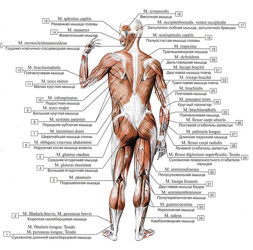
Fig. 215. Muscles of the human body, posterior view:
1 — Fibularis longus; Tendon; Peroneus longus; Tendon; 2 — Fibularis brevis; Peroneus brevis; 3 — Plantaris; 4 — Gluteus maximus; 5 — Gluteus medius: 6— Extemal oblique; 7— Latissimusdorsi; 8— Serratus anterior; 9— Teres major; 10— Infraspinatus; 11 — Teres minor; 12— Brachioradialis; 13 — Sternocleidomastoid; 14 — Masseter; 15 — Splenius capitis; 16 — Temporalis; Temporal muscle; 17 — Occipitofrontalis, occipital belly; 18 — Semispinalis capitis; 19 — Trapezius; 20 — Deltoid; 21 — Biceps brachii; 22 — Triceps brachii; 23 — Brachialis; 24 — Pronator teres; 25 — Brachioradialis; 26 — Flexor carpi ulnaris; 27 — Palmaris longus; 28 — Flexor carpi radialis; 29 — Flexor digitorum superficialis, Tendon; 30— Semitendinosus; 31 — Biceps femoris; 32— Semimembranosus; 33 — Gastrocnemius; 34 — Soleus
Last update: 08/08/2026
Editorial and Educational Adaptation: This material has been compiled based on the primary/original source text. The project team performed an editorial review, corrected technical inaccuracies, structured sections, and adapted the content for an educational format.
What was processed:
- elimination of formatting defects (OCR errors, structural breaks, corrupted characters);
- editorial organization of content;
- standardization of terminology in accordance with academic sources;
- verification of factual statements against the original source text.
All mentions of the author, publication year, and origin of the primary text have been preserved in accordance with the source.