Practical Guide to Zoology: A Textbook - T. A. Dauda 2014
Multicellular Animals
Annelids
Class Leeches
Medical leech (Hirudo medicinalis).
The medicinal leech is an inhabitant of freshwater bodies (swamps, ponds, lakes, backwaters). It feeds exclusively on Blood, attacking humans and mammalian animals that visit its habitat. With each feeding, it is capable of sucking a significant amount of blood. It lays its egg cocoons in the soil along the shoreline, from which tiny leeches emerge. The juveniles feed on the blood of fish and amphibians. The medicinal leech is of great practical significance: it is used in human and veterinary medicine in a living state as a therapeutic agent, and also serves as a valuable raw material for The production of the drug hirudin. In medicine, it is employed in almost all fields—therapeutics, gynecology, surgery, dermatology, ophthalmology, and others.
Materials and equipment. Due to its widespread medical use, medicinal leeches can be purchased at a pharmacy. However, they are also easy to collect in the wild. In the warm season, leeches can be readily caught using a simple hand net in stagnant Water bodies with muddy bottoms. Medicinal leeches survive well in captivity if kept in a cool place within an opaque container tightly covered with a cloth. It is beneficial to place a small amount of horsehair in the water, against which the leeches can clean off their mucus. Maintaining 10 worms requires at least 1 L of water; the water should be changed every 2–4 days, and the container thoroughly washed. Leeches are capable of fasting for long periods. Nevertheless, when kept long-term, they must be periodically fed with clots of fresh blood (obtained from a slaughterhouse) or provided with a live frog placed in the container for a time.
Before studying the external anatomy or performing a dissection, as well as prior to fixation for histological purposes, leeches are killed by immersion in weak (10–15%) alcohol or an aqueous solution of chromic acid. Alternatively, a small amount of chloroform can be added to the water to anesthetize the leeches. Initially, the animal swims actively and regurgitates blood, after which it calms down and becomes motionless. Following this, the abundant mucus covering the body should be thoroughly wiped off with a towel. The Study of external Morphology is best conducted on freshly killed specimens. The methodology for preparing histological sections, fixing them, and staining is provided in the “Large Practical Course on Invertebrate Zoology” (see the Recommended reading list).
Required equipment and tools: Microscope; dissecting magnifier; hand lens; sheets of white paper; dissection instruments (tray, scalpel, scissors, safety razor blade, forceps, dissecting needles, pins); jars containing live adult medicinal leeches (both starved and engorged); a wet mount of a dissected medicinal leech showing the Digestive System; a wet mount of a dissected leech showing the reproductive, excretory, and nervous systems; leeches killed with ether or 10% alcohol.
Assignment. Examine and sketch the External Structure of the medicinal leech. Dissect the leech and examine its digestive, nervous, circulatory, reproductive, and excretory systems.
Observation of a living specimen. Observe the live leeches kept in jars of water. Place a leech on a piece of paper and observe its locomotion patterns and changes in body shape. Compare the body shapes of leeches swimming versus resting at the bottom of the jar, as well as starved versus engorged specimens. The length of a medicinal leech reaches up to 10–20 cm. Note that the body is somewhat flattened in the dorsoventral direction (Fig. 66). However, body length and shape can vary significantly due to the contraction of its highly developed musculature.
Examine the coloration of the leeches: the main coloration is olive-green or greenish-gray, though Various Forms of the medicinal leech (such as the apothecary or medicinal varieties) exhibit different shades. Reddish-yellow stripes run along the greenish-gray dorsum (a characteristic feature of medicinal leeches). The ventral side varies in color among different forms. The exterior of the leech's body is covered by a cuticle that is periodically shed. At the bottom of the jar containing the leeches, one can sometimes notice the shed cuticle appearing as a brownish-white film.
Class="center">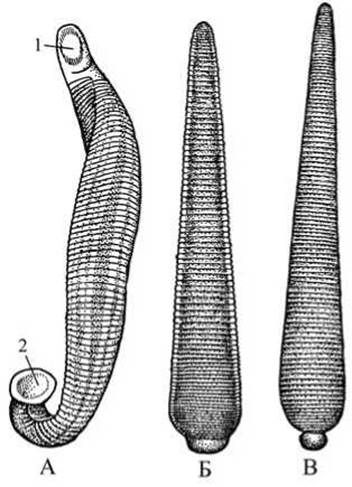
Fig. 66 Leeches:
A — medicinal (Hirudo medicinalis): 1 — oral aperture within the anterior sucker; 2 — posterior sucker; B — horse leech (Limnatis nilotica); C — European medicinal horse leech (Haemopis sanguisaga).
Note that The surface of the leech's body is moist and slippery; this is because numerous Skin glands located in the epithelium beneath the cuticle secrete a large amount of mucus. It is clearly visible that the body of leeches is composed of rings. The external annulation does not correspond to the internal segmentation: there are 102 external rings in total, whereas the number of internal segments is 26. Secondary annulation provides body flexibility.
Locate the anterior sucker with the Mouth opening at the anterior end of the body. At the posterior end, the larger posterior sucker is visible, which serves as an organ of attachment. Using a magnifying Glass, locate 5 pairs of eyes on the DORSAL SIDE OF the anterior end, which are especially distinct in pigmented specimens.
On the ventral side, closer to the anterior end, locate the two genital pores—male and female (situated posteriorly). The anus, located on the dorsal side near the Base of the posterior sucker, is difficult to discern even with a magnifying glass. The openings of the excretory Organs (on the ventral side of the body) are faintly distinguishable. Parapodia and setae are absent in leeches.
Sketch the external Anatomy of the leech from the dorsal view: indicate the eyes, posterior sucker, and annulation.
Dissection (Fig. 67). Study the Internal Organs concurrently on a dissected leech and using wet preparations. Place the ether-anesthetized leeches into dissecting trays, wipe them free of mucus, and pin them down with two pins, ventral side down, while attempting to stretch the body lengthwise. Insert the pin anchoring the anterior end slightly off the body's midline so as not to damage the supraesophageal ganglion and the leech's jaws. Insert the other pin into the posterior sucker.
Make an incision along the dorsal side of the body using a sharp scalpel or safety razor blade (do not use scissors!). The incision must not be too deep to avoid damaging the gut wall. If the leech has ingested a large amount of blood, it must be squeezed out through the incisions in the intestine by running the handle of the scalpel along the body, and the water in the dissecting tray should be changed several times. Evert the edges of the incision outwards—unlike the dissection of an earthworm, this cannot be done immediately; they must be carefully separated from the gut using the tip of the scalpel while constantly lifting the body wall with forceps. Beneath the cuticle lies a thin epithelium, followed by circular, longitudinal, and dorsoventral Muscles. The body cavity of leeches is a primary body cavity (pseudocoelom). Unlike earthworms, it is filled with parenchyma; only remnants of the secondary body cavity (coelom) persist in the form of lacunae and clefts between the organs.
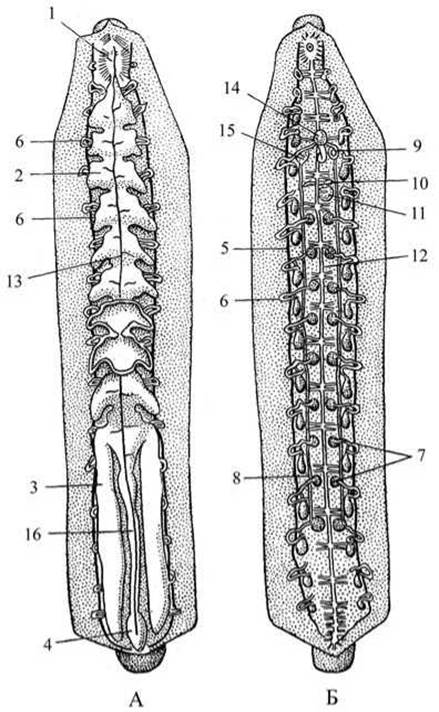
Fig. 67 Dissected medicinal leech:
A — digestive system; B — excretory, reproductive, and nervous systems: 1 — Pharynx; 2 — lateral caeca of the intestine; 3 — tenth pair of lateral intestinal caeca; 4 — hindgut; 5 — lateral lacuna; 6 — metanephridial tubules; 7 — Testes; 8 — vas deferens; 9 — copulatory organ; 10 — Ovary; 11 — Vagina; 12 — ventral nerve cord; 13 — dorsal lacuna; 14 — Prostate Gland; 15 — epididymides (testicular appendages); 16 — intestine.

Fig. 68 Jaw apparatus of the medicinal leech:
A — anterior end of the body from the ventral side; B — wound in human skin inflicted by a leech; C — anterior end of the body dissected from the ventral side; D — LATERAL VIEW OF a jaw at high magnification; 1 — oral sucker; 2 — mouth opening; 3 — jaws; 4 — pharynx; 5 — Esophagus.
Respiration in leeches occurs through the skin, which is richly supplied with a capillary network.
Carefully teasing away the parenchyma, expose the intestine. Change the water in the tray and proceed to study the digestive system.
Medical leeches are typical hematophages. Adult leeches feed exclusively on the blood of humans and vertebrate animals.
Locate the mouth opening deep within the oral sucker (Fig. 68). Examine three semicircular jaws near the ventral side of the mouth opening, with edges lined by small, sharp Teeth (80–90 in number).
Using its jaws, the leech rapidly punctures—or rather, saws through—the skin of not only humans but also animals (such as cows, horses, and wild boars). The mouth opening leads into a short, muscular pharynx that acts as a pump.
Locate the midgut just behind the pharynx. It develops from the endoderm, whereas the anterior region (mouth opening, pharynx) and the posterior region of the digestive tract (hindgut) are of ectodermal origin.
Note that glands—the so-called "Salivary Glands" that secrete hirudin—are located among the muscles around the pharynx and the anterior part of the midgut. Hirudin prevents Blood Coagulation, allowing the leech to ingest large amounts of blood while causing physiological shifts in the body of the human or animal serving as the temporary host. The ducts of these glands open into the pharynx.
Examine the initial (Stomach) section of the midgut; it forms a narrow, distensible tube divided by constrictions into a series of chambers with lateral pouch-like evaginations. This voluminous and distensible gut, consisting of 10 pairs of caeca, stores a significant amount of blood ingested by the leech. Thanks to hirudin, the blood in this section of the midgut does not coagulate and is not digested; it serves as a blood reservoir, enabling the engorged leech to survive for long periods (up to 1–1.5 years) without food.
Pay attention to the final, 10th pair of caeca. The following, shorter section of the midgut (the absorptive intestine) is separated from The Stomach section by a sphincter, which allows blood to enter it in small portions. Here, the blood clots, is digested, and is absorbed. Locate the hindgut situated posterior to the absorptive intestine, which opens via the anus. This section of the alimentary canal accumulates fecal matter that is periodically expelled into the water.
Make a drawing of the digestive System of the leech.
Parting the Muscle fibers extending from the pharynx to the body wall, use a magnifying glass to locate the whitish suprapharyngeal ganglion. Cut the pharynx anterior to it with scissors and, carefully trimming with a scalpel and teasing with forceps, remove the entire intestine. Change the water in the dissecting dish and proceed to examine the other Organ Systems of the dissected leech. Locate the ventral nerve cord beneath the intestine. By lifting it with a dissecting needle, examine the individual nodes—ganglia with commissures—along the ventral nerve cord.
Unlike in earthworms, the Circulatory system OF the leech is open (lacunar). It consists of two lateral lacunae, a dorsal lacuna, and a ventral lacuna, as well as a complex system of smaller lacunae and sinuses through which blood circulates. Observing the circulatory system without special preparation is difficult. The dorsal lacuna may have been severed during the dissection of the leech, the ventral one contains the nerve cord, and the lateral lacunae can be observed while studying the excretory system.
Locate the excretory organs—17 pairs of nephridia—along the sides of the nerve cord. The nephridia consist of convoluted renal tubules that expand into a Urinary Bladder before reaching the exterior. The nephridial funnels are not visible; they open into the lateral lacunae. The renal tubules and urinary bladders are white, clearly distinguishable, and can be counted using a magnifying glass.
Examine the 9 pairs of whitish, rounded testes clearly visible in the middle region of the body between the nerve cord and the nephridia. The vasa efferentia are not distinguishable. They empty into the vasa deferentia, which run parallel to the ventral nerve cord. The vasa deferentia are white and in many cases stand out clearly in the dissected leech. Near the anterior end of the body, the vasa deferentia coil spirally to form the epididymides and then unite into a common unpaired duct that terminates in the copulatory organ. Below this, a pair of small, spherical Ovaries can be seen, followed by the vagina. The oviducts, extending from the ovaries and joining at the vagina, are not visible.
Examine the preserved mount of the dissected leech with the intestine removed.
Make a drawing of the dissected leech showing the reproductive, nervous, and excretory systems.
Horse leech (Limnatis nilotica).
The horse leech is found in freshwater bodies of Central Asia, Kazakhstan, and Transcaucasia (Armenia, Georgia, Azerbaijan). The horse leech (Limnatis nilotica) is the CAUSATIVE AGENT OF limnatiasis, a disease affecting humans and animals. The infection is severe and often fatal. The Limnatis leech feeds exclusively on blood by attaching to the mucous membranes of its host. Unlike the medicinal leech, Limnatis feeds frequently—sometimes multiple times a day—in small portions. The hosts of Limnatis include humans and animals (horses, mules, donkeys, camels, dogs, and cattle and small ruminants) that it attacks while drinking water.
Most commonly, Limnatis attaches to the mucosa of the Lips, Gums, pharynx, base of the Tongue, palate, inner surface of the Cheeks, and esophageal mucosa. These leeches may also attach around the glottis, in the Larynx, in the nasal passages, and less frequently on the conjunctiva of the eyes.
The Pathogenesis of the disease caused by the horse leech stems, firstly, from the mechanical impact of the parasite and, secondly, from the loss of large quantities of blood resulting from the leech's feeding and the injection of hirudin into the host's bloodstream. When a leech remains in the respiratory tract for an extended period, the engorged parasite obstructs breathing and can cause death by asphyxiation. Blood loss weakens the host Organism. At the sites of parasite attachment, alongside bleeding, edema, Swelling, and hyperemia of the mucous membrane are observed.
Duck leech (Protoclepsis sp.).
Duck leeches (Protoclepsis tesselata and the less common Pr. maculosa) (see Fig. 69) inhabit freshwater, overgrown or overgrown-over water bodies with slow-flowing water. They are dangerous parasites of agricultural waterfowl, attacking ducklings and goslings primarily. The avian disease caused by these leeches is called protoclepsiosis. It is a severe avian affliction that frequently results in death. Affected birds exhibit labored breathing associated with the presence of the leech in the respiratory tract. In cases of heavy infestation, choking fits recur, leading to death. The infected bird frequently shakes its HEAD and sneezes. In some cases, birds experience emaciation, feather loss, and death from exhaustion. When leeches crawl from the nasal cavities deeper toward the Ethmoid bone, running birds fall onto their backs, throw their heads back, and die. When the conjunctival sac of the eye is affected, the eye appears inflamed, half-closed, and watery, and the cornea loses its transparency and turns white. Sometimes the eye increases in volume and protrudes from the Orbit.

Fig. 69 Duck leeches:
A — Protoclepsis tesselata; B — Protoclepsis maculosa; 1 — body extended; 2 — body contracted.
Last update: 13/08/2026
Editorial and Educational Adaptation: This material has been compiled based on the primary/original source text. The project team performed an editorial review, corrected technical inaccuracies, structured sections, and adapted the content for an educational format.
What was processed:
- elimination of formatting defects (OCR errors, structural breaks, corrupted characters);
- editorial organization of content;
- standardization of terminology in accordance with academic sources;
- verification of factual statements against the original source text.
All mentions of the author, publication year, and origin of the primary text have been preserved in accordance with the source.